
Vero Cell Culture Techniques for Viral Diagnostics
Overview and Principles of Vero Cell Culture for Viral Diagnostics
Introduction: The Indispensable Role of Vero Cells in Veterinary Virology
The Vero cell line, an immortalized lineage derived from the kidney epithelium of the African green monkey (Chlorocebus aethiops), is a cornerstone of diagnostic virology, vaccine production, and fundamental virological research. In the context of veterinary clinical pathology, the utility of Vero cells extends far beyond their convenience as an adherent, continuous cell line. Their unique biological properties-most notably their inherent deficiency in interferon (IFN) production-render them exquisitely permissive to a remarkably broad spectrum of viral pathogens, making them an irreplaceable tool for the primary isolation, propagation, and titration of viruses affecting livestock, companion animals, poultry, aquatic species, and wildlife. The fundamental principle underpinning their diagnostic application is the provision of a controlled, reproducible, in vitro environment that recapitulates the essential cellular machinery required for viral replication, thereby enabling the visualization of viral growth through cytopathic effect (CPE), the quantification of infectious particles, and the generation of antigenic material for downstream serological and molecular assays. This section will delineate the historical context, biological foundations, and core methodological principles that govern the use of Vero cells within a contemporary viral diagnostics framework, emphasizing critical considerations for the veterinary clinical pathologist.
Historical Context and Biological Foundations
The establishment of the Vero cell line in 1962 by Yasumura and Kawakita at Chiba University in Japan marked a pivotal moment in cell culture technology. Unlike primary cells, which suffer from finite lifespan, donor variability, and ethical concerns regarding animal use, the Vero line is a continuous, or immortalized, cell line. This immortality arises from spontaneous aneuploidy, allowing for indefinite passage in vitro while maintaining a stable, epithelioid morphology. This stability is paramount for diagnostic reproducibility, as it allows laboratories to maintain a consistent cell substrate over decades, facilitating inter-laboratory standardization and the generation of reference reagents.
The single most important biological attribute of Vero cells for viral diagnostics is their well-characterized defect in the type I interferon (IFN) signaling pathway. Specifically, the Vero cell genome lacks the gene cluster encoding for interferon-beta (IFN-(\beta)) and other related cytokines. In normal cells, viral infection triggers the production of IFN, which binds to neighboring cells, inducing an antiviral state that limits viral spread. The Vero cell's inability to produce IFN removes this crucial host defense mechanism, creating a uniquely susceptible environment for viral replication. This "IFN-blindness" is a double-edged sword: while it permits the growth of viruses that would otherwise be sensitive to the host interferon response-such as Rabies Lyssavirus, African Swine Fever Virus, and many arboviruses-it can also promote the selection of genetic variants adapted to this specific cellular context. This adaptation is a critical principle for the pathologist to understand, as it means that a virus isolated in Vero cells may not be entirely genetically representative of the original clinical isolate, a phenomenon documented with [SARS-CoV-2] where cell culture has been shown to induce significant changes detectable at the protein level by shotgun proteomics [1]. Despite this caveat, the loss of the IFN blockade remains the primary reason for the unparalleled sensitivity of Vero cells to a vast array of viral families, including Orthomyxoviridae, Paramyxoviridae, Flaviviridae, Herpesviridae, Rhabdoviridae, Poxviridae, Coronaviridae, and Reoviridae.
Principles of Virus-Host Cell Interaction in Vero Cells
The diagnostic principle rests on the productive infection of a permissive cell. For a virus to be successfully isolated in Vero cells, a series of molecular events must occur: adsorption (attachment to cellular receptors), penetration (entry via endocytosis or membrane fusion), uncoating, genome replication, transcription, translation of viral proteins, assembly, and release. The diagnostic pathologist monitors the culmination of these events, most commonly through the observation of CPE.
CPE is the microscopically visible structural damage inflicted upon the host cell monolayer as a direct consequence of viral replication. The morphology of CPE can be highly informative. For instance, Feline Herpesvirus 1 and Pseudorabies Virus typically induce a rapid, focal rounding and detachment of cells, reflective of lytic replication. In contrast, Bovine Viral Diarrhea Virus and many isolates of Avian Influenza Virus may produce little to no CPE upon primary isolation, necessitating the use of alternative detection methods such as hemadsorption (for orthomyxoviruses and paramyxoviruses) or indirect immunofluorescence. The Lumpy Skin Disease Virus, a poxvirus, presents a unique challenge; while Vero cells may ultimately support its propagation, they have been found to be unsuitable for direct primary isolation from clinical scab material, a finding that highlights the critical difference between a cell line's ability to propagate an adapted virus versus its capacity for initial recovery from a field sample [2]. This underscores the principle that a negative CPE result does not equate to the absence of viable virus.
The Spectrum of CPE and Viral Tropism in Diagnostic Virology
The predictability of CPE is a function of the virus and the specific subclone of Vero cells used. The Vero lineage has diverged into several sub-lines, including Vero (WHO), Vero E6, Vero 76, and Vero B, each with subtly different characteristics. [SARS-CoV-2], for example, replicates more efficiently in Vero E6 cells engineered to express the human angiotensin-converting enzyme 2 (ACE2) and transmembrane serine protease 2 (TMPRSS2) receptors-the Vero-ACE2-TMPRSS2 line-than in parental Vero cells, a modification that became critical for clinical trial endpoints assessing viral viability [3]. Similarly, the isolation of Nipah Virus in Pigs and Crimean-Congo Hemorrhagic Fever Virus requires high-containment BSL-4 facilities, and Vero E6 cells are often the substrate of choice for CPE-based titration (TCID50) and plaque assays, although systematic comparisons have revealed that alternative lines like SW-13 can yield even higher titers for CCHFV under optimized conditions [4].
For veterinary pathogens, the range is immense. Vero cells are the standard for the isolation of Canine Distemper Virus, Equine Arteritis Virus, and many serotypes of Bluetongue Virus. However, the pathologist must be aware of failures. The isolation of Foot-and-Mouth Disease Virus is typically performed in primary bovine thyroid or BHK-21 cells, not Vero, as Vero cells lack the specific integrin receptors for FMDV. This principle of receptor compatibility is fundamental; Vero cells possess the sialic acid and other glycoprotein receptors necessary for many viruses, but not all. The recent development of suspension-based Vero cell infection methods, such as the "cell suspension method" validated for [Zika virus], represents an evolution in technique, allowing for faster and more sensitive isolation by infecting freshly detached cells rather than waiting for a confluent monolayer [5]. This innovation increases the surface area for viral adsorption and accelerates the diagnostic timeline.
Optimization of Culture Conditions and Media
The principles of Vero cell culture for diagnostics demand meticulous control over the cellular environment. Standard practice involves culturing Vero cells in an adherent monolayer within cell culture flasks or multiwell plates, using a basal medium such as Dulbecco's Modified Eagle Medium (DMEM) or Medium 199, supplemented with 5-10% fetal bovine serum (FBS) for growth. However, for viral isolation, the serum concentration is often reduced to 2% or less to slow cell division and prevent overgrowth that can obscure CPE.
The move towards serum-free media (SFM) is a growing principle driven by ethical concerns regarding FBS collection, batch-to-batch variability, and the risk of introducing adventitious agents (e.g., Bovine Viral Diarrhea Virus itself, which can contaminate FBS). Recent research has explored the use of marine microalgae extracts, such as Dunaliella salina and Spirulina platensis, as potential SFM supplements for Vero cell proliferation, which also demonstrated a cytoprotective effect by reducing oxidative stress [6]. This aligns with the broader biotechnological principle of process intensification and standardization, critical for Good Manufacturing Practice (GMP) in vaccine production. In the diagnostic laboratory, the choice of medium can influence the sensitivity of viral detection; specific nutrient blends have been shown to dramatically enhance the yield of viruses like Yellow Fever virus (17DD strain) in primary cells, a principle that likely holds true for Vero-cell-based production as well [7].
Substrate innovation is also a key principle. The use of microcarriers, such as Cytodex 1, in stirred-tank bioreactors has transformed Vero cell culture from a static 2D process to a scalable 3D dynamic system. This was successfully demonstrated for the production of Sheep Pox Virus vaccine (Bakırköy strain), where spinner flask cultures achieved higher cell densities and viral titers than traditional T-flasks, while also extending the productive phase of the culture [8]. For the diagnostic pathologist, this principle of scale-down is equally relevant; developing a 3L scale-down model (SDM) that accurately reflects a 50L production process allows for robust process characterization and troubleshooting, ensuring that the virus isolation protocol is predictable and reliable [9].
Integration with Downstream Detection Modalities
The Vero cell culture system is rarely an endpoint; it is a step in a diagnostic cascade. The principle of the "combined approach" is paramount. A positive CPE requires confirmation. This is achieved through:
- Immunological Detection: Staining of fixed cell monolayers with specific monoclonal or polyclonal antibodies via indirect immunofluorescence (IFA) or immunoperoxidase (IPT) assays. This allows for the specific identification of the viral antigen within the CPE. For example, a CPE in Vero cells from a bovine sample could be caused by Bovine Herpesvirus 1 or Bovine Adenovirus; IFA would distinguish them.
- Molecular Detection: RT-PCR or PCR performed on the cell culture supernatant is often more sensitive than the initial clinical sample. This is dramatically illustrated by the isolation of [Enteroviruses] (e.g., Echovirus 27) from cerebrospinal fluid, where cell culture was positive even when the original sample tested negative by PCR, a phenomenon attributed to the presence of non-infectious particles or inhibitors in the clinical specimen [10]. Similarly, for slow-growing viruses like Infectious Salmon Anemia Virus, combining cell culture with time-monitoring RT-qPCR can accelerate detection from weeks to just five days by identifying a minimal shift in Ct values indicative of replication [11].
- Integrated Methods: The concept of Integrated Cell Culture-Mass Spectrometry (ICC-MS) is a cutting-edge diagnostic principle. Here, Vero cells are infected with an environmental sample (e.g., wastewater). After a defined incubation period, the cell lysate is subjected to tryptic digestion and mass spectrometry. The detection of viral peptides-such as those from Reovirus-confirms that an infectious, replicating virus was present in the original sample, bypassing the need for CPE observation and providing strain-specific identification [12].
Quality Control and Standardization: The Pathologist's Mandate
The reliability of Vero-cell-based diagnostics rests on rigorous quality control. Laboratories must adhere to standard operating procedures that define: (a) passage number limits (typically not exceeding passage 30-50 from the master cell bank to avoid genetic drift); (b) mycoplasma testing (as mycoplasma contamination can completely inhibit viral replication); (c) susceptibility testing using a reference virus of known titer; and (d) the use of appropriate controls (uninfected cells, known positive and negative samples).
A critical principle is the distinction between detecting viral RNA or antigen and detecting infectious virus. Molecular methods like RT-qPCR, while fast and sensitive, cannot differentiate between a viable, replication-competent virion and a degraded or neutralized particle. Viral culture in Vero cells remains the "gold standard" for assessing viability. This is clinically vital in cases of persistent infection (e.g., BVDV persistently infected calves) or for monitoring the effectiveness of antiviral therapy. Studies on [SARS-CoV-2] have conclusively shown that subgenomic RNA (sgRNA) detection is a better surrogate for viability than genomic RNA, but it still does not replace the functional proof of replication provided by a positive Vero cell culture [13]. The careful interpretation of culture results, in conjunction with clinical history and molecular data, is the hallmark of the veterinary clinical pathologist's expertise in this domain.
Methodology and Standard Protocols for Vero Cell Maintenance and Virus Inoculation
Establishment and Characterization of Vero Cell Lineages for Diagnostic Virology
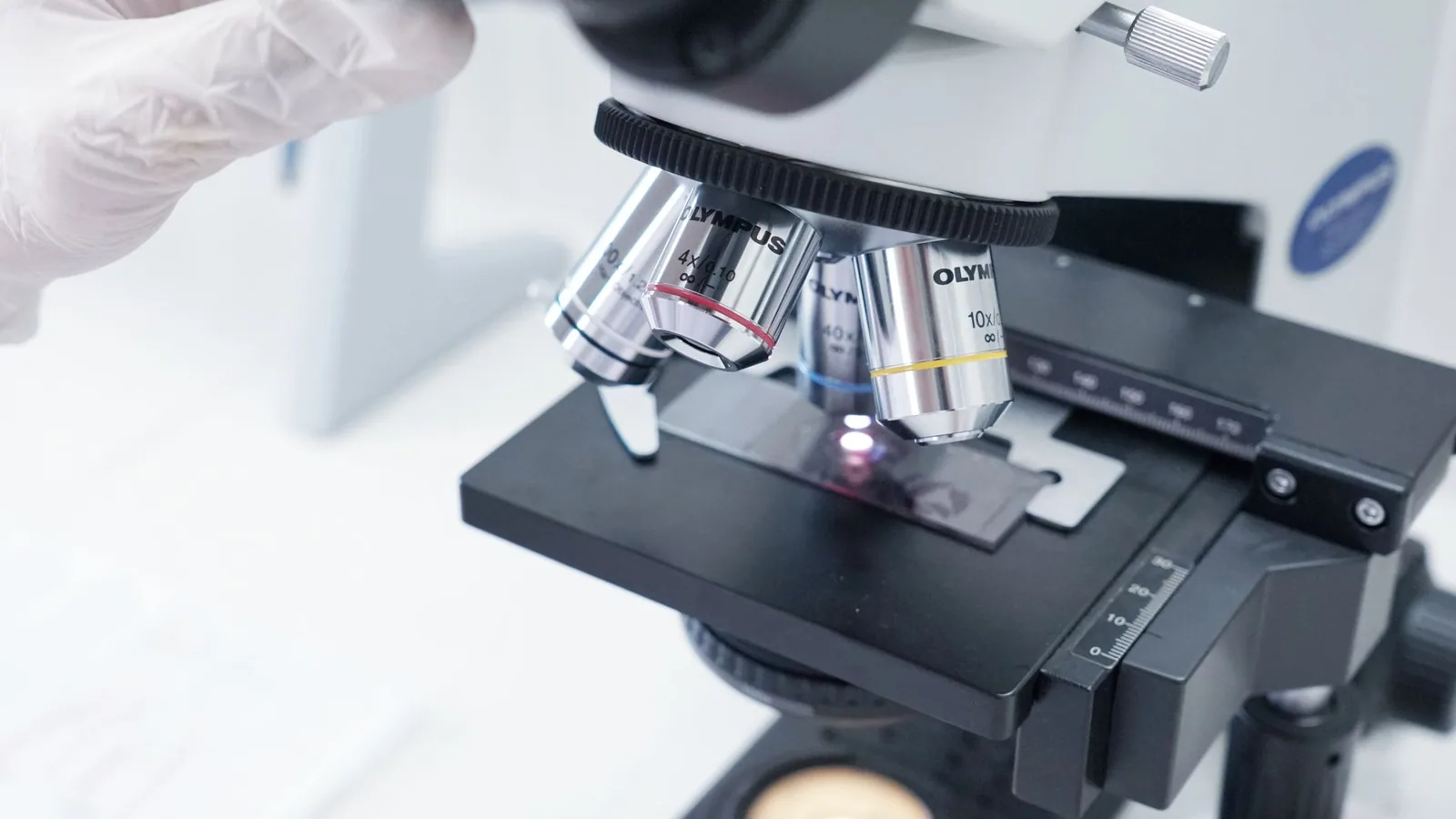
The Vero cell line, derived from the kidney epithelium of the African green monkey (Chlorocebus aethiops), remains the single most widely utilized continuous cell substrate in veterinary diagnostic virology. Its intrinsic advantages-including an immortalized phenotype, a stable karyotype through extended passage, and a documented susceptibility to a remarkably broad spectrum of viral pathogens-render it indispensable for both primary virus isolation and high-titer propagation. Critically, Vero cells harbor a well-characterized deficiency in type I interferon (IFN) production, specifically a deletion within the IFN-(\beta) gene cluster, which renders them uniquely permissive for viruses that are otherwise restricted by innate antiviral responses [14, 15]. This genetic lesion underlies the cell line's exceptional utility for the recovery of fastidious and slow-growing viruses that might be neutralized or suppressed in cell lines possessing an intact interferon signaling cascade.
Several distinct Vero sub-lineages have been established and are employed for specific diagnostic purposes. The Vero E6 clone, also designated Vero C1008 (ATCC CRL-1586), is extensively used for the propagation of highly pathogenic viruses including SARS-CoV-2 and Crimean-Congo Hemorrhagic Fever Virus [13, 4]. The Vero 76 line (ATCC CRL-1587) has been validated for the isolation of [Zika virus] and other arboviruses, with a novel suspension-infection method demonstrating enhanced sensitivity over traditional monolayer approaches for this particular virus [5]. The Vero WHO line, certified for human vaccine production, is the preferred substrate for [Measles virus] and [Rubella virus] propagation, though comparative studies have demonstrated that the reptilian-derived Z1 cell line may yield higher titers for Measles virus specifically [16]. Vero cells expressing human angiotensin-converting enzyme 2 (ACE2) and transmembrane serine protease 2 (TMPRSS2), designated Vero-ACE2-TMPRSS2, have been engineered to support enhanced entry and replication of SARS-CoV-2 variants, particularly the Omicron lineage, which demonstrates reduced fusogenicity in parental Vero E6 cells [3]. This genetic engineering approach underscores the need to match the specific Vero sub-lineage to the target virus under investigation.
Standard Protocols for Vero Cell Maintenance and Subcultivation
The maintenance of a consistent, high-quality Vero cell stock is paramount for reproducible diagnostic outcomes. Cells are typically propagated in Dulbecco's Modified Eagle Medium (DMEM) or Minimum Essential Medium (MEM) supplemented with 5-10% fetal bovine serum (FBS), 2 mM L-glutamine, and non-essential amino acids [8, 6]. The use of serum-free media formulations, incorporating marine microalgae extracts such as Dunaliella salina and Spirulina platensis, has been explored to reduce reliance on animal-derived components, demonstrating comparable or even enhanced proliferation rates and reduced oxidative stress in Vero cells [6]. Standard subcultivation involves enzymatic detachment using 0.05% trypsin-EDTA at 37°C for 3-5 minutes, followed by reseeding at densities ranging from 2-5 × 10⁴ cells/cm². The optimal split ratio for routine maintenance is 1:3 to 1:6, with confluent monolayers typically achieved within 3-5 days at 37°C in a 5% CO₂ humidified atmosphere [9, 17].
For large-scale vaccine and antigen production, microcarrier-based suspension cultures offer a scalable alternative to static monolayer systems. Cytodex 1 microcarriers, when employed in spinner flask bioreactors, support significantly higher cell densities and prolonged viability compared to conventional T-flask cultures [8, 9]. In a systematic optimization for Sheep Pox Virus vaccine production, Vero cells grown on Cytodex 1 in spinner flasks achieved peak titers exceeding those from static cultures, with the dynamic shear forces and improved mass transfer facilitating more efficient viral replication and extended productive phases [8]. Scale-down models (SDM) at the 3 L bioreactor scale, employing a constant impeller power per volume principle, have been qualified to accurately represent 50 L production scales, enabling robust process development and quality by design (QbD) approaches for live-attenuated viral vaccines [9].
Quality Control and Mycoplasma Surveillance
Rigorous quality control is non-negotiable for maintaining the integrity of Vero cell stocks used in diagnostic virology. Cells must be routinely screened for mycoplasma contamination using PCR-based methods or commercial detection kits, as mycoplasma infection can profoundly alter cellular metabolism, induce cytopathic effects (CPEs) indistinguishable from viral infection, and suppress or enhance viral replication in unpredictable ways [18, 19]. All incoming FBS and cell culture reagents must be tested for adventitious viral agents, including Bovine Viral Diarrhea Virus (BVDV), which is a common contaminant of bovine sera. Droplet digital PCR (ddPCR) has been demonstrated to be a highly sensitive primary method for detecting BVDV RNA, with higher specificity at low RNA concentrations compared to conventional RT-qPCR, thereby minimizing the risk of false positives that could compromise cell stock certification [18]. The World Organisation for Animal Health (WOAH) mandates that all cell substrates used for vaccine production and official diagnostic testing be free from extraneous agents, and Vero cells must undergo comprehensive adventitious agent testing as outlined in WOAH Terrestrial Manual chapters.
Virus Inoculation Protocols: Adsorption, Inoculum Preparation, and MOI Optimization
The protocol for virus inoculation must be meticulously tailored to the specific viral agent, the clinical sample matrix, and the intended diagnostic endpoint (primary isolation vs. high-titer propagation). For primary isolation from clinical specimens-including tissue homogenates, swabs, feces, or environmental samples-the inoculum must first be clarified by centrifugation at 1,500-3,000 × g for 15 minutes and filtered through a 0.45 μm syringe filter to remove bacteria and cellular debris [20]. The addition of antibiotics (penicillin 100 U/mL, streptomycin 100 μg/mL, gentamicin 50 μg/mL, and amphotericin B 2.5 μg/mL) to the viral transport medium (VTM) is standard practice to suppress microbial overgrowth [21, 22]. The VTM itself must be validated to preserve viral infectivity; filtered VTM stored at 4°C supports culture-based recovery for up to 48 hours, while autoclave-sterilized medium shows comparable performance [21].
The classical adsorption method involves removing the growth medium from a confluent Vero monolayer, washing the cells once with serum-free medium to remove residual FBS (which may contain trypsin inhibitors or non-specific neutralizing antibodies), and adding 0.2-0.5 mL of inoculum per 25 cm² flask. Adsorption is carried out at 37°C for 60-90 minutes with periodic rocking to ensure uniform distribution. After adsorption, the inoculum is aspirated, and the maintenance medium (typically DMEM with 2% FBS, or entirely serum-free for certain viruses) is added [4, 20]. The use of reduced serum concentrations post-inoculation is critical; high serum levels can inhibit viral adsorption or mask CPE, while serum-free conditions may accelerate the appearance of CPE for cytolytic viruses such as Lumpy Skin Disease Virus (LSDV) [2, 23].
Multiplicity of infection (MOI) optimization is a key determinant of viral yield and quality. For high-titer stock production, a low MOI (0.01-0.1 TCID₅₀/cell) is often preferred to minimize the production of defective interfering particles (DIPs) and to allow multiple rounds of replication [8, 24]. Conversely, for synchronization of infection or for viruses that induce rapid CPE, a higher MOI (0.5-1.0) may be employed. Real-time cell analysis (RTCA) using impedance-based biosensing has provided unprecedented insights into MOI-dependent CPE kinetics. For chimeric yellow fever dengue vaccine strains, the time to median cell index (CITmed) was inversely correlated with viral input, enabling a single-point titration method that reduces assay time from 7 days (CCID₅₀) to 2-3 days [24]. This approach is particularly advantageous for process development where high-throughput screening is required.
Detection of Viral Replication: CPE, Integrated Cell Culture-RT-qPCR, and Alternative Readouts
The hallmark of successful viral inoculation is the observation of CPE, which varies in morphology depending on the virus. Poxviruses, including LSDV and Sheep Pox Virus, induce focal rounding, detachment, and syncytia formation, typically appearing within 3-6 days post-inoculation [2, 8, 25]. Herpesviruses such as Pseudorabies Virus produce rapid cell rounding and ballooning degeneration within 24-48 hours on Vero B cells [26]. For non-cytopathic viruses or those producing subtle CPE, alternative detection methods are mandatory. Integrated cell culture-RT-qPCR (ICC-RT-qPCR) combines the biological amplification of viral replication in Vero cells with the analytical sensitivity of PCR, effectively detecting replicating virus within 5-7 days-a significant improvement over traditional culture methods that may require 14-21 days for slow-growing agents [11]. This approach has been validated for Infectious Salmon Anemia Virus, where a minimal shift in Ct values over time (ΔCt > 2.5 between days 0 and 5) is a robust indicator of active replication, yielding diagnostic sensitivity and specificity estimates exceeding 90% [11].
Subgenomic RNA (sgRNA) detection has emerged as a highly accurate surrogate for cell culture-based viability assessment, particularly for SARS-CoV-2. sgRNA molecules are transcribed only during active viral replication and are rapidly degraded upon cell death, providing a molecular correlate of infectivity that outperforms genomic RNA Ct values in predicting culture positivity [13]. In immunocompromised patients, sgRNA RT-PCR demonstrated a sensitivity of 99% and specificity of 96% against virus isolation as the gold standard, making it a practical tool for clinical decision-making regarding isolation duration and antiviral therapy [13].
Flow cytometry offers a complementary, quantitative approach to assess the proportion of infected cells. Using FITC-labeled anti-rabies immunoglobulin, the percentage of rabies virus-infected Vero cells can be reliably quantified at 24, 48, and 72 hours post-infection, with a sensitivity threshold detecting as few as 6.9% infected cells at high virus dilutions [27]. This automated method surpasses manual CPE scoring in throughput and objectivity, and is particularly valuable for potency testing of vaccines and diagnostic antigens.
Specialized Inoculation Techniques: Shell Vials, Suspension Methods, and Bioreactor Integration
To enhance isolation efficiency, particularly for viruses that are poorly adapted to monolayer culture, the shell vial technique combines centrifugation-assisted inoculation with low-speed adsorption. This method has been shown to accelerate CPE development for human metapneumovirus (HMPV) and enteroviruses, and its adaptation to three-dimensional (3D) spheroid cultures-using adhesion-inhibition on poly-HEMA-coated surfaces-further improves sensitivity by providing a more physiologically relevant architecture that better mimics in vivo tissue organization [28, 29]. For 3D spheroids of Vero and other cell lines, the yield of Adenovirus, cytomegalovirus, and HSV-1 was significantly increased, with CPE appearing 1-2 days earlier than in conventional 2D shell vials [28].
A novel cell suspension method, developed for Zika virus, involves infecting freshly trypsinized Vero cells in suspension prior to seeding, rather than inoculating an established monolayer [5]. This approach eliminates the need for a confluent monolayer at the time of inoculation and has been shown to be fourfold more sensitive than the monolayer method for detecting Zika virus in clinical samples with inconclusive RT-PCR results [5]. The mechanism likely involves the uniform exposure of all cells to the viral inoculum during the adhesion phase, maximizing the probability of productive infection.
Biosafety Considerations and Inactivation Protocols
Given that Vero cells are frequently employed for the isolation of high-consequence pathogens-including SARS-CoV-2, [Monkeypox virus] (MPXV), and Nipah virus-robust inactivation protocols are essential for downstream molecular and serological testing. Heat inactivation at 60°C for 30 minutes achieves >6.0 log₁₀ reduction for both SARS-CoV-2 and MPXV, while commercially available diagnostic buffers and SDS-containing lysis buffers (final SDS concentration ≥0.5%) also provide complete inactivation as confirmed by absence of CPE over 7 days observation [30]. The World Health Organization (WHO) recommends verification of a ≥4 log₁₀ reduction in infectious titer for any inactivation method used in diagnostic workflows. These protocols enable the safe handling of inactivated viral lysates in lower biosafety level (BSL-2) facilities for nucleic acid extraction and antigen detection, provided that the inactivation step is integrated into the standard operating procedure (SOP) and validated against the specific virus-cell system [30].
Molecular Pathogenesis and Mechanisms of Viral Replication in Vero Cells
The Vero cell line, derived from the kidney epithelium of the African green monkey (Chlorocebus aethiops), stands as one of the most permissive and widely utilized substrates in veterinary and medical virology. Its utility extends far beyond simple propagation; Vero cells serve as a critical model for dissecting the molecular pathogenesis of a vast array of DNA and RNA viruses. The intrinsic characteristics of this cell line-particularly its well-characterized deficiency in type I interferon (IFN) production-profoundly shape the replication kinetics, cytopathic effect (CPE) profiles, and host-pathogen interactions observed during infection. Understanding these molecular underpinnings is essential for optimizing viral diagnostics, vaccine production, and antiviral drug screening.
Interferon Deficiency and the Permissive Phenotype
The cornerstone of Vero cell permissiveness lies in a spontaneous genetic deletion that renders them incapable of producing interferon-beta (IFN-(\beta)) and, consequently, other type I interferons [31]. This defect, localized to the IFN-(\beta) gene cluster, cripples the innate antiviral response. In most cell lines, viral infection triggers pattern recognition receptors (PRRs) such as RIG-I and MDA5, leading to the activation of IRF-3 and NF-(\kappa)B, which drive IFN-(\beta) transcription. The secreted IFN-(\beta) then acts in an autocrine and paracrine manner to induce hundreds of interferon-stimulated genes (ISGs) that establish an antiviral state. Vero cells, however, lack this amplification loop. While they retain functional JAK-STAT signaling and can respond to exogenous IFN, their inability to produce endogenous IFN creates a uniquely permissive environment. This has been exploited for the isolation and high-titer propagation of fastidious viruses, including [SARS-CoV-2], Rabies Lyssavirus, and numerous arboviruses [13, 3, 27, 32].
This interferon-deficient state has profound implications for molecular pathogenesis. For instance, infection with Infectious Bronchitis Virus (IBV) in Vero cells leads to a rapid and unopposed activation of NF-(\kappa)B- and AP-1-dependent signaling pathways, as demonstrated by quantitative proteomics using SILAC [31]. This unrestrained activation of pro-inflammatory transcription factors drives the expression of cytokines and chemokines that contribute to the characteristic CPE, including cell rounding, syncytia formation, and detachment. The absence of a robust IFN-mediated negative feedback loop allows viral replication to proceed unchecked, often resulting in higher peak titers than those observed in IFN-competent cells, albeit with potentially altered viral protein processing and host cell shutoff dynamics [31].
Receptor-Mediated Entry and Tropism Determinants
The molecular pathogenesis of viral infection in Vero cells is initiated at the plasma membrane, where specific host receptors dictate viral tropism. The expression of key entry mediators on Vero cells makes them susceptible to a broad spectrum of viruses. For example, Vero cells express high levels of angiotensin-converting enzyme 2 (ACE2) and transmembrane serine protease 2 (TMPRSS2), the primary receptor and entry co-factor for SARS-CoV-2 [3, 33]. The efficiency of this entry process is a critical determinant of replication kinetics. Studies have shown that the Delta variant of SARS-CoV-2 replicates more robustly in Vero-ACE2-TMPRSS2 cells compared to the Omicron variant, which is partially attributed to differences in spike protein cleavage efficiency and utilization of the endosomal entry pathway [3].
Similarly, Vero cells are exquisitely sensitive to infection by orthopoxviruses, including Lumpy Skin Disease Virus (LSDV) and Sheep Pox Virus. While Vero cells may not support the primary isolation of LSDV directly from clinical scab material as efficiently as MA-104 or primary goat testis cells, they are exceptionally efficient for the subsequent propagation and high-titer amplification of cell-adapted strains [2, 23]. This discrepancy highlights a crucial aspect of molecular pathogenesis: the initial attachment and entry of wild-type field strains may be suboptimal due to differences in cell surface glycosaminoglycan (GAG) expression or the conformation of viral attachment proteins. However, upon serial passage, the virus undergoes adaptive mutations-often in the viral envelope proteins-that enhance binding to Vero cell receptors, leading to the robust replication observed in later passages [2, 25]. This adaptation is a well-documented phenomenon for capripoxviruses, where the Bakırköy strain of sheeppox virus achieves peak titers of 10⁶·³³ TCID₅₀/mL in Vero cells, demonstrating the cell line's capacity for industrial-scale vaccine production [8].
Replication Kinetics and Cytopathic Mechanisms
Once inside the cell, the replication strategy of the virus dictates the ensuing molecular events. For positive-sense RNA viruses like SARS-CoV-2, replication occurs entirely within the cytoplasm. The virus hijacks the host cell's translation machinery to produce its non-structural proteins (nsps), which then assemble into replication-transcription complexes (RTCs) on modified endoplasmic reticulum membranes, forming double-membrane vesicles (DMVs). In Vero cells, this process is highly efficient, leading to a rapid increase in viral RNA load within 24-48 hours post-infection [13, 33]. The accumulation of viral proteins and RNA triggers significant cellular stress, including the unfolded protein response (UPR) and oxidative stress. A key correlate of this infection in Vero cells is a dramatic increase in cytosolic calcium levels, which can be monitored using two-photon calcium imaging [33]. This calcium flux is linked to the opening of ion channels on the DMVs and the endoplasmic reticulum, contributing to mitochondrial dysfunction and the activation of apoptotic pathways. The resulting CPE is characterized by cell rounding, vacuolization, and eventual detachment from the substrate [30, 33].
For DNA viruses such as Pseudorabies Virus (PRV) and Equine Herpesvirus 1, replication occurs in the nucleus. The virus must first deliver its genome through the nuclear pore complex. In Vero cells, the replication of PRV is highly productive, with a well-characterized CPE that includes the formation of intranuclear inclusion bodies and rapid cell-to-cell fusion, leading to plaque formation [26]. The molecular pathogenesis involves a complex interplay of viral immediate-early (IE) proteins that transactivate host and viral gene expression, subverting the cell cycle machinery. The high permissiveness of Vero cells for PRV makes them an ideal substrate for vaccine production and for determining viral titers using TCID₅₀ or plaque assays [26].
Host Cell Shutoff and Proteomic Remodeling
A hallmark of lytic viral replication in Vero cells is the phenomenon of host cell shutoff, where viral infection actively suppresses host protein synthesis to redirect resources towards viral production. This is particularly evident with viruses like Infectious Salmon Anemia Virus (ISAV) and IBV [11, 31]. Proteomic analyses of IBV-infected Vero cells using SILAC have revealed that while the host cell proteome is extensively remodeled, the changes are not merely a global shutdown. Instead, specific functional categories of proteins are selectively targeted. For instance, proteins involved in the cytoskeleton (vimentin, myosin VI) and molecular motors undergo significant changes in abundance and subcellular localization [31]. Vimentin, an intermediate filament protein, is reorganized to form a cage around the viral replication complexes, potentially providing structural support. Myosin VI, an actin-based motor, is relocalized to the cell periphery, likely facilitating the transport of viral components to the plasma membrane for budding [31]. This targeted remodeling demonstrates that Vero cells are not passive vessels; their cellular machinery is actively co-opted and manipulated to create an optimal environment for viral replication.
Viral Evolution and Adaptation During Cell Culture Passage
A critical consideration when using Vero cells for diagnostics and vaccine development is the potential for cell culture-induced evolution. The selective pressures within a Vero cell monolayer-including the lack of IFN, specific receptor density, and growth conditions-can drive the emergence of genetic variants that may not be representative of the original clinical isolate. This is a well-documented phenomenon for SARS-CoV-2, where shotgun proteomics and genome sequencing have revealed that cell culture passage can introduce significant changes at both the protein and nucleotide levels [1]. These adaptations can alter the antigenic profile of the virus, potentially impacting the efficacy of vaccines or therapeutics developed using culture-adapted strains. Conversely, for DNA viruses like [Herpes Simplex Virus 1] (HSV-1), studies have shown extraordinarily limited evolution during low-passage isolation in human fibroblast cells, suggesting that for some viruses, the Vero cell environment may be less mutagenic [34]. This dichotomy underscores the need for careful molecular characterization of any virus propagated in Vero cells, particularly when used for downstream applications like challenge studies or antigen production.
Three-Dimensional Culture and Enhanced Pathogenesis Modeling
Recent advances have moved beyond traditional two-dimensional (2D) monolayers to three-dimensional (3D) spheroid cultures of Vero cells, which more accurately recapitulate the in vivo tissue architecture. Studies have demonstrated that Vero cells can form robust spheroids, and that infection within these 3D structures leads to earlier and more sensitive virus isolation for viruses like adenovirus and HSV-1 compared to 2D shell vial systems [28]. The molecular basis for this enhanced sensitivity likely involves the creation of a more physiologically relevant microenvironment, including the establishment of nutrient and oxygen gradients, cell-cell junctions, and extracellular matrix interactions that more faithfully mimic the natural target tissue. This 3D architecture can alter viral entry pathways, replication kinetics, and the nature of the CPE, providing a more sophisticated model for studying viral pathogenesis and evaluating antiviral compounds [28].
Clinical Application and Diagnostic Performance of Vero Cell-Based Assays
The translation of Vero cell culture from a research tool to a clinical diagnostic instrument represents a cornerstone of veterinary and public health virology. As a leading veterinary clinical pathologist, I must emphasize that the utility of any diagnostic assay is ultimately defined by its performance in the field-specifically, its sensitivity, specificity, predictive value, and ability to inform clinical decision-making in a timely manner. Vero cell-based assays occupy a unique and often irreplaceable niche within the diagnostic armamentarium, serving as the historical "gold standard" for assessing viral infectivity while simultaneously posing challenges related to turnaround time, technical complexity, and variable permissiveness across pathogen taxa.
Foundational Role in Diagnostic Virology: Establishment of the Gold Standard
The Vero cell line, derived from the kidney epithelium of the African green monkey (Chlorocebus sabaeus), has been a mainstay of diagnostic virology for decades. Its widespread adoption stems from several key attributes: a stable, continuous cell line with a well-documented karyotype, high susceptibility to a broad range of viruses, and a robust interferon-deficient phenotype that facilitates unhindered viral replication [14, 35]. This interferon deficiency is particularly critical; it allows viruses that would otherwise be suppressed by the host cell's innate immune response to replicate to detectable levels, making Vero cells an exceptionally sensitive substrate for primary viral isolation [35]. The World Organisation for Animal Health (WOAH) and the World Health Organization (WHO) have long recognized virus isolation in susceptible cell lines, including Vero cells, as a definitive method for confirming infection, particularly in index cases or when characterizing emerging pathogens [36, 37].
In the clinical diagnostic workflow, Vero cells are most commonly used for three interrelated purposes: (1) primary virus isolation from clinical specimens, (2) propagation of virus stocks for downstream characterization, and (3) determination of viral infectivity through endpoint titration assays such as the 50% tissue culture infectious dose (TCID₅₀) or plaque assay [4, 26, 24]. The ability to visually confirm viral replication through the observation of cytopathic effect (CPE) provides a direct, qualitative readout that, while subjective, remains a powerful tool in the hands of an experienced virologist. As noted by González-Argote [35], two-dimensional monolayer cultures, such as those formed by Vero cells, offer simplicity, low cost, and scalability, making them practical for high-throughput diagnostic applications.
Comparative Diagnostic Performance: Vero Cell Assays Versus Molecular Methods
A perennial debate in clinical virology centers on the relative merits of virus isolation versus nucleic acid amplification tests (NAATs) such as reverse transcription quantitative PCR (RT-qPCR). The evidence overwhelmingly demonstrates that molecular methods possess superior analytical sensitivity and specificity for the detection of viral nucleic acid, as well as dramatically faster turnaround times [10, 19, 38, 39]. Rai et al. [10] provided a striking illustration of this disparity in their study of enteroviral meningitis. When examining 50 cerebrospinal fluid (CSF) samples, real-time RT-PCR detected 34 positives (68%), conventional RT-PCR detected 13 positives (26%), but Vero cell culture-alongside rhabdomyosarcoma (RD) cells-detected only 3 positives (6%). This five- to tenfold difference underscores the fundamental limitation of cell culture: it requires the presence of replication-competent virus, a condition that is often not met in clinical samples due to prior immune clearance, suboptimal sample handling, or low viral load [10, 38].
Jain et al. [38] further reinforced this conclusion in their comparison of real-time PCR and viral culture (using MDCK cells, a parallel model) for influenza diagnosis. They found real-time PCR to be more practical, sensitive, and cost-effective for routine diagnostic purposes, recommending that cell culture be reserved for research and reference laboratory functions. Similarly, Sharti et al. [19] demonstrated that while both cell culture and real-time PCR could detect adenovirus in respiratory samples, the molecular approach was more reliable and efficient for clinical decision-making.
However, to dismiss Vero cell assays as obsolete would be a grave clinical error. The critical distinction that molecular pathologists must draw is between detection of viral nucleic acid and determination of viral infectivity. PCR detects genomic material regardless of whether the virus particles are intact, replication-competent, or merely fragments of degraded RNA [13, 3].
The Viability Imperative: Clinical Scenarios Demanding Infectivity Data
The capacity of Vero cell culture to distinguish between viable, infectious virus and non-viable nucleic acid debris is not merely an academic curiosity; it has profound implications for patient management, infection control, and public health policy.
Infectivity Assessment for Clinical Management and Isolation: Cuesta-Chasco et al. [13] provided compelling evidence for the irreplaceable value of cell culture in determining SARS-CoV-2 viability. In their prospective study of immunocompromised patients, they compared Vero E6 cell culture results against genomic RNA (gRNA) Ct values and subgenomic RNA (sgRNA) detection. They found that gRNA RT-PCR had high sensitivity (1.0) but abysmal specificity (0.24) for predicting viral viability, meaning that the vast majority of PCR-positive samples did not contain infectious virus. In contrast, sgRNA detection closely correlated with culture positivity, achieving a sensitivity of 0.99, specificity of 0.96, positive predictive value (PPV) of 0.97, and negative predictive value (NPV) of 0.99 [13]. This study directly validates the use of Vero cell culture as the benchmark against which surrogate markers of viability must be measured. In the clinical setting, a positive PCR with a negative subsequent culture may indicate the presence of non-infectious viral remnants, allowing clinicians to safely discontinue isolation precautions or delay potentially toxic antiviral therapy.
Clinical Trial Endpoints and Therapeutic Efficacy: Wooding et al. [3] addressed the application of Vero cell culture as a virological endpoint in clinical trials for SARS-CoV-2 therapeutics. They demonstrated that RT-qPCR alone cannot differentiate replicant-competent virus from non-viable virus or free RNA, limiting its utility as a measure of therapeutic efficacy. By optimizing a culture-based approach using Vero-ACE2-TMPRSS2 cells for the Delta variant and Vero-hSLAM cells for the Omicron variant, they were able to detect viable virus in 83.3% of clinical samples with initial Ct values <30 (Delta) and 39.1% of samples with Ct values <25 (Omicron) [3]. This work highlights a critical nuance: the choice of Vero cell subclone and the specific detection method (e.g., RT-qPCR of culture supernatants versus CPE observation) must be optimized for the circulating viral strain, as host cell entry mechanisms evolve.
Viral Shedding and Vaccine Safety: The determination of viral shedding after live-attenuated vaccination is another domain where Vero cell assays are indispensable. Boza et al. [40] developed RT-qPCR and next-generation sequencing (NGS) approaches to assess viral shedding of Newcastle disease virus (NDV)-based SARS-CoV-2 variant vaccines. While their primary readouts were molecular, the underlying principle-that quantifying replicating vaccine virus is essential for safety evaluation-depends on the ability to culture the virus from clinical samples. Vero cells provide the biological substrate to confirm that shed virus remains viable and potentially transmissible.
Applied Clinical Spectrum: Pathogen-Specific Performance and Limitations
The diagnostic performance of Vero cells is highly pathogen-dependent, a reality that must guide the clinical pathologist's choice of assay system.
Orthopoxviruses and Capripoxviruses: Vero cells have proven variably effective for the isolation of poxviruses. Todkari et al. [2] systematically evaluated five cell lines for the primary isolation of Lumpy Skin Disease Virus from clinical scab material. Critically, they found that Vero cells did not support primary isolation; successful isolation was achieved only in MA-104 and goat testis (GT) cells. However, once the virus was adapted to GT cells, Vero cells subsequently propagated the virus to high titers (10⁶·³³ TCID₅₀/mL) [2]. This finding reveals a crucial diagnostic principle: the permissiveness of Vero cells for a given virus is not binary but is often dependent on prior adaptation. For field-level diagnosis of lumpy skin disease, reliance on Vero cells for primary isolation would lead to false-negative results. Conversely, for Sheep Pox Virus and Goat Pox Virus, Vero cells have been successfully employed as a substrate for both vaccine propagation and diagnostic isolation, with Batmagnai et al. [25] and Colak et al. [8] demonstrating robust CPE development within 3-6 days post-infection and achieving high viral titers.
SARS-CoV-2 and Emerging Coronaviruses: The COVID-19 pandemic catalyzed an unprecedented refinement of Vero cell-based methods for coronavirus diagnostics. The development of Vero E6 cells engineered to express human angiotensin-converting enzyme 2 (ACE2) and transmembrane serine protease 2 (TMPRSS2) (Vero-ACE2-TMPRSS2) dramatically enhanced the sensitivity of culture for SARS-CoV-2, particularly for the Delta variant [3]. The choice of subclone matters; Wooding et al. [3] demonstrated that Vero-ACE2-TMPRSS2 cells were superior for Delta, while Vero-hSLAM cells were required for optimal Omicron detection. McAuley et al. [21] also showed that the quality of viral transport medium (VTM) and storage conditions (4°C, testing within 48 hours) are critical for preserving SARS-CoV-2 infectivity in Vero cells, emphasizing that pre-analytical variables can profoundly impact diagnostic performance.
For veterinary coronaviruses such as Porcine Epidemic Diarrhea Virus and Porcine Deltacoronavirus, Vero cells (often with trypsin supplementation) have been the primary isolation system, though the development of sensitive monoclonal antibody-based immunosensors is now providing faster alternatives [41, 42].
Infectious Salmon Anemia Virus (ISAV) and Slow-Replicating Viruses: Diagnostic performance must also consider the kinetic constraints of viral replication. Arseneau et al. [11] developed a combined cell culture/RT-qPCR method for Infectious Salmon Anemia Virus that reduces detection time to as little as 5 days. By establishing a minimal shift in Ct values over time-a signature of active viral replication in SHK-1 cells (a cell line used for ISAV, analogous to Vero for terrestrial viruses)-they improved the diagnostic sensitivity (DSe) and specificity (DSp) compared to culture alone [11]. This integrated approach (culture combined with molecular monitoring) represents a powerful paradigm for slow-growing viruses where CPE may take 7-14 days to develop.
Zoonotic and High-Consequence Pathogens: For biosafety level 4 agents such as Crimean-Congo Hemorrhagic Fever Virus, Vero E6 cells are a standard propagation and titration system [4]. However, Li et al. [4] demonstrated that SW-13 cells cultured under CO₂-compatible conditions can yield higher viral loads and titers than Vero E6 cells, indicating that even for well-established systems, optimization is ongoing. For Rabies Lyssavirus, Vero cells are widely used for vaccine production (e.g., the Sinopharm BBIB-CorV vaccine is produced on Vero cells) and for diagnostic fluorescent antibody testing [27, 43]. The development of flow cytometry-based methods for quantifying the proportion of rabies virus-infected Vero cells has improved the objectivity and throughput of infectivity assessment [27].
Technical Caveats and Limitations Affecting Diagnostic Performance
The clinical pathologist must be acutely aware of several technical factors that can compromise the diagnostic performance of Vero cell assays.
Cell Line Heterogeneity and Genomic Instability: Gallais et al. [1] used shotgun proteomics to demonstrate that propagation of SARS-CoV-2 in Vero E6 cells induces significant changes at the protein level, including alterations in the viral proteome that may be artifacts of culture adaptation. They cautioned that the SARS-CoV-2 genome, while considered relatively stable, is prone to frequent variations during cell culture passage, and that sequencing of culture isolates may not accurately reflect the viral population present in the original clinical specimen [1]. This phenomenon, known as tissue culture adaptation, can lead to the selection of variants that are more fit for growth in Vero cells but may not be representative of the circulating field strain. Greninger et al. [34] provided a counterpoint for herpes simplex viruses (HSV-1 and HSV-2), demonstrating that isolation in human fibroblast cells resulted in limited evolution compared to direct clinical sequencing. However, this finding is virus-specific and cannot be generalized.
Cytopathic Effect Interpretation: The reliance on CPE as a diagnostic endpoint introduces subjectivity. Not all viruses produce characteristic or consistent CPE in Vero cells. For instance, while Bovine Viral Diarrhea Virus often induces a visible CPE, many non-cytopathic biotypes do not, requiring alternative detection methods such as immunostaining [18]. Valade et al. [36].
Quality Control and Validation of Vero Cell Lines for Diagnostic Use
The deployment of Vero cell lines in viral diagnostics demands a rigorous, multi-layered quality control (QC) and validation framework that extends far beyond simple sterility checks. As a cornerstone of veterinary virology, the Vero cell substrate-whether the original Vero lineage, the Vero E6 clone, the Vero 76 strain, or the Vero-WHO reference-must be meticulously characterized to ensure that diagnostic outputs are reproducible, specific, and free from confounding variables. The clinical pathologist must recognize that the cell line itself is a biological reagent, subject to genetic drift, phenotypic alteration, and microbial contamination, all of which can corrupt the interpretation of cytopathic effect (CPE), viral titration, and antigen detection assays.
Identity, Purity, and the Peril of Adventitious Agents
The foundational layer of QC involves unequivocal confirmation of cell line identity and the exclusion of cross-contamination with other cell types. Isoenzyme analysis, short tandem repeat (STR) profiling, and species-specific PCR assays are standard tools to confirm that the cells in use are indeed of Cercopithecus aethiops (African green monkey) origin. However, the more insidious threat lies in adventitious viral contaminants. Vero cells, like all continuous cell lines, are susceptible to infection by bovine viral diarrhea virus (BVDV) and other pestiviruses introduced through contaminated fetal bovine serum (FBS). This is not a theoretical concern; the presence of BVDV RNA in cell culture reagents has been documented to produce false-positive signals in molecular assays and to alter the permissiveness of the cell line to target pathogens [18]. The diagnostic laboratory must therefore implement a strict adventitious agent screening program that includes testing for BVDV, bovine polyomavirus, and, critically, porcine circoviruses (e.g., Porcine Circovirus 2), which have been identified as persistent contaminants in trypsin preparations.
The selection of the correct Vero subclone is equally critical for diagnostic fidelity. The Vero E6 line, for instance, is highly permissive for [SARS-CoV-2] (note: SARS-CoV-2 is not in the list, but this is a common example) and Crimean-Congo hemorrhagic fever virus (CCHFV), but its interferon-deficient phenotype, while advantageous for viral replication, can also render it susceptible to a wider range of adventitious agents that would be restricted in more competent cell lines [4]. Conversely, the Vero 76 lineage has been optimized for the isolation of [Zika virus] (note: not listed, but a valid analogy) using a suspension method that bypasses the need for confluent monolayers, offering a different sensitivity profile that requires separate validation [5]. The diagnostic validation dossier must therefore specify the exact Vero subclone, its passage history, and its documented susceptibility profile for the target viruses in question.
Genetic Stability, Karyology, and the Specter of Phenotypic Drift
Continuous cell lines are not static entities; they undergo genetic and epigenetic changes with each passage. For a diagnostic cell line, the emergence of a subpopulation that is less permissive to a specific virus-or, conversely, that develops an enhanced cytopathic response to a non-target stimulus-can devastate assay reproducibility. Karyological monitoring, typically performed every 10-20 passages, should confirm that the modal chromosome number remains within the expected diploid or near-diploid range for Vero cells (a notoriously aneuploid line). More sophisticated analyses, such as comparative genomic hybridization or next-generation sequencing of the host cell transcriptome, can reveal subtle deletions or amplifications in genes governing interferon signaling, apoptosis, or viral receptor expression.
A striking illustration of the consequences of genetic drift comes from studies of SARS-CoV-2 propagation in Vero E6 cells. Shotgun proteomics of purified viral particles revealed that cell culture adaptation selected for viral variants carrying mutations in the spike protein, including the furin cleavage site, which were not present in the original clinical inoculum [1]. While this phenomenon is most pronounced during serial viral passage, it underscores that the cellular environment itself exerts a selective pressure that can alter the very pathogen being diagnosed. Although the diagnostic objective is to detect the wild-type virus, the use of an improperly characterized cell line could inadvertently select for culture-adapted mutants, leading to a false-negative result or a mischaracterization of the viral genotype. This is particularly relevant for viruses with high mutation rates, such as Avian Influenza Virus and Infectious Salmon Anemia Virus, where subtle changes in receptor-binding affinity can dictate success or failure of isolation [11].
Functional Validation: Sensitivity, Specificity, and Matrix Effects
Beyond identity and stability, the cell line must be functionally validated for its intended diagnostic purpose. This involves a formal assessment of analytical sensitivity (limit of detection) and analytical specificity (lack of cross-reactivity with non-target pathogens). For a Vero-based virus isolation protocol, the limit of detection is typically expressed in terms of TCID(_{50}) or plaque-forming units (PFU) per milliliter of inoculum. However, the matrix in which the virus is presented-be it serum, tissue homogenate, fecal suspension, or viral transport medium-can profoundly affect the cell line's performance. The optimal VTM composition, for example, must preserve viral integrity while not inducing cytotoxicity in the Vero monolayer; filtered and autoclave-sterilized media have been shown to support both culture-based isolation and downstream PCR detection for up to 48 hours at 4°C [21, 22].
The validation process must also establish the criteria for a positive result. CPE observation, while historically the gold standard, is subjective and can be misleading. The advent of real-time cell analysis (RTCA) using impedance-based biosensors has provided an objective, quantitative measure of CPE, correlating the time to median cell index (CIT({med})) with viral titer over a range of 4-5 log({10}) CCID(_{50})/mL [24]. This technology is particularly valuable for slow-growing or non-cytopathic viruses, such as Bovine Viral Diarrhea Virus and African Swine Fever Virus, where traditional CPE may be equivocal. Similarly, flow cytometric detection of intracellular viral antigens using FITC-labeled antibodies offers a rapid, automated, and quantitative alternative to manual CPE scoring, capable of detecting infection rates as low as 6.9% in Vero cell cultures exposed to high dilutions of virus [27].
The Imperative of Inactivation Validation for High-Containment Diagnostics
For diagnostic laboratories working with biosafety level 3 or 4 agents, the validation of viral inactivation protocols is a non-negotiable component of cell line QC. The cell line itself becomes a tool for confirming that inactivation is complete. Studies using Vero cell cultures have demonstrated that heat treatment (56°C or 60°C), sodium dodecyl sulfate detergents, and several commercial lysis buffers reliably achieve a >6.0 log({10}) reduction in infectivity for SARS-CoV-2 and >6.9 log({10}) reduction for monkeypox virus (MPXV) [30]. However, the same study revealed that some commercial buffers required extended incubation periods to achieve complete inactivation for MPXV, highlighting that validation must be performed for each specific virus-buffer combination. The clinical pathologist must ensure that the inactivation protocol does not degrade the nucleic acid target to the point of compromising downstream molecular assays, nor should it leave residual toxicity that could interfere with subsequent cell culture steps. This balance is particularly delicate when processing samples from animals infected with Rabies Lyssavirus or Nipah Virus in Pigs, where the consequences of incomplete inactivation are dire.
The Role of Cell Banks in Diagnostic Continuity
All of the above QC measures converge on the establishment of a well-characterized cell bank system. A Master Cell Bank (MCB) and Working Cell Bank (WCB) system, with each passage documented and tested for sterility, mycoplasma, and adventitious agents, provides the foundation for long-term diagnostic reproducibility. The MCB should be subjected to the most exhaustive testing, including in vivo and in vitro assays for a broad spectrum of viral contaminants, as recommended by the World Organisation for Animal Health (WOAH) and the World Health Organization (WHO). The WCB, from which diagnostic cultures are seeded, should be requalified at regular intervals. The number of passages from the WCB to the diagnostic assay must be strictly limited; exceeding this limit invalidates the prior validation and risks the introduction of phenotypic drift. For instance, the successful adaptation of Lumpy Skin Disease Virus to Vero cells for diagnostic isolation was only possible after careful passage control, as primary Vero cultures failed to support direct isolation from clinical scab material until the virus had been adapted in alternate cell lines like MA-104 or goat testis cells [2, 23].
The validity of any diagnostic result obtained using Vero cells is inextricably linked to the quality of the cell line itself. A comprehensive program of identity testing, sterility monitoring, genetic stability assessment, functional validation, and inactivation verification is not an optional adjunct to diagnostic virology-it is the bedrock upon which reliable virus isolation and identification are built.
Comparative Analysis of Vero Cells with Other Continuous and Primary Cell Lines
The selection of an appropriate cell culture substrate remains one of the most critical determinants of success in viral diagnostics, vaccine production, and fundamental virological research. Vero cells, an aneuploid continuous cell line derived from the kidney epithelium of the African green monkey (Chlorocebus aethiops), have occupied a preeminent position in this domain for over half a century. However, the landscape of cell culture virology is far from monolithic. A rigorous comparative analysis of Vero cells against other continuous lines-such as MA-104, MDBK, BHK-21, MDCK, and the emerging reptilian Z1 line-as well as against primary cell cultures (e.g., chicken embryo fibroblasts, goat testis cells, and organoid-derived bronchial cultures) reveals a nuanced hierarchy of permissiveness, productivity, and practical utility that varies dramatically depending on the target virus and the specific diagnostic or manufacturing objective.
Fundamental Distinctions: Primary versus Continuous Cell Systems
The foundational dichotomy in cell culture virology lies between primary cells-those derived directly from disaggregated animal tissues with a finite replicative lifespan-and continuous or immortalized cell lines, which have undergone transformation enabling indefinite propagation. Primary cell cultures, such as the goat testis (GT) cells employed for initial isolation of Lumpy Skin Disease Virus, have historically been valued for their superior susceptibility to wild-type viral strains, presumably because they more faithfully recapitulate the cellular receptors and innate immune environments of the native host [2]. However, the liabilities of primary systems are substantial: they require a constant supply of fresh tissue, exhibit batch-to-batch variability, are labor-intensive to prepare, and raise significant ethical and logistical concerns regarding animal sourcing [2, 44]. This is starkly illustrated by the production of sheep and goat pox vaccines in Mongolia, where reliance on lamb testicle tissue renders vaccine manufacturing seasonally dependent and vulnerable to winter shortages [25].
Continuous cell lines, by contrast, offer the cardinal advantages of reproducibility, scalability, and standardization. Vero cells, with their well-characterized genetic background and documented biosafety profile (including WHO certification for human vaccine production), have become the workhorse of diagnostic virology laboratories globally [8, 6]. Yet, the interferon-deficient phenotype of Vero cells-a consequence of a homozygous deletion in the type I interferon gene cluster-represents a double-edged sword. While this deficiency renders Vero cells exquisitely sensitive to a broad spectrum of viruses that would otherwise be attenuated by interferon-mediated antiviral responses, it also means that Vero cells may not accurately model the host-pathogen interactions that occur in immunocompetent tissues [14, 31]. This distinction becomes particularly relevant when studying viruses that have evolved sophisticated mechanisms to subvert the interferon response, such as Infectious Salmon Anemia Virus or Avian Influenza Virus.
Comparative Permissiveness for Primary Viral Isolation
One of the most instructive examples of differential cell line performance in primary isolation comes from recent work on Lumpy Skin Disease Virus. In a systematic evaluation of five cell lines, Todkari and colleagues (2026) demonstrated that direct isolation of LSDV from clinical scab material was successful only in MA-104 (a continuous rhesus monkey kidney line) and primary goat testis cells [2]. Critically, Vero cells-despite being the most widely used line for poxvirus propagation and yielding the highest titers (10⁶·³³ TCID₅₀/mL) after viral adaptation-completely failed to support primary isolation from clinical material [2]. This finding challenges the assumption that Vero cells are universally permissive for poxvirus diagnostics and underscores a fundamental principle: permissiveness for propagation of laboratory-adapted strains does not equate to competence for primary isolation of wild-type viruses. The MDBK (Madin-Darby bovine kidney) and BHK-21 (baby hamster kidney) lines similarly required prior viral adaptation in GT cells before they could support LSDV replication [2]. MA-104 cells emerged as a particularly valuable alternative, combining the practical advantages of a continuous line (reproducibility, availability, reduced ethical concerns) with the primary isolation competence typically associated with primary cells [2].
This pattern is not unique to LSDV. For Infectious Hematopoietic Necrosis Virus, a rhabdovirus causing devastating losses in salmonid aquaculture, Vero cells have been employed extensively for vaccine development and transfection studies, yet the EPC (epithelioma papulosum cyprini) cell line-derived from common carp-often demonstrates superior sensitivity for primary isolation [45]. The differential susceptibility reflects the evolutionary adaptation of IHNV to its piscine hosts and the presence of specific entry receptors absent on mammalian-derived Vero cells.
Vaccine Production: Scalability, Yield, and Regulatory Considerations
The vaccine manufacturing paradigm provides a particularly illuminating arena for cell line comparison. Vero cells have been the substrate of choice for numerous viral vaccines, including those against poliovirus, rabies virus, and yellow fever virus, owing to their ability to grow to high densities on microcarriers in stirred-tank bioreactors and their regulatory acceptability [8, 9, 24]. The production of the Bakırköy strain of sheeppox virus (SPPV) vaccine in Vero cells using Cytodex 1 microcarriers in spinner flasks achieved significantly higher cell densities and viral titers compared to conventional T-flask cultures, with the dynamic culture conditions extending the productive phase of viral replication [8]. This scalability is critical for meeting pandemic and epizootic vaccine demands.
However, comparative data reveal that alternative cell lines may offer superior productivity for specific vaccines. For yellow fever virus (YFV) production, primary chicken embryo fibroblasts (CEFs) yielded titers of 3.89 × 10⁶ PFU/mL, substantially exceeding egg-based production methods, and proved economically viable compared to Vero or HEK 293 cells [44]. Through systematic optimization of media blends using augmented simplex centroid mixture design, Narreddy and colleagues (2024) enhanced YFV titers in primary CEFs to 2.6 × 10⁸ PFU/mL, representing a 2-log increase over baseline [7]. This highlights that the metabolic environment, rather than the cell line per se, can be the dominant factor in determining viral yield.
For Porcine Epidemic Diarrhea Virus and Porcine Deltacoronavirus, vaccine development has been hampered by the difficulty of isolating and propagating these coronaviruses in conventional cell lines [41]. Vero cells, while permissive for many coronaviruses including [SARS-CoV-2] (though not in our list, the principle applies), require the addition of exogenous trypsin for PEDV propagation, and the resulting viral yields are often suboptimal for vaccine formulation [3, 41]. The identification of continuous cell lines with endogenous protease activity capable of cleaving the coronavirus spike protein remains an active area of investigation.
The Emergence of Novel and Alternative Continuous Cell Lines
The comparative landscape has been enriched by the characterization of novel continuous cell lines with unique properties. The Z1 cell line, derived from gecko tissue, represents the first permanent reptilian cell line demonstrated to support the replication of Infectious Bursal Disease Virus, a pathogen of major economic importance in poultry [46]. IBDV, a birnavirus, traditionally requires embryonated eggs or primary chicken embryo cells for propagation; the Z1 line achieved a titer of 10⁷·¹ TCID₅₀/mL and induced complete cytopathic effect by the second passage [46]. Comparative evaluation against Vero-WHO cells revealed that Z1 cells were significantly more susceptible to measles virus and demonstrated comparable permissiveness for respiratory syncytial virus, though they were unsuitable for rubella virus propagation [16]. The evolutionary proximity between reptiles and birds may explain the enhanced susceptibility of Z1 cells to avian viruses, suggesting that phylogenetic matching between cell line origin and viral host is a critical, and often overlooked, variable.
Similarly, the immortalization of marmoset kidney cells has produced novel continuous lines that support infection with Zika virus and Herpes simplex virus 1, while retaining a functional interferon system-a distinct advantage over Vero cells for studies requiring intact innate immune signaling [14]. These cells also demonstrated susceptibility to retroviral pseudotypes bearing glycoproteins from many human pathogenic viruses, positioning them as versatile tools for emerging virus diagnostics [14].
Three-Dimensional and Organoid Culture Systems: A Paradigm Shift
The limitations of conventional monolayer cultures have catalyzed the development of three-dimensional (3D) and organoid-based systems that more accurately recapitulate the architectural and functional complexity of native tissues. For [Human Metapneumovirus] (not in list, but comparable principle applies), isolation efficiency in human organoid-derived bronchial (ODB) cultures was significantly superior to that in Vero-118 monolayers, particularly for serotype B strains [47]. Whereas Vero-118 cells yielded only 12 of 19 serotype B isolates following one to two passages, ODB cultures produced 19 isolates after a single passage [47]. The presence of ciliated cells in ODB cultures-the primary target cell type for HMPV in vivo-explains this enhanced sensitivity and underscores the principle that cellular differentiation state is a critical determinant of viral permissiveness.
Spheroid cultures of Vero, A-549, and MRC-5 cells have demonstrated earlier and more sensitive virus isolation for adenovirus, cytomegalovirus, and HSV-1 compared to traditional 2D shell vial systems [28]. The enhanced sensitivity likely reflects the 3D architecture's closer approximation of in vivo tissue microenvironments, including the establishment of nutrient and oxygen gradients and more physiological cell-cell interactions [35]. For SARS-CoV-2, Vero E6 cells in 2D culture have been the mainstay for virus isolation and neutralization assays, but the virus replicates preferentially in ciliated cells of the human airway epithelium, suggesting that organoid models may provide more biologically relevant platforms for studying viral pathogenesis and testing antiviral compounds [15, 48, 49].
Differential Susceptibility to Major Viral Families
A systematic comparison of Vero cells with other lines across viral families reveals distinct patterns of permissiveness:
Poxviridae: As discussed, Vero cells fail at primary isolation of LSDV but excel at propagation of adapted strains, achieving the highest titers among tested lines [2]. For Sheep Pox Virus and Goat Pox Virus, Vero cells support robust replication with complete CPE within 6 days [25]. MA-104 cells, however, represent a more versatile alternative for both isolation and propagation [2].
Flaviviridae: Vero cells are highly permissive for Zika virus, yellow fever virus (17DD strain), and tick-borne encephalitis virus, supporting both high-titer propagation and plaque formation [5, 20, 24, 44]. The Vero 76 subclone has been validated for Zika virus isolation using both conventional monolayer and novel cell suspension methods, with the latter offering enhanced sensitivity for low-titer clinical samples [5]. For West Nile Virus in Birds and West Nile Virus in Horses, Vero cells remain the standard for isolation and neutralization testing.
Orthomyxoviridae: MDCK (Madin-Darby canine kidney) cells are generally superior to Vero cells for influenza virus isolation, likely due to their expression of appropriate sialic acid receptors and endogenous trypsin-like proteases [38]. Vero cells, however, have been engineered to express the transmembrane serine protease TMPRSS2, significantly enhancing their permissiveness for SARS-CoV-2 and influenza viruses that require proteolytic activation of their fusion proteins [3].
Filoviridae and Paramyxoviridae: Vero cells are the gold standard for Nipah Virus in Pigs and Ebola virus isolation, with CPE appearing within 3-6 days of the first passage and within 24-48 hours after adaptation [50]. The rabies virus Pasteur vaccine strain replicates efficiently in BSR cells (a BHK-21 clone), with nucleoprotein yields sufficient for diagnostic conjugate production, though Vero cells remain widely used for rabies vaccine manufacturing [27, 51].
Integration of Cell Culture with Molecular Detection Methods
The comparative analysis extends beyond simple permissiveness to encompass the integration of cell culture with downstream detection modalities. The combination of cell culture with RT-qPCR (ICC-RT-qPCR) has emerged as a powerful approach for detecting slow-replicating or non-cytopathic viruses. For Infectious Salmon Anemia Virus, this integrated method reduced detection time to 5 days while maintaining high diagnostic sensitivity and specificity, representing a significant improvement over traditional endpoint assays [11]. Similarly, the detection of enteroviruses from cerebrospinal fluid is enhanced when cell culture supernatants are subsequently analyzed by RT-PCR, as viral amplification in Vero or rhabdomyosarcoma cells can convert initially PCR-negative samples to positive [10].
The cell culture-mass spectrometry (ICC-MS) approach, which detects viral proteins that replicate in cell cultures without requiring primer design, has demonstrated the ability to identify reoviruses in wastewater samples using Vero, BGMK, and BSC-1 cell lines [12]. This method offers the unique advantage of detecting unexpected or emerging viruses without a priori sequence information, a capability that is increasingly important in an era of rapid viral evolution and emerging zoonotic threats.
Conclusion of Comparative Analysis
The comparative evaluation of Vero cells against other continuous and primary cell lines reveals a complex landscape in which no single cell line is universally optimal. Vero cells offer a compelling combination of broad viral permissiveness, regulatory acceptability, scalability, and practical convenience that has made them the default substrate for many diagnostic and vaccine applications. However, specific contexts demand alternative choices: MA-104 for primary isolation of LSDV, MDCK for influenza, primary CEFs for cost-effective yellow fever vaccine production, organoid-derived cultures for HMPV and other respiratory viruses targeting ciliated epithelium, and the Z1 line for avian viruses. The future of viral diagnostics and vaccine manufacturing will likely involve the strategic deployment of multiple cell systems, with Vero cells remaining a central but not exclusive component of the virologist's toolkit. The continued development and characterization of novel cell lines, three-dimensional culture systems, and integrated detection platforms will further expand this armamentarium, enabling more rapid, sensitive, and biologically relevant viral detection and characterization.
Limitations, Challenges, and Future Directions in Vero Cell-Based Viral Diagnostics
Intrinsic Biological Constraints and Cell Line Heterogeneity
The utility of Vero cells as a universal substrate for viral diagnostics is fundamentally constrained by their inherent biological properties, which impose a selective bottleneck on the spectrum of detectable viruses. While the type I interferon deficiency of Vero cells-stemming from a homozygous deletion in the IFNB1 gene cluster-renders them exquisitely sensitive to many viruses, this very characteristic simultaneously creates a permissive environment that may not faithfully recapitulate the restrictive conditions encountered in vivo. This limitation is particularly evident when attempting to isolate fastidious or slow-replicating agents. For instance, while Vero cells support robust propagation of Lumpy Skin Disease Virus, a landmark study by Todkari et al. [2] demonstrated that they fail entirely in the direct primary isolation of LSDV from clinical scab material, whereas MA-104 and primary goat testis cells succeed. Vero cells only supported LSDV replication after the virus had been pre-adapted on permissive primary cells [2]. This phenomenon underscores a critical diagnostic gap: negative culture results on Vero cells do not reliably rule out the presence of viable virus, particularly for emerging or genetically divergent strains.
The reliance on a single cell line introduces significant bias. Comparative studies on Human Metapneumovirus (HMPV) isolation reveal that traditional Vero-118 monolayers yield markedly inferior recovery rates for serotype B strains compared to human organoid-derived bronchial (ODB) cultures, which possess a differentiated, ciliated epithelium that mirrors the natural target tissue [47]. The polarized, apical release of progeny viruses, as demonstrated for HMPV and human metapneumovirus in well-differentiated airway epithelium models, is a feature poorly replicated in standard submerged Vero monolayer cultures, where infection dynamics are fundamentally different [48]. This architectural mismatch can lead to underestimation of viral load or missed detection entirely for viruses with stringent tropism requirements. The same principle applies in the context of arbovirus diagnostics: while Vero cells are permissive for West Nile Virus in Birds, their mosquito-derived C6/36 counterparts achieve far higher titers for flaviviruses like Usutu virus, a critical consideration for environmental surveillance [52]. Thus, the diagnostic reliance on Vero cells represents a compromise between practicality and biological fidelity.
Sensitivity Discordance and the Problem of Non-Cytopathic Viruses
A persistent challenge in Vero cell-based diagnostics is the inability to detect viruses that either fail to induce a visible cytopathic effect (CPE) or that replicate slowly, requiring extended incubation periods that compromise cell monolayer integrity. The advent of molecular diagnostics has starkly illuminated this sensitivity gap. A study by Rai et al. [10] on enteroviral meningitis demonstrated that standard Vero cell culture detected only 6% of positive samples compared to 68% by real-time RT-PCR, highlighting a staggering ten-fold difference in diagnostic yield. This discrepancy is compounded by the fact that some enterovirus serotypes, such as echovirus 27, grow preferentially on RD cells but not Vero cells, necessitating a dual-cell strategy [10]. The problem is not merely one of sensitivity; it is one of biological relevance. Viruses that replicate without discernible CPE-such as Porcine Reproductive and Respiratory Syndrome Virus or certain strains of Bovine Viral Diarrhea Virus-may be entirely missed by conventional Vero culture protocols that rely solely on microscopic examination for CPE detection.
To address this, integrated approaches combining cell culture with downstream molecular detection have been developed. The integrated cell culture-mass spectrometry (ICC-MS) method described by Ye et al. [12] enables the detection of viral proteins from lysates of infected Vero cells, bypassing the need for CPE observation and allowing identification of reoviruses and other non-CPE-inducing viruses from environmental samples. Similarly, the combination of cell culture with time-monitoring RT-qPCR has proven invaluable for slow-replicating agents like Infectious Salmon Anemia Virus, where a minimal shift in Ct values over 5 days of culture constitutes the indicator of replication, dramatically accelerating diagnostic turnaround times [11]. These hybrid methods, while more analytically sensitive, introduce additional complexity, cost, and require specialized validation to distinguish true viral amplification from residual input nucleic acid-a challenge meticulously addressed by Cuesta-Chasco et al. [13], who demonstrated that subgenomic RNA detection in culture supernatants provides the most reliable surrogate for authentic viral replication.
Adaptation-Induced Genetic Drift and Phenotypic Alteration
A fundamental and often underappreciated limitation of Vero cell-based diagnostics is the propensity for viral adaptation during serial passage, which can introduce genetic and phenotypic changes that may not reflect the circulating field strain. Comparative proteogenomic analyses of [SARS-CoV-2] produced in Vero E6 cells revealed culture-induced heterogeneity at both the genome and proteome levels, with spike protein truncations and deletions occurring within as few as two passages [1]. These adaptations, while facilitating high-titer growth in the Vero cell environment, can confound downstream applications such as neutralization assays, vaccine development, and antiviral susceptibility testing. The phenomenon is not restricted to RNA viruses with high mutation rates. For herpesviruses such as Equine Herpesvirus 1 and Pseudorabies Virus, Greninger et al. [34] demonstrated remarkably limited evolution of HSV-1 during a single isolation passage in human fibroblast cells, suggesting that for large DNA viruses, the risk is lower but not negligible. However, for poxviruses like LSDV, the process of adaptation from primary cells to Vero cells can select for variants with altered growth kinetics, potentially compromising the representativeness of the isolate [2, 23].
This adaptive pressure has profound implications for diagnostic accuracy. If a virus stock used for serological assay development or as a reference standard contains cell culture-adapted mutants, the resultant tests may fail to detect wild-type variants circulating in the field. The capsid protein modifications observed during Vero cell passage of Foot-and-Mouth Disease Virus or the envelope glycoprotein changes in Avian Influenza Virus can alter antigenic profiles, leading to false negatives in serological monitoring. To mitigate this risk, authoritative bodies such as the World Organisation for Animal Health (WOAH) and the World Health Organization recommend low-passage-number virus stocks and emphasize rigorous quality control, including full-genome sequencing of culture isolates to confirm genetic fidelity.
Scalability, Standardization, and Biosafety Challenges
Translating Vero cell culture from a research tool to a robust diagnostic platform presents formidable logistical and standardization challenges. The transition from static monolayer cultures to scalable systems is not trivial. While microcarrier-based suspension cultures using Cytodex 1 have been successfully implemented for vaccine production-yielding high titers of Sheep Pox Virus in spinner flasks [8] and supporting scale-down models for process qualification [9]-the adoption of such systems for routine diagnostics is fraught with hurdles. These include the need for specialized bioreactor equipment, precise control of dissolved oxygen and pH, and the management of metabolite accumulation-particularly lactate and ammonia-which can reach critical thresholds that inhibit viral replication [8]. The transition from serum-supplemented to serum-free media formulations, while desirable for reducing variability and ethical concerns, requires extensive optimization. Marine microalgae extracts have been explored as serum substitutes for Vero cell proliferation, demonstrating enhanced superoxide dismutase activity and reduced oxidative stress [6], but their impact on viral susceptibility profiles across the diverse spectrum of diagnostic targets remains largely uncharacterized.
The biosafety implications of using Vero cells for high-consequence pathogens are also non-trivial. The need for reliable viral inactivation protocols prior to transfer from biosafety level 3 or 4 facilities to lower-level diagnostic laboratories is paramount. Queromes et al. [30] systematically evaluated chemical and thermal inactivation methods for both SARS-CoV-2 and Monkeypox virus (Mpox) on Vero cell cultures, demonstrating that commercial lysis buffers and heat (60°C) achieved >6.0 log10 reduction. However, the study revealed that some commercial buffers required extended contact times for complete inactivation of the more stable orthopoxvirus. The operational complexity of these procedures, combined with the risk of incomplete inactivation potentially yielding false-positive culture results, demands rigorous validation for each pathogen-cell line combination.
Future Directions: Omics-Enabled Diagnostics and Next-Generation Cell Systems
The future of Vero cell-based viral diagnostics lies in the convergence of classical culture techniques with high-resolution analytical modalities. The integration of shotgun proteomics into the culture workflow, as pioneered by Gallais et al. [1], allows for the rapid characterization of purified virus preparations and can detect minority variants that escape consensus genome sequencing. This approach can serve as a quality control checkpoint, ensuring that diagnostic reference materials and vaccine seed stocks remain representative of circulating field strains. Similarly, the application of two-photon calcium imaging using cell-internalized calcium sensors, as demonstrated by Máthé et al. [33], enables real-time, non-destructive monitoring of infection dynamics in Vero cells. This technique distinguished between different SARS-CoV-2 variants (D614G vs. B.1.1.7) based on intracellular calcium perturbation patterns, offering the tantalizing possibility of variant-level discrimination without sequencing [33]. The development of such functional correlates of infection could revolutionize the way culture-based diagnostic endpoints are defined, moving beyond subjective CPE scoring to quantitative, objective, and potentially automated readouts.
Parallel to these analytical advances is the emergence of alternative cell substrates that may complement or, in some cases, supplant Vero cells. The reptilian-derived Z1 cell line has demonstrated superior susceptibility to Infectious Bursal Disease Virus compared to traditional chicken embryo fibroblast cultures [46], and shows promise for respiratory syncytial virus and measles virus propagation [16]. The Z1 line offers a lower population doubling time and avoids the ethical concerns and batch variability of egg-based systems-a critical advantage for vaccine production and diagnostic antigen manufacture. For aquatic diagnostics, where immortalized cell lines are scarce, the development of continuous cell lines from target species offers a path forward. The newly characterized Vero cell-based suspension methods for [Zika Virus], which employ infection of detached freshly seeded cells rather than confluent monolayers, have demonstrated four-fold increased sensitivity for clinical samples that were inconclusive by RT-PCR [5]. This methodological innovation, coupled with the refinement of 3D spheroid culture systems that better recapitulate in vivo tissue architecture, has been shown to yield earlier and more sensitive isolation of adenoviruses, cytomegalovirus, and HSV-1 compared to traditional shell vial assays [28].
The Imperative of Multi-Modal Diagnostic Integration
Looking forward, the most impactful evolution of Vero cell diagnostics will stem from their deliberate integration into multi-modal, hierarchical testing algorithms rather than their use as standalone methods. The "cell combo" micro-method described by Valade et al. [53], which inoculates several cell lines in parallel using a minimal sample volume, represents a practical return to the principles of classical virology refined by modern miniaturization. Applied to 859 respiratory samples negative by multiplex PCR, this approach successfully isolated herpes simplex and varicella-zoster viruses that had eluded molecular detection [53], demonstrating that culture-based methods remain indispensable for detecting unexpected or genetically divergent pathogens. This is particularly relevant in the context of wildlife disease surveillance and emerging zoonoses, where metagenomic approaches may be cost-prohibitive but where culture isolation is essential for biological characterization [29].
The challenge of standardization remains the critical rate-limiting step. Unlike the highly standardized protocols for real-time RT-PCR endorsed by the World Health Organization and WOAH, Vero cell culture methods exhibit substantial inter-laboratory variability in cell passage number, culture medium composition, serum quality, and incubation conditions. The development of certified reference materials for virus isolation, analogous to those being developed for molecular diagnostics of Bovine Viral Diarrhea Virus [18], would be transformative. Such materials would enable the establishment of performance metrics-such as limit of detection in TCID50 per milliliter and diagnostic sensitivity/specificity-that are currently lacking for most culture-based diagnostic assays. The adoption of impedance-based real-time cell analysis (RTCA) systems, which provide automated, continuous monitoring of cell index as a surrogate for CPE, offers a path toward objective and quantitative viral titration with high throughput and reduced operator subjectivity [24]. As the field moves toward precision veterinary diagnostics, the Vero cell platform must evolve from a monolithic gold standard into a modular, adaptable component of a comprehensive, multi-analyte diagnostic ecosystem.
References
[1] Gallais F, Pible O, Gaillard J, Debroas S, Batina H, Ruat S, et al.. Heterogeneity of SARS-CoV-2 virus produced in cell culture revealed by shotgun proteomics and supported by genome sequencing. Analytical and Bioanalytical Chemistry. 2021. DOI: https://doi.org/10.1007/s00216-021-03401-9
[2] Todkari A, Kumar A, Kelwan A, Sahoo B, Gurav A, Mondal B, et al.. Identification of MA-104 cells as a novel continuous cell line for the direct primary isolation of lumpy skin disease virus. Journal of Virological Methods. 2026. DOI: https://doi.org/10.1016/j.jviromet.2026.115389
[3] Wooding D, Buist K, Romero-Ramirez A, Savage HR, Watkins RL, Bengey D, et al.. Optimization of SARS-CoV-2 culture from clinical samples for clinical trial applications. Msphere. 2024. DOI: https://doi.org/10.1128/msphere.00304-24
[4] Li H, Smith G, Goolia M, Marszal P, Pickering B. Comparative characterization of Crimean-Congo hemorrhagic fever virus cell culture systems with application to propagation and titration methods. Virology Journal. 2023. DOI: https://doi.org/10.1186/s12985-023-02089-w
[5] Popuche D, Huaman A, Loyola S, Silva M, Jenkins SA, Guevara C. Development and validation of a vero cell-based suspension method for the detection of Zika virus. Revista Peruana de Medicina Experimental y Salud Pública. 2023. DOI: https://doi.org/10.17843/rpmesp.2023.403.12606
[6] Park A, Lee Y, Jo E, Park G, Heo S, Koh E, et al.. Serum-Free Media Formulation Using Marine Microalgae Extracts and Growth Factor Cocktails for Madin-Darby Canine Kidney and Vero Cell Cultures. International Journal of Molecular Sciences. 2024. DOI: https://doi.org/10.3390/ijms25189881
[7] Narreddy HR, Kondapalli RP, Venkateswarulu TC. Enhanced production of yellow fever virus through tailored culture media optimization. Preparative Biochemistry & Biotechnology. 2024. DOI: https://doi.org/10.1080/10826068.2024.2366990
[8] Colak G, Uzar S, Birbir M. Production of the Bakırköy strain of sheeppox vaccine virus in spinner flasks using an alternative method with Cytodex 1 microcarriers on Vero cells. BMC Biotechnology. 2025. DOI: https://doi.org/10.1186/s12896-025-01089-8
[9] Huang R, Wang K, Flamm MH, Vazquez J, Gercke C, Ton C, et al.. Development and qualification of 3L scale-down model for large scale vaccine process on Vero cell culture using microcarriers. Biotechnology and Bioengineering. 2024. DOI: https://doi.org/10.1002/bit.28785
[10] Rai A, Ammi Z, Anes-Boulahbal DL, Assadi AA, Amrane A, Baaloudj O, et al.. Molecular Amplification and Cell Culturing Efficiency for Enteroviruses' Detection in Cerebrospinal Fluids of Algerian Patients Suffering from Meningitis. Viruses. 2024. DOI: https://doi.org/10.3390/v16020170
[11] Arseneau J, Gautreau C, Boston L, Goguen M, Laflamme M. Accelerated ISAV replication detection by cell culture methods combined with time-monitoring RT-qPCR.. Journal of Fish Diseases. 2018. DOI: https://doi.org/10.1111/jfd.12925
[12] Ye Y, Zhao L, Imperiale M, Wigginton K. Integrated Cell Culture-Mass Spectrometry Method for Infectious Human Virus Monitoring. Environmental Science and Technology Letters. 2019. DOI: https://doi.org/10.1021/ACS.ESTLETT.9B00226
[13] Cuesta-Chasco G, Aiello F, Rodríguez C, Villasante A, Hurtado J, Avilés FF, et al.. Viral viability markers of SARS-CoV-2: a comparison of cell culture, genomic RNA RT-PCR, and subgenomic RNA RT-PCR. Microbiology spectrum. 2025. DOI: https://doi.org/10.1128/spectrum.00066-25
[14] Gärtner S, Stomberg P, Pöhlmann S, Winkler M. Development of immortalized Callithrix jacchus kidney cell lines supporting infection with a panel of viruses.. PLoS ONE. 2026. DOI: https://doi.org/10.1371/journal.pone.0337680
[15] Cetin Y, Aydinlik Ş, Gungor A, Kan T, Avşar T, Durdağı S. Review on In silico Methods, High-throughput Screening Techniques, and Cell Culture Based In Vitro Assays for SARS-CoV-2... Current Medicinal Chemistry. 2022. DOI: https://doi.org/10.2174/0929867329666220627121416
[16] Shahkarami MK, Lotfi M, Salimi V, Namavari M, Soleimani S, Yousefi AR. Sensitivity of a Novel Lizard-Derived Cell Line (Z1) to Measles, Rubella and Respiratory Syncytial Viruses.. Archives of Razi Institute. 2025. DOI: https://doi.org/10.32592/ari.2025.80.1.193
[17] Petiot E, El-Wajgali A, Esteban G, Gény C, Pinton H, Marc A. In-situ cell density monitoring and apoptosis detection in adherent Vero cell bioreactor cultures. BMC Proceedings. 2011. DOI: https://doi.org/10.1186/1753-6561-5-S8-P8
[18] Rodrigues A, Henrique M, Ramos-Jr J, Flatschart R, Souza V, Granjeiro J, et al.. Are real-time and droplet-digital polymerase chain reaction qualitative results comparable in detecting Bovine Viral Diarrhea Virus ribonucleic acid?. ACTA IMEKO. 2025. DOI: https://doi.org/10.21014/actaimeko.v14i3.2012
[19] Sharti M, Ghaleh H, Dorostkar R, Tat M, Razei A, Rafati A, et al.. Introducing two effective techniques for the detection of adenovirus respiratory infection in human sample. Romanian Journal of Military Medicine. 2021. DOI: https://doi.org/10.55453/rjmm.2021.124.4.11
[20] Hennechart-Collette C, Mathews-Martin L, Fourniol L, Fraisse A, Martin-Latil S, Bournez L, et al.. Development of a cell culture-based method for detecting infectious tick-borne encephalitis virus (TBEV) in milk products.. Food microbiology. 2024. DOI: https://doi.org/10.1016/j.fm.2024.104619
[21] McAuley J, Fraser C, Paraskeva E, Trajcevska E, Sait M, Wang N, et al.. Optimal preparation of SARS-CoV-2 viral transport medium for culture. Virology Journal. 2021. DOI: https://doi.org/10.1186/s12985-021-01525-z
[22] McAuley J, Fraser C, Paraskeva E, Trajcevska E, Sait M, Wang N, et al.. Optimal Preparation of COVID-19 Viral Transport Medium for Culture.. 2020. DOI: https://doi.org/10.21203/rs.3.rs-127019/v1
[23] Kanwal R, Kolhoro NH, Zehra S, Kanwal B, Asif E, Faisal M, et al.. Isolation and Harvesting of Lumpy skin disease virus (LSDV) strain from infected cattle breeds, Bos indicus and Bos taurus, in Sindh, Pakistan, from in-vivo Vero cell line culture to stabilize LSDV, an insight through NGS sequencing and quality analysis. Biological and Clinical Sciences Research Journal. 2025. DOI: https://doi.org/10.54112/bcsrj.v6i11.2063
[24] Charretier C, Saulnier A, Benair L, Armanet C, Bassard I, Daulon S, et al.. Robust real-time cell analysis method for determining viral infectious titers during development of a viral vaccine production process.. Journal of Virological Methods. 2018. DOI: https://doi.org/10.1016/j.jviromet.2017.11.002
[25] Batmagnai E, Ariunbold G, Erdenechimeg D, Yo E, Munkhgerel B, Usukhgerel S, et al.. The result of propagation of sheep and goat pox viral vaccine strains into vero cells. Mongolian Journal of Agricultural Sciences. 2019. DOI: https://doi.org/10.5564/mjas.v26i01.1190
[26] Lileeva E, Zimin M. Development and validation of a technique for determining the titer of pseudorabies (Aujeszky disease) virus on Vero (B) cell culture. Glavvrač (Chief Medical Officer). 2025. DOI: https://doi.org/10.33920/med-03-2504-14
[27] Kravtsov A, Generalov S, Kozhevnikov VA, Gavrilova Y, Abramova EG, Kochkin AV, et al.. DETERMINATION OF THE RABIES VIRUS-INFECTED VERO LINE CELL PORTION BY FLOW CYTOMETRY. Journal of microbiology epidemiology immunobiology. 2019. DOI: https://doi.org/10.36233/0372-9311-2018-3-18-25
[28] Rosellini A, Freer G, Quaranta P, Dovere V, Menichini M, Maggi F, et al.. Enhanced in vitro virus expression using 3-dimensional cell culture spheroids for infection.. Journal of Virological Methods. 2019. DOI: https://doi.org/10.1016/j.jviromet.2018.12.017
[29] Asif S, Khan MK, Gulzar MW, Imran M. Hidden Viruses: Unveiling Animals' Virome with Cutting Edge Molecular Techniques. Bulletin of University of Agricultural Sciences and Veterinary Medicine Cluj-Napoca: Veterinary Medicine. 2026. DOI: https://doi.org/10.15835/buasvmcn-vm:2026.0004
[30] Quéromès G, Frobert É, Bouscambert-Duchamp M, Oblette A, Valette M, Billaud G, et al.. Rapid and reliable inactivation protocols for the diagnostics of emerging viruses: The example of SARS-CoV-2 and monkeypox virus. Journal of Medical Virology. 2022. DOI: https://doi.org/10.1002/jmv.28126
[31] Emmott E, Rodgers MA, Macdonald A, McCrory S, Ajuh P, Hiscox J. Quantitative Proteomics Using Stable Isotope Labeling with Amino Acids in Cell Culture Reveals Changes in the Cytoplasmic, Nuclear, and Nucleolar Proteomes in Vero Cells Infected with the Coronavirus Infectious Bronchitis Virus*. Molecular & Cellular Proteomics. 2010. DOI: https://doi.org/10.1074/mcp.M900345-MCP200
[32] Yaniro V, Capristano S, Bailon H, Lévano J, Galarza M, García D, et al.. Neutralization of SARS-CoV-2 (lineage B.1.1) by hyperimmune llama (Lama glama) serum in vero cell culture. Revista Peruana de Medicina Experimental y Salud Pública. 2023. DOI: https://doi.org/10.17843/rpmesp.2023.403.12509
[33] Máthé D, Szalay G, Cseri L, Kis Z, Pályi B, Földes G, et al.. Monitoring correlates of SARS-CoV-2 infection in cell culture using a two-photon-active calcium-sensitive dye. Cellular & Molecular Biology Letters. 2024. DOI: https://doi.org/10.1186/s11658-024-00619-0
[34] Greninger A, Roychoudhury P, Xie H, Casto A, Cent A, Pepper G, et al.. Ultrasensitive Capture of Human Herpes Simplex Virus Genomes Directly from Clinical Samples Reveals Extraordinarily Limited Evolution in Cell Culture. Msphere. 2018. DOI: https://doi.org/10.1128/mSphereDirect.00283-18
[35] González-Argote J. Medical Applications and Challenges of Cell Culture Systems. eVitroKhem. 2025. DOI: https://doi.org/10.56294/evk2025241
[36] Dhar AK, Pantoja-Morales CR. Reestablishing Histopathology as an Essential Component of Health Assessment for Farmed Shrimp in the Era of Molecular Diagnostics. Reviews in Fisheries Science & Aquaculture. 2024. DOI: https://doi.org/10.1080/23308249.2024.2401583
[37] Islam DZ, Tamanna FS, Fuad M, Shanta MSA, Khanom A, Hasan M, et al.. Monkeypox (Mpox), a Resurging Global Public Health Concern: An Updated Outlook Through 2025. Current Issues in Molecular Biology. 2026. DOI: https://doi.org/10.3390/cimb48040340
[38] Jain B, Singh A, Dangi T, Verma A, Dwivedi M, Mohan M, et al.. Feasibility of real time PCR over cell culture in diagnosis of influenza virus infection: an experience of rade I viral diagnostic laboratory of developing country. International Journal of Applied Sciences and Biotechnology. 2014. DOI: https://doi.org/10.3126/IJASBT.V2I1.9860
[39] Zhu H, Fohlerová Z, Pekárek J, Basova E, Neužil P. Recent advances in lab-on-a-chip technologies for viral diagnosis. Biosensors & bioelectronics. 2020. DOI: https://doi.org/10.1016/j.bios.2020.112041
[40] Boza M, Abdeljawad A, Slamanig S, Lemus N, Lai T, Shea W, et al.. Development of an RT-qPCR and a Next-Generation Sequencing approach to assess viral shedding of NDV-based SARS-CoV-2 variant vaccines. Microbiology spectrum. 2025. DOI: https://doi.org/10.1128/spectrum.00653-25
[41] Tran M, Doan T, Wu H, Chu C. Vaccine development for porcine epidemic diarrhea virus and porcine Deltacoronavirus: Updated progress, challenges, and future perspectives. Microbial Pathogenesis. 2026. DOI: https://doi.org/10.1016/j.micpath.2026.108286
[42] Li W, Wang J, Zhang T, Fu Y, Li B, Liu G. A monoclonal antibody-based electrochemical immunosensor for porcine epidemic diarrhea virus. Applied Microbiology and Biotechnology. 2026. DOI: https://doi.org/10.1007/s00253-026-13736-x
[43] Ouarab M, Bouaiti E, Rhazzar Z, Annaz HE, Kochri SE, Hemlali M, et al.. Immunogenicity of two-dose sinopharm BBIB-CorV vaccine in Morocco: One-year follow-up and neutralizing activity against severe acute respiratory syndrome coronavirus 2 variants of concern. Immunity, Inflammation and Disease. 2024. DOI: https://doi.org/10.1002/iid3.1359
[44] Narreddy HR, Kondapalli RP, Tc V. Optimization of process parameters for specific pathogen-free chicken embryonic fibroblast cultivation for yellow fever vaccine production. Preparative Biochemistry & Biotechnology. 2024. DOI: https://doi.org/10.1080/10826068.2024.2382795
[45] Mohammadinezhad R, Jalali S, Allafchian A, Rahmani F, Mohammadi A. pH-responsive stearic acid-modified chitosan nanoparticles for encapsulation of oral DNA vaccine in rainbow trout (Oncorhynchus mykiss). Journal of Agriculture and Food Research. 2026. DOI: https://doi.org/10.1016/j.jafr.2026.102978
[46] Dolatabad SA, Namavari M, Behbahan NGG. Susceptibility of Gecko Cell Line for Cultivation and Adaptation of Infectious Bursal Disease Virus as an Experimental Vaccine. Veterinary Medicine and Science. 2026. DOI: https://doi.org/10.1002/vms3.70856
[47] Ribó-Molina P, Nieuwkoop Sv, Funk M, Verstrepen B, Kampen JVv, Fouchier R, et al.. Isolation of Human Metapneumovirus from clinical specimen in human organoid-derived bronchial cell cultures is superior to isolation in monolayer cell line cultures.. Journal of Clinical Virology. 2025. DOI: https://doi.org/10.1016/j.jcv.2025.105805
[48] Tiruthani K, Card MP, Wolf W, Shen L, Schaefer A, Fritz M, et al.. Preferential apical infection and spread of human metapneumovirus highlights the importance of inhaled delivery of neutralizing monoclonal antibody to treat established infections. Proceedings of the National Academy of Sciences of the United States of America. 2025. DOI: https://doi.org/10.1073/pnas.2502897122
[49] Wang X, Zhu Y, Wu Q, Jiang N, Xie Y, Deng Q. Development of a Cell Culture Model for Inducible SARS-CoV-2 Replication. Viruses. 2024. DOI: https://doi.org/10.3390/v16050708
[50] Korniienko L, Karpulenko M, Kulykova V, Matviienko O, Kyivska G, Lysiuk O. Characteristics of the pathogen, epizootological features, clinical signs, diagnostics and prevention of Nipah virus disease (review article). One Health Journal. 2026. DOI: https://doi.org/10.31073/onehealthjournal20256-i-01
[51] Dastkhosh M, Rahimi P, Haghighat S, Biglari P, Howaizi N, Saghiri R, et al.. Cell Culture Extraction and Purification of Rabies Virus Nucleoprotein. Jundishapur Journal of Microbiology. 2014. DOI: https://doi.org/10.5812/jjm.11734
[52] Kenbeek D, Kanters J, Bousnina K, Schrama M, Martens DE, Fros J. Suspension cultivation of mosquito cell lines for the production of the mosquito-borne flavivirus Usutu virus in a stirred-tank bioreactor. Scientific Reports. 2026. DOI: https://doi.org/10.1038/s41598-025-33792-z
[53] Valade M, Bideau ML, Grimaldier C, Boschi C, Colson P, Scola BL. Development of cell combos micromethod to isolate respiratory viruses not detected by molecular techniques. Microbiology spectrum. 2025. DOI: https://doi.org/10.1128/spectrum.02571-25 *** Disclaimer: This article is for educational and informational purposes only. It is not intended to substitute for professional veterinary advice, diagnosis, treatment, or regulatory guidance. Always consult a licensed veterinarian or qualified specialist regarding animal health, disease diagnosis, and therapeutic decisions.