
Foot-and-Mouth Disease Virus
Overview and Taxonomy of Foot-and-Mouth Disease Virus
Classification
Foot-and-mouth disease virus (FMDV) is the etiological agent of one of the most economically consequential infectious diseases of livestock, recognized globally by the World Organisation for Animal Health (WOAH) as a notifiable transboundary pathogen. Taxonomically, FMDV is classified within the family Picornaviridae, genus Aphthovirus [1, 2]. The genus Aphthovirus currently includes four species: Foot-and-mouth disease virus, Equine rhinitis A virus (ERAV), Bovine rhinitis A virus, and Bovine rhinitis B virus [3]. FMDV is distinguished from other picornaviruses by its exceptional acid lability, the virion dissociates into pentameric subunits at pH below 6.8, a feature critical for uncoating and also exploited in vaccine production [3, 4]. Within the species Foot-and-mouth disease virus, seven immunologically distinct serotypes are recognized: O, A, C, Asia 1, and Southern African Territories (SAT) 1, SAT 2, and SAT 3 [1, 5]. These serotypes do not confer cross-protection, meaning that infection or vaccination with one serotype leaves an animal susceptible to others, a central challenge for disease control. Each serotype encompasses a highly diverse array of topotypes (geographically restricted phylogenetic clusters), genetic lineages, and antigenic variants, driven by the error-prone nature of the viral RNA-dependent RNA polymerase (3Dpol) and strong selective pressures from host immunity and vaccination [6, 7, 8].
Genomic Organization and Virion Structure
FMDV possesses a single-stranded, positive-sense RNA genome of approximately 8.5 kb, which is infectious upon entry into a susceptible host cell [3, 2]. The genome is organized with a long, highly structured 5′ untranslated region (UTR) containing an internal ribosome entry site (IRES), followed by a single open reading frame (ORF) encoding a polyprotein of approximately 2,330 amino acids, and a short 3′ UTR terminating in a poly(A) tail [2]. Co- and post-translational cleavage by viral proteases, primarily the Leader protease (Lpro) and the 3C protease (3Cpro), processes the polyprotein into four structural viral proteins (VP1, VP2, VP3, VP4) and at least ten nonstructural proteins (Lpro, 2A, 2B, 2C, 3A, 3B1-3, 3Cpro, 3Dpol), along with several cleavage intermediates [9, 10, 2].
The icosahedral capsid, approximately 30 nm in diameter, is composed of 60 copies each of VP1, VP2, VP3, and VP4, assembled into pentameric subunits [11, 4]. VP4 is internal and myristoylated, while VP1, VP2, and VP3 form the outer surface and harbor the major antigenic sites. A critical feature is the VP1 GH loop, a flexible, surface-exposed loop containing a conserved arginine-glycine-aspartic acid (RGD) tripeptide motif that mediates cell entry by binding integrin receptors (primarily αvβ6) on the host cell surface [12, 13]. This RGD motif is also a dominant neutralizing epitope, placing it under intense immune selection [8]. The capsid is inherently unstable: the interfaces between pentameric subunits are held together by tenuous noncovalent interactions, and the virus is exquisitely sensitive to acidic pH, which triggers dissociation and release of the viral RNA for replication [4]. Understanding these structural dynamics has informed the rational design of stabilized empty capsid vaccine candidates [14].
Serotype Diversity and Global Distribution
The seven FMDV serotypes exhibit markedly uneven global distribution, shaped by historical livestock trade, ecological niches, and vaccination programs [1, 5]. Serotypes O and A are the most prevalent worldwide, circulating in Asia, Africa, and parts of South America, and are responsible for the majority of outbreaks [6, 5]. Serotype O itself is subdivided into numerous topotypes (e.g., Euro-SA, ME-SA, SEA, EA-1, EA-2, WA), with lineages such as O/ME-SA/PanAsia, O/SEA/Mya-98, and O/ME-SA/Ind-2001 having caused widespread epidemics across continents [6, 7, 15, 16]. The Ind-2001 lineage, for instance, emerged from the Indian subcontinent and spread to the Middle East, North Africa, Southeast Asia, and even reached South Africa in 2000-2001 [17, 15, 18]. More recently, the O/EA-2 topotype was detected for the first time in Namibia in 2021, originating from Zambia, representing a significant incursion into the Southern African virus pool [17].
Serotype A is similarly complex, with topotypes including ASIA, AFRICA, and EURO-SA. The emergence of the A/ASIA/G-VII lineage in the Middle East between 2015 and 2016, closely related to strains from the Indian subcontinent, highlighted how intensive vaccination can drive antigenic drift in critical epitopes [19]. Serotype Asia 1 is largely restricted to Asia, with sporadic incursions into Southeast Asia; for example, Myanmar detected Asia 1 in 2017 after a 12-year absence, linked to viruses circulating in Bangladesh and India [20].
The SAT serotypes are predominantly confined to sub-Saharan Africa, where they circulate in both domestic livestock and wildlife (especially African buffalo, Syncerus caffer), which act as maintenance hosts [1, 21, 22]. SAT1 was reported in Nigeria in 2015 after a 35-year hiatus, representing a novel topotype that underscores the dynamic and poorly understood ecology of FMDV in West Africa [22]. SAT2 caused major epidemics in Egypt and the Palestinian Autonomous Territories in 2012, crossing from Africa into the Middle East [6]. SAT3 is the least reported serotype and is almost exclusively found in African buffalo [23].
Serotype C holds a unique historical position: it was the first FMDV serotype to become extinct in the field. No confirmed outbreaks have occurred globally since 2004 (Brazil and Kenya), and international surveillance has not detected it for over 15 years despite thousands of annual clinical submissions [24]. The disappearance of serotype C is attributed to a combination of reduced circulation, cessation of vaccination against it, and the inherent risks of laboratory escape now representing the greatest threat for its re-emergence [24]. This extinction demonstrates that sustained control and surveillance can eliminate a serotype, offering hope for global FMD eradication efforts.
Evolutionary Dynamics and Antigenic Variation
FMDV exists as a quasispecies, a highly heterogeneous population of related but not identical genomes within a single host, driven by the low fidelity of 3Dpol (estimated error rate ~10⁻⁴ substitutions per nucleotide per replication) [25]. This genetic plasticity enables rapid adaptation to host immune pressures, cell culture conditions, and antiviral interventions. Deep sequencing of intra-host viral populations during the 2007 UK outbreak revealed distinct sub-consensus variants at different lesion sites within the same animal, indicating compartmentalized evolution [25]. Such within-host diversity fuels the emergence of antigenic variants capable of escaping vaccine-induced immunity.
Antigenic evolution is most pronounced in the capsid-encoding regions, particularly within the VP1 GH loop and other surface-exposed loops (e.g., VP2 and VP3). The RGD motif itself is conserved for receptor binding, but flanking residues and other epitopes undergo frequent substitution [12, 8]. Serotype O, for instance, has undergone continuous antigenic drift, with new lineages (e.g., O/ME-SA/Ind-2001d) periodically emerging and spreading transnationally [7, 15]. The antigenic phenotypes can be tracked using antigenic phylogenies derived from serological cross-reactivity data, which often correlate poorly with genetic distances; thus, vaccine strain selection requires empirical testing rather than sequence homology alone [8]. For SAT1, antigenic sites have been validated using monoclonal antibody escape mutants, revealing a complex interplay of residues across VP1, VP2, and VP3 [8].
Recombination further contributes to FMDV evolution. Experimental coinfections of cattle with two serotypes have demonstrated that interserotypic recombination can occur within days in the upper respiratory tract of superinfected carriers, producing dominant chimeric genomes containing the capsid of one serotype and the nonstructural backbone of another [26]. This finding implicates persistently infected carrier animals, a hallmark of FMDV biology, as potential mixing vessels for novel strain emergence [27, 26].
The taxonomy of FMDV is defined by a hierarchical structure of serotypes, topotypes, and lineages, shaped by a combination of high mutation rates, recombination, host ecology, and anthropogenic factors such as trade and vaccination. Understanding this complex evolutionary landscape is essential for designing effective surveillance, vaccine strain selection, and global control strategies under the Progressive Control Pathway for FMD (PCP-FMD) advocated by FAO and WOAH [1, 6, 5].
Genome Organization and Viral Protein Functions
Foot-and-mouth disease virus (FMDV), the etiological agent of one of the most economically devastating livestock diseases globally, is the prototypic member of the genus Aphthovirus within the family Picornaviridae [1, 2]. The virus is classified by the World Organisation for Animal Health (WOAH) as a notifiable pathogen due to its immense capacity for transboundary spread and severe impact on food security and international trade [28, 29]. The FMDV genome is a single-stranded, positive-sense RNA molecule of approximately 8,500 nucleotides, a structure that is both compact and exquisitely efficient, encoding a wealth of functions through a single, large open reading frame (ORF) flanked by highly structured untranslated regions (UTRs) [3, 2]. Understanding this genomic architecture and the precise functions of each encoded viral protein is fundamental to elucidating the molecular mechanisms of FMDV pathogenesis, immune evasion, and replication, thereby informing the rational design of vaccines and antivirals.
Genomic Architecture: The 5' and 3' Untranslated Regions
The FMDV genome is characterized by a complex 5' untranslated region (5' UTR) that is essential for viral RNA replication and translation. This region, spanning several hundred nucleotides, contains multiple structural domains, including a long homopolymeric poly(C) tract, a pseudoknot region, a cis-acting replication element (CRE), and an internal ribosome entry site (IRES) [2]. The IRES is a critical RNA structure that allows for cap-independent translation initiation of the viral polyprotein, a strategy that enables FMDV to hijack the host translational machinery while simultaneously evading host cap-dependent translation shut-off mechanisms [2, 30]. The 3' UTR, while shorter, is equally vital, containing a poly(A) tail and secondary structures that are indispensable for negative-strand RNA synthesis, serving as a template for the viral RNA-dependent RNA polymerase (RdRp) [2]. The interplay between these non-coding elements and viral non-structural proteins orchestrates the temporal and spatial regulation of the viral life cycle.
Polyprotein Processing and the Viral Proteases
The single ORF is translated into a large polyprotein, which is co- and post-translationally processed by two viral proteases, the Leader protease (Lpro) and the 3C protease (3Cpro), into a suite of mature structural and non-structural proteins [3, 2]. Lpro is a thiol protease that is unique to aphthoviruses and is the first protein to be translated. It has been extensively documented as a multifunctional virulence factor, primarily responsible for cleaving the host translation initiation factor eIF4G, thereby shutting off cap-dependent host protein synthesis while allowing viral IRES-driven translation to proceed [10, 30]. Beyond translation shut-off, Lpro is a master antagonist of the host innate immune response. It cleaves the adaptor protein G3BP1 and G3BP2, thereby inhibiting the formation of stress granules, which are cytoplasmic foci with antiviral properties [31]. Lpro directly targets key innate immune sensors. It cleaves melanoma differentiation-associated gene 5 (MDA5) at a conserved sequence within the helicase motif VI, effectively crippling the RIG-I-like receptor (RLR) signaling pathway and reducing type I interferon (IFN) induction [32]. Most recently, Lpro has been shown to target the cGAS/STING DNA-sensing pathway, cleaving porcine cGAS at a specific motif (KVKNNLKRQ, residues 322-330) to subvert this antiviral response, demonstrating a previously unappreciated role for an RNA virus in targeting DNA-sensing pathways [33]. This pleiotropic nature of Lpro underscores its central role in FMDV’s immune evasion strategy.
The 3Cpro is a chymotrypsin-like cysteine protease responsible for the majority of cleavages within the viral polyprotein, processing the P1 (structural), P2, and P3 (non-structural) regions [2]. Its proteolytic activity is essential for generating the individual proteins necessary for capsid assembly, replication complex formation, and host modulation. However, 3Cpro is equally critical as a virulence factor. It cleaves numerous host proteins involved in signaling and innate immunity. For instance, 3Cpro degrades the autophagy-related protein complex ATG5-ATG12, thereby suppressing autophagy and the downstream activation of the NF-κB and IRF3 signaling pathways, which would otherwise promote antiviral IFN-β expression [34]. Additionally, 3Cpro also cleaves the cGAS sensor at a distinct site (Q140/T141) from Lpro, demonstrating a sophisticated, redundant mechanism to ensure the antagonism of this host defense pathway [33]. The combined actions of Lpro and 3Cpro create a highly effective barrier against the host’s innate antiviral response.
Structural Proteins: Capsid Architecture and Receptor Binding
The P1 region of the polyprotein is processed by 3Cpro to yield the four structural proteins: VP1, VP2, VP3, and VP4. These proteins assemble into an icosahedral capsid of 60 copies each, which encloses the viral RNA [2]. The external surface of the capsid is primarily composed of VP1, VP2, and VP3, with VP4 residing internally in contact with the RNA core. The major determinant of host cell tropism is a highly conserved arginine-glycine-aspartic acid (RGD) tripeptide motif located within a prominent, flexible loop on VP1, known as the GH loop [3, 12]. This RGD motif is recognized by a subset of integrin receptors on the host cell surface, primarily αvβ6, which is expressed on epithelial cells, the primary target of FMDV in vivo [3, 12]. The interaction between the RGD loop and the integrin, visualized at high resolution by cryo-electron microscopy, involves the integrin adopting a fully open, active conformation and engaging both the RGD and downstream hydrophobic residues of the GH loop [12]. This binding event triggers viral internalization via clathrin-mediated endocytosis. In cell culture-adapted strains, alternative attachment mechanisms, such as binding to heparan sulfate proteoglycans, often arise through mutations that confer basic residues on the capsid surface, though such viruses are typically attenuated in natural hosts [11, 12]. The capsid undergoes acid-induced dissociation within endosomes to release the viral RNA, and specific pH-sensitive residues, particularly in a quadrangular region of the capsid, are critical for this uncoating process [4].
Beyond receptor binding, the structural proteins also play direct roles in modulating host cellular processes. FMDV capsid protein VP2 has been shown to induce autophagy through a signaling cascade involving the EIF2S1-ATF4 pathway and interaction with the heat shock protein HSPB1 [35]. This autophagy induction is, paradoxically, beneficial for viral replication, as its inhibition reduces FMDV yields [35]. Similarly, the small internal capsid protein VP4 has been identified as a potent immune antagonist. It induces the lysosomal degradation of NME1, a negative regulator of p53, thereby impairing p53-mediated transcription of interferon-inducible antiviral genes such as ISG20 and IRF9 ([36]. This highlights how even the least exposed capsid protein has evolved to subvert host defenses.
Non-Structural Proteins: Replication, Pathogenesis, and Immune Subversion
The P2 and P3 non-structural proteins (NSPs) orchestrate the viral replication complex and are central to the intricate interplay between FMDV and its host. The P2 region yields proteins 2A, 2B, and 2C. The 2A peptide is a short, self-cleaving element that mediates the separation of the P1 and P2 regions during polyprotein processing [2]. The 2B protein is a multifaceted virulence factor with viroporin activity. It forms transmembrane pores in the endoplasmic reticulum (ER) membrane, leading to increased cytosolic Ca²⁺ concentration, disruption of ER homeostasis, and induction of autophagy [37]. 2B also directly antagonizes the RLR-mediated interferon response by interacting with and reducing the protein expression of both RIG-I and MDA5, the key sensors of viral RNA [38, 39]. This suppression of IFN-β induction occurs upstream of TBK1 and IRF3 phosphorylation [39]. The third P2 protein, 2C, possesses ATPase and helicase activities and is essential for RNA replication, although its specific role in host immune modulation is less well-defined compared to 2B [2].
The P3 region encodes proteins 3A, three copies of VPg (3B1, 3B2, 3B3), 3Cpro (discussed above), and 3Dpol (the RdRp). The 3A protein is a key determinant of host range and virulence. It is targeted to ER and Golgi membranes and is involved in the formation of the replication complex [2]. Importantly, 3A exerts a potent immunosuppressive effect by upregulating the autophagy-related protein LRRC25, which in turn targets the adaptor protein G3BP1 for degradation [40]. This degradation of G3BP1 inhibits the RIG-I-like helicase signaling pathway, representing a novel link between autophagy and innate immune suppression [40]. Clinical isolates of FMDV, particularly those from pigs, often contain deletions within the 3A protein, which correlate with altered virulence and host tropism [2, 41]. The three VPg proteins (3B1-3) are small, genomically-linked proteins that are covalently attached to the 5' end of the viral RNA and serve as primers for RNA synthesis [2]. The 3Dpol, the RNA-dependent RNA polymerase, is the core enzymatic engine for replication, transcribing the positive-sense genome into a negative-sense intermediate and then into new positive-sense genomes [13, 2]. The 3Dpol gene is a common target for molecular diagnostics, including real-time RT-PCR assays, due to its highly conserved sequence across all seven serotypes [13, 42, 43].
Conclusion of Functional Integration
The FMDV genome, despite its small size, encodes a sophisticated arsenal of structural and non-structural proteins that work in a coordinated manner to hijack cellular machinery, dismantle host innate immunity, and ensure efficient replication. From the initial translation shut-off and stress granule disruption by Lpro, to the replication complex scaffolding by 3A and 2C, and the direct targeting of RLRs, cGAS/STING, and autophagy pathways by Lpro, 3Cpro, 2B, and VP4, the virus employs a multifaceted strategy of immune evasion [33, 40, 32, 30, 34, 39]. This deep understanding of genome organization and protein function has already enabled the rational design of stable empty capsid-based vaccines and the identification of critical targets for antiviral drug development, underscoring the immense translational value of basic molecular virology [3, 14].
Molecular Pathogenesis: Immune Evasion and Host-Virus Interactions
Foot-and-mouth disease virus (FMDV), the etiological agent of one of the most economically devastating livestock diseases globally, exemplifies a masterful capacity for subverting host antiviral defenses. Belonging to the genus Aphthovirus within the Picornaviridae family, FMDV possesses a compact, positive-sense, single-stranded RNA genome that encodes a surprisingly sophisticated arsenal of non-structural proteins (NSPs) dedicated to dismantling the innate immune response. This section provides an exhaustive analysis of the molecular pathogenesis of FMDV, focusing on the intricate mechanisms by which the virus evades host immunity and the complex interplay that defines the outcome of infection. The World Organisation for Animal Health (WOAH) lists FMD as a notifiable disease, underscoring its transboundary significance and the critical need to understand its pathogenic strategies for developing effective countermeasures.
Targeted Degradation of Innate Immune Sensors and Signaling Hubs
The cornerstone of the host antiviral response is the detection of pathogen-associated molecular patterns (PAMPs) by pattern recognition receptors (PRRs), which triggers a signaling cascade culminating in the production of type I and type III interferons (IFNs). FMDV has evolved a multi-pronged strategy to neutralize these sensors, primarily through the proteolytic actions of its two viral proteases: the Leader protease (Lpro) and the 3C protease (3Cpro) [9, 10, 30].
Disarming the RIG-I-Like Receptor (RLR) Family: The RLR family, including retinoic acid-inducible gene I (RIG-I) and melanoma differentiation-associated gene 5 (MDA5), are critical cytosolic sensors for RNA viruses. FMDV employs redundant and convergent mechanisms to silence these pathways. Lpro directly cleaves MDA5 at a conserved RGRAR sequence within its helicase motif VI, a cleavage site shared with LGP2, another RLR family member [32]. This cleavage significantly reduces IFN induction, a finding confirmed by comparing wild-type FMDV with a non-cleavable MDA5 mutant that retained antiviral activity [32]. Concurrently, FMDV uses its 2B protein to target RIG-I. Zhu et al. (2016) demonstrated that while FMDV infection increases RIG-I transcription, the protein level is dramatically reduced by the concerted action of Lpro, 3Cpro, and notably, the 2B protein [38]. The 2B-mediated reduction is specific to FMDV and operates independently of proteasomal, lysosomal, or caspase pathways, requiring specific C-terminal residues (105-144) for both interaction with RIG-I and its subsequent downregulation [38]. The 2B protein also interacts with and suppresses the expression of both RIG-I and MDA5, thereby inhibiting the phosphorylation of TBK1 and IRF3, which are downstream signaling nodes essential for IFN-β induction [39].
Subverting the cGAS-STING DNA-Sensing Pathway: Although primarily an RNA virus, FMDV must contend with the cGAS-STING pathway, which can be activated by mitochondrial DNA released during infection or by reverse-transcribed viral DNA [33]. Sanz et al. (2024) uncovered a novel antagonism mechanism, demonstrating that FMDV targets the cytosolic DNA sensor cGAS itself for degradation [33]. Remarkably, the virus employs both Lpro and 3Cpro in a redundant fashion to cleave swine cGAS at specific sites (Q140/T141 for 3Cpro and a KVKNNLKRQ motif at residues 322-330 for Lpro) [33]. This dual-protease assault effectively dampens the cGAS/STING-dependent antiviral response, highlighting the critical role this pathway plays in restricting FMDV replication, as evidenced by increased viral titers upon chemical inhibition or depletion of cGAS [33].
Disruption of Stress Granule Formation and Autophagic Pathways
Beyond direct signaling inhibition, FMDV actively manipulates cellular homeostatic processes, such as stress granule (SG) formation and autophagy, that have potent antiviral functions.
Cleavage of G3BP1/2 to Abolish Stress Granules: The integrated stress response leads to the formation of SGs, which are cytoplasmic aggregates of stalled translation initiation complexes that have antiviral properties. FMDV Lpro is a potent antagonist of SG formation. Visser et al. (2018) demonstrated that Lpro cleaves the essential SG scaffold proteins G3BP1 and G3BP2, thereby preventing their aggregation into functional SGs [31]. This cleavage occurs without affecting the upstream activation of PKR or eIF2α phosphorylation, indicating that Lpro acts downstream of the signaling cascade, specifically dismantling the scaffold [31]. This activity is conserved among aphthoviruses, as the Lpro of equine rhinitis A virus (ERAV) exerts a similar effect [31]. In a parallel but distinct mechanism, the 3A protein of FMDV also targets G3BP1. Yang et al. (2020) showed that 3A upregulates the autophagy-related protein LRRC25, which in turn mediates the degradation of G3BP1, further inhibiting RIG-I-like helicase signaling [40]. This illustrates a sophisticated cross-talk where FMDV co-opts the autophagy machinery to disable innate immune signaling.
Exploiting and Antagonizing Autophagy: The role of autophagy in FMDV infection is complex and biphasic. Early in infection, FMDV induces a complete autophagic process that facilitates viral replication, as inhibition of autophagy significantly decreases viral production [35]. The capsid protein VP2 plays a direct role in this induction by interacting with heat shock protein family B member 1 (HSPB1) to activate the EIF2S1-ATF4 pathway, leading to autophagy [35]. However, as infection progresses, FMDV must prevent the lysosomal degradation of viral components. This is achieved through the action of 3Cpro, which degrades the essential autophagy elongation complex ATG5-ATG12 [34]. Fan et al. (2017) demonstrated that ATG5-ATG12 positively regulates antiviral NF-κB and IRF3 signaling, and its degradation by 3Cpro not only suppresses autophagy but also dampens the host innate immune response [34]. This dual regulatory logic is further refined by the viroporin activity of the 2B protein, which induces intense autophagy, disrupting ER and Golgi homeostasis to favor viral replication and release [37]. The host protein Sec62 attempts to counter this by activating the IRE1α-JNK pathway to restore ER homeostasis and promote autophagic flux towards viral clearance, but its expression is reduced in later stages of infection [44].
Antagonism of Interferon Induction and Signaling
The cumulative effect of the aforementioned immune evasion tactics is the potent suppression of the IFN system. The viral proteases Lpro and 3Cpro are central to this, targeting multiple steps in the IFN induction cascade. They cleave key signaling molecules, such as the adaptor protein MAVS/VISA, and transcription factors like IRF3/7 [10, 30]. A particularly elegant mechanism involves the 3A protein, which not only degrades G3BP1 but also upregulates LRRC25 to inhibit RLH signaling [40]. The VP4 capsid protein induces lysosomal degradation of NME1 (nucleoside diphosphate kinase 1), a tumor suppressor that enhances p53-mediated transcription of interferon-inducible antiviral genes like ISG20 and IRF9 [36]. By impairing this NME1-p53 axis, FMDV indirectly dampens the expression of key antiviral effectors [36]. The 2B protein acts as a general viroporin, disrupting membrane integrity and protein trafficking, which contributes to the inhibition of cytokine secretion and the host translation shutoff, further crippling the IFN response [38, 39].
Host-Virus Interactions: Persistence, Recombination, and Cell-to-Cell Spread
The host-virus interaction extends beyond acute immune evasion to encompass mechanisms of persistence, genetic variation, and non-lytic spread, all of which have profound pathogenic implications.
The Carrier State and Superinfection Recombination: A hallmark of FMDV pathogenesis is the establishment of a persistent, subclinical "carrier state" in ruminants, lasting longer than 28 days post-infection [27]. While carriers are infectious in vitro, their contagiousness to naïve contact animals remains a major conundrum, a topic of intense debate since the 1950s [27]. These persistently infected animals act as mixing vessels for viral evolution. Arzt et al. (2021) demonstrated that carriers can be superinfected with a heterologous FMDV serotype, leading to the rapid emergence of dominant interserotypic recombinant genomes in the upper respiratory tract [26]. These recombinants, with capsid-coding regions from one serotype and nonstructural regions from another, were detected within 10 days, emphasizing the potential for carriers to generate novel viral strains with unpredictable antigenic and pathogenic properties [26]. This finding directly links the molecular pathogenesis of persistence to the epidemiological phenomenon of viral emergence and extinction in endemic settings [7].
Exosome-Mediated Non-Lytic Transmission: FMDV has evolved a strategy to circumvent neutralizing antibodies during cell-to-cell spread. Zhang et al. (2019) discovered that exosomes isolated from FMDV-infected cells contain full-length viral genomic RNA and partial viral proteins [45]. These exosomes can transmit infection to naïve cells, and critically, this exosome-mediated infection is not fully blocked by FMDV-specific neutralizing antibodies [45]. This provides a novel immune evasion mechanism that allows the virus to spread in the face of a robust humoral response, potentially contributing to the establishment of the carrier state or recrudescence of infection.
Manipulation of Innate Immune Cell Function: In vivo, FMDV infection profoundly modulates the function of innate immune cells. The virus can induce lymphopenia and interfere with dendritic cell (DC) maturation and function [30]. The Lpro is a major virulence factor in this context, as a recombinant FMDV lacking Lpro is attenuated in vivo and elicits a stronger IFN response and DC activation [30]. The structural protein VP2 also plays a role by inducing autophagy, which creates a favorable microenvironment for replication within infected cells [35]. These interactions are not merely antiviral countermeasures but are fundamental to the immunopathogenesis of the disease, shaping the balance between protective immunity and severe pathology.
The molecular pathogenesis of FMDV is defined by a highly redundant and evolutionarily refined network of immune evasion strategies. The virus employs its limited proteome to simultaneously target multiple PRR pathways, dismantle antiviral granules, manipulate cellular degradation systems like autophagy, and exploit non-lytic spread mechanisms. This sophisticated interplay dictates the trajectory of infection from acute, severe disease to the enigmatic carrier state, with profound implications for viral evolution and the global control of this devastating disease.
Epidemiology and Transmission Dynamics
Foot-and-mouth disease virus (FMDV) remains one of the most economically devastating pathogens affecting global livestock production, with its epidemiology characterized by complex interplay between viral serotype diversity, host susceptibility, environmental persistence, and anthropogenic factors. The disease is endemic across vast swathes of Africa, Asia, and parts of South America, while numerous countries have achieved and maintain FMD-free status through rigorous surveillance, vaccination, and trade restrictions [1, 6, 5]. Understanding the transmission dynamics of FMDV requires a multi-scale analysis encompassing molecular determinants of infectivity, host-level pathogenesis, population-level contact structures, and environmental drivers of viral persistence.
Global Distribution and Serotype Dynamics
The global distribution of FMDV is partitioned into seven distinct serotypes (O, A, C, Asia-1, SAT1, SAT2, and SAT3), each exhibiting characteristic geographic ranges and evolutionary trajectories [1, 24, 5]. Serotype O is the most prevalent worldwide, responsible for the majority of outbreaks across Asia, Africa, and the Middle East, while serotype A shows similarly broad but more sporadic distribution [6, 5]. The Southern African Territories (SAT) serotypes are predominantly confined to sub-Saharan Africa, where they circulate in both domestic livestock and African buffalo (Syncerus caffer) populations, which serve as maintenance hosts [22, 23]. Serotype Asia-1 is largely restricted to the Indian subcontinent and parts of Southeast Asia, though sporadic incursions into new territories underscore its pandemic potential [20, 5].
Perhaps the most remarkable epidemiological event in FMDV history has been the apparent extinction of serotype C. First identified in Europe during the 1920s, serotype C was historically less common than serotypes O and A, causing sporadic outbreaks across Europe, South America, Asia, and East Africa [24]. Since 2004, when the last confirmed outbreaks occurred in Brazil and Kenya, no serotype C cases have been confirmed globally despite intensive surveillance involving over 2,000 clinical submissions per year to reference laboratories [24]. This extinction event provides unique insights into the vulnerability of FMDV lineages to elimination through coordinated control measures, though the continued use of serotype C in vaccines and laboratory stocks poses a persistent risk of re-emergence through accidental release [24].
The epidemiological landscape of FMDV is characterized by periodic emergence and transboundary spread of novel lineages. The O/ME-SA/Ind-2001 lineage, historically endemic to the Indian subcontinent, has demonstrated remarkable invasive capacity since 2008, spreading westward into Saudi Arabia, Libya, Tunisia, and Algeria, and eastward into Southeast Asia, including Lao PDR, Vietnam, Myanmar, and China [6, 15, 16]. Phylogenetic analyses indicate that this lineage underwent at least two independent introductions into Southeast Asia, with viruses from Lao PDR and Vietnam clustering with Indian strains from 2013, while Myanmar isolates showed closer affinity to contemporaneous Bangladeshi viruses [15]. Similarly, the A/ASIA/G-VII lineage, originating in the Indian subcontinent, caused widespread outbreaks across the Middle East during 2015-2016, with evidence of antigenic drift in critical capsid protein VP1 sites potentially driven by intensive vaccination programs [19].
Transmission Routes and Mechanisms
FMDV transmission occurs through multiple pathways, with direct contact between infected and susceptible animals representing the primary route in most epidemiological contexts [28, 29]. Infected animals shed virus in prodigious quantities through respiratory aerosols, saliva, urine, feces, milk, and semen, with peak excretion occurring during the acute phase of infection before and immediately after the appearance of clinical signs [28, 46]. The basic reproduction number (R₀) for direct transmission under experimental conditions has been estimated at values substantially above 1, indicating the high transmissibility that underpins FMDV's capacity for explosive outbreaks [29].
Airborne transmission represents a particularly challenging epidemiological feature, characterized as a "low probability-high consequence" event that requires specific meteorological and virological conditions to facilitate long-distance spread [28]. Aerosolized FMDV is generated primarily through respiratory exhalation from infected animals, with pigs being particularly efficient aerosol generators due to their high tidal volume and viral excretion levels [28]. The survival of aerosolized virus depends critically on relative humidity (RH) and temperature; virus persists longest under conditions of high RH (>60%) and low temperatures, while rapid inactivation occurs at low RH and elevated temperatures [28, 47]. Mathematical modeling incorporating atmospheric dispersion parameters has demonstrated that under favorable conditions, airborne FMDV can travel distances exceeding 60 km over land and potentially hundreds of kilometers across water bodies, complicating quarantine and movement restriction measures [28].
Environmental transmission through contaminated fomites, premises, and vehicles constitutes a significant indirect transmission route. Experimental studies have demonstrated that environmental contamination alone can sustain FMDV outbreaks, with an estimated R₀ of 1.65 for environmental transmission in cattle under controlled conditions [46]. Critically, infectious virus was detected in the environment before clinical signs appeared in infected cattle, and viral decay rates indicated persistence for up to 14 days under experimental conditions [46]. These findings have profound implications for outbreak control, emphasizing the need for rigorous decontamination protocols and extended quarantine periods beyond the resolution of clinical disease.
The survival of FMDV in the environment is highly dependent on physicochemical conditions. A comprehensive meta-analysis of 20 studies examining environmental persistence revealed that temperature alone does not adequately predict survival; rather, interactions between RH and temperature exert dominant effects [47]. At 16°C and 86% RH, virus survival at day 50 was approximately 90%, whereas at the same temperature but 37.5% RH, survival approached zero [47]. Survival on vegetation was notably higher than on inanimate surfaces, with >70% survival at day 75 under conditions exceeding 20°C with high RH [47]. These findings are particularly relevant for tropical endemic regions where high temperatures persist year-round but RH varies seasonally, potentially explaining the observed seasonality of outbreaks in some settings.
Feed-borne transmission has emerged as a recognized risk pathway, particularly relevant for transboundary introduction of FMDV into previously free regions. Experimental contamination of commercial pig feed and feed ingredients demonstrated that FMDV can remain infectious for durations compatible with transoceanic transport [48]. Viability varied by virus strain, feed product, and storage temperature, with soybean meal showing prolonged infectivity compared to pelleted whole feed; both evaluated strains remained viable for at least 37 days in soybean meal stored at 4°C or 20°C [48]. The minimum infectious dose for pigs consuming contaminated feed ranged from 10⁶·² to 10⁷ TCID₅₀, depending on virus strain and exposure duration, though variations in feeding conditions and biophysical characteristics of different strains may alter infection probability [48]. Pre-treatment of feed with formaldehyde- or lactic acid-based additives mitigated infection risk, suggesting potential mitigation strategies for high-risk import pathways [48].
Exosome-mediated transmission represents a recently elucidated mechanism with significant epidemiological implications. FMDV-infected cells release exosomes containing full-length viral genomic RNA and partial viral proteins, which can transmit infection to naïve cells and suckling mice [45]. Critically, exosome-mediated infection was not fully blocked by FMDV-specific neutralizing antibodies, suggesting a potential immune evasion mechanism that could facilitate spread within partially immune populations [45]. This finding challenges traditional assumptions about the efficacy of humoral immunity in preventing transmission and may explain observed transmission events in vaccinated herds.
The Carrier State and Subclinical Infection
The existence of a prolonged, subclinical carrier state in ruminants represents one of the most contentious and epidemiologically significant aspects of FMDV biology. Following acute infection, a proportion of ruminants, particularly cattle and buffalo, may harbor infectious virus in the oropharynx for extended periods, with documented persistence exceeding 12 months [27, 49]. The carrier state is defined by the detection of infectious FMDV or viral RNA in oropharyngeal fluid beyond 28 days post-infection, in the absence of clinical signs [27].
The epidemiological significance of carriers remains a subject of intense debate. Despite substantial experimental evidence demonstrating that carriers harbor infectious virus, transmission from carriers to susceptible in-contact animals has rarely been demonstrated under controlled conditions [27]. This "carrier conundrum" has profound implications for trade policy and disease control strategies, particularly for countries seeking to regain FMD-free status following outbreaks [27]. However, recent experimental evidence suggests that carriers may serve as "mixing vessels" for the emergence of novel FMDV strains through superinfection and recombination. Cattle persistently infected with one FMDV serotype that were subsequently superinfected with a heterologous serotype developed dominant interserotypic recombinant viruses within 10 days in the upper respiratory tract [26]. These recombinants contained capsid coding regions from the superinfecting virus and nonstructural coding regions from the persistent virus, demonstrating that carriers can generate novel viral genomes with potentially altered antigenic and pathogenic properties [26].
Field studies in endemic settings have revealed a high prevalence of subclinical infections that are epidemiologically distinct from the classical carrier state. In a longitudinal study of vaccinated Asian buffalo in Pakistan, 60% of animals had detectable FMDV RNA in at least one oropharyngeal fluid sample over 12 months, with evidence of new infections, persistent infections, and serial infections with different strains [49]. Similarly, in Vietnam, 22.3% of sampled ruminants showed serological evidence of prior infection, with 10.8% of these classified as persistent asymptomatic carriers [50]. These findings indicate that subclinical infections are far more common than clinical disease in endemic settings, contributing to viral maintenance and complicating control efforts.
Host Species and Transmission Dynamics
Different livestock species exhibit distinct roles in FMDV epidemiology, reflecting species-specific differences in susceptibility, viral excretion patterns, and clinical manifestation. Cattle are highly susceptible and excrete large quantities of virus, making them efficient amplifiers and transmitters [28, 29]. Pigs are particularly important in the epidemiology of FMDV due to their capacity for massive aerosol generation; infected pigs can excrete up to 10⁸ TCID₅₀ per animal per day in respiratory aerosols, far exceeding cattle and sheep [28]. This makes pig holdings potential "superspreader" sites that can seed airborne transmission over considerable distances.
Small ruminants (sheep and goats) often exhibit mild or subclinical disease, making them particularly dangerous as undetected virus carriers that can facilitate long-distance spread through trade movements [21, 29]. African buffalo serve as the primary wildlife reservoir for SAT serotypes in sub-Saharan Africa, maintaining virus circulation independent of domestic livestock populations [22, 23]. The role of wildlife in FMDV epidemiology varies by region and serotype, with evidence suggesting that wildlife-livestock interfaces in Africa and Asia contribute to viral persistence and genetic diversity [23, 51].
Viral Evolution and Emergence Dynamics
The epidemiological behavior of FMDV is fundamentally shaped by its evolutionary dynamics. As an RNA virus with a highly error-prone RNA-dependent RNA polymerase, FMDV exists as a quasispecies within infected hosts, with extensive intra-host genetic diversity that provides raw material for adaptation [7, 25]. Deep sequencing of FMDV populations from infected cattle revealed complex swarm structures with multiple sub-consensus variants, including variants shared between animals and distinct populations evolving independently at different lesion sites within the same host [25].
Metapopulation dynamics drive the emergence and extinction of FMDV lineages at regional scales. Analysis of co-circulating lineages within the South Asian virus pool demonstrated that new variants periodically emerge from Southern Asia, precipitating waves of virus incursions that systematically travel in a westerly direction [7]. Transmission in different host species regulates the evolutionary space of virus serotypes, with host species-specific selective pressures shaping antigenic and receptor-binding properties [7]. The World Organisation for Animal Health (WOAH) and the Food and Agriculture Organization (FAO) have established global surveillance networks to monitor these evolutionary dynamics, recognizing that antigenic drift necessitates periodic updates of vaccine strains to maintain protective efficacy [6, 8].
Risk Factors and Spatial Epidemiology
Numerous risk factors modulate FMDV transmission at local, regional, and global scales. Animal movement, particularly through informal trade networks, represents the primary mechanism for long-distance virus spread [15, 29]. In Bangladesh, outbreak investigations identified sharing of common grazing lands, absence of vaccination, cattle-wildlife interactions, and cross-border animal movements as key risk factors [51]. In Ethiopia, crossbred cattle, older animals (>2 years), and herds co-mingled with small ruminants showed higher seroprevalence, reflecting both management practices and differential exposure risks [21].
Spatial heterogeneity in transmission risk is evident at multiple scales. In Vietnam, buffalo had higher exposure risk than cattle, while beef cattle showed higher carrier prevalence, suggesting species-specific differences in susceptibility to persistent infection [50]. Commune-level spatial heterogeneity in exposure risk was observed, likely reflecting local variations in animal density, movement patterns, and biosecurity practices [50]. The detection of FMDV RNA in environmental samples from live goat markets in Nepal demonstrates that livestock markets serve as important hubs for virus amplification and dissemination, with 4.1% of environmental swabs positive for FMDV RNA across multiple sampling visits [52].
Implications for Control and Surveillance
The epidemiological complexity of FMDV necessitates integrated control strategies that address multiple transmission pathways and host species. The demonstrated capacity for environmental transmission to sustain outbreaks [46], the potential for feed-borne introduction [48], and the existence of subclinical infections in vaccinated populations [49] all challenge traditional assumptions about disease control. The progressive control pathway for FMD, coordinated by the FAO and WOAH, emphasizes the need for risk-based surveillance, strategic vaccination, and movement controls tailored to local epidemiological conditions [6, 5].
Advances in molecular diagnostics have enhanced surveillance capabilities, with field-deployable real-time RT-PCR and loop-mediated isothermal amplification (LAMP) assays enabling rapid detection in resource-limited settings [43, 53, 54, 55]. Environmental sampling methods, including swabbing of surfaces and collection of oral fluids using cotton ropes, provide non-invasive approaches for monitoring virus circulation in endemic areas and during outbreak responses [52, 56, 57]. These tools, combined with genomic epidemiology and mathematical modeling, offer the potential for more targeted and effective control interventions that account for the full complexity of FMDV transmission dynamics.
Host Range, Clinical Signs, and Pathology
Host Range and Species Susceptibility
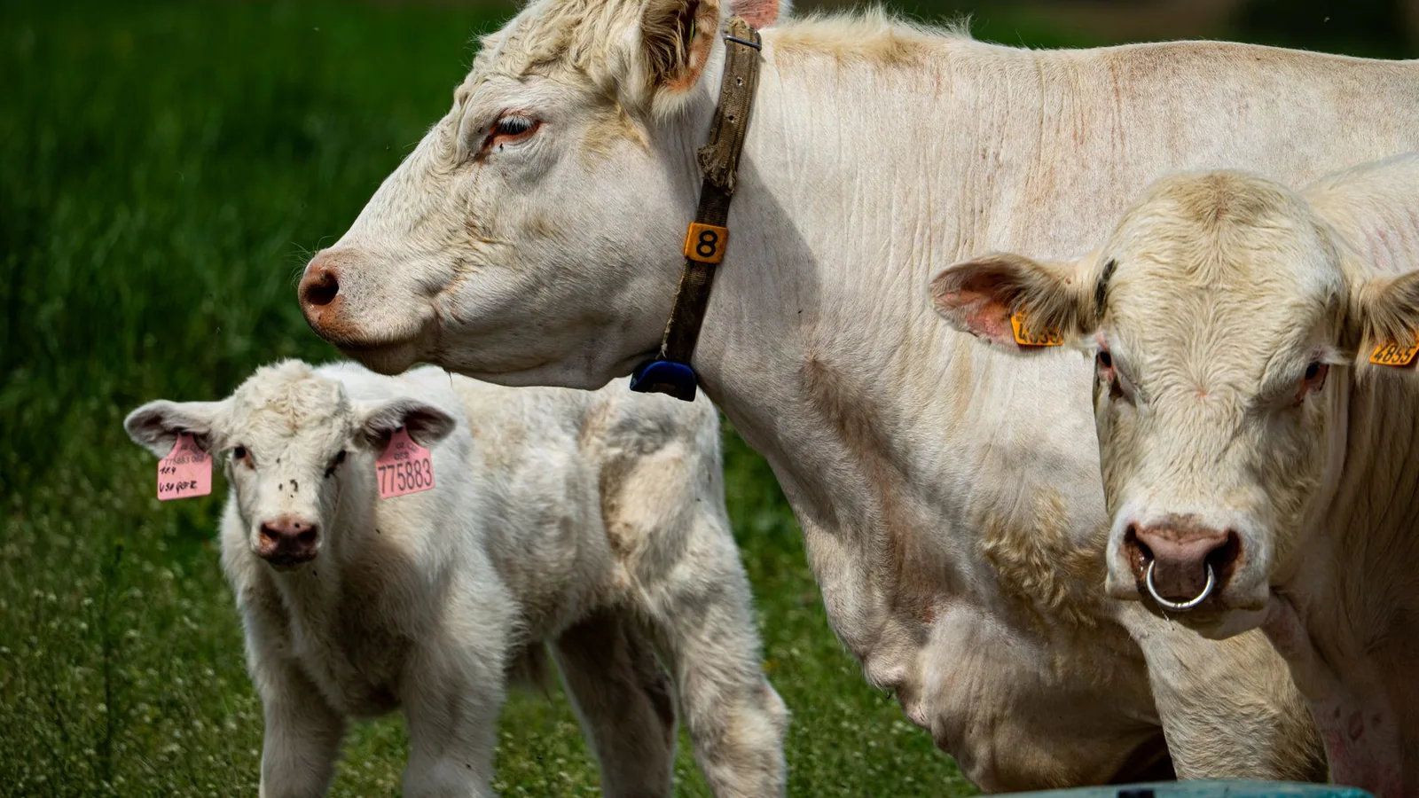
Foot-and-mouth disease virus (FMDV) exhibits a remarkably broad host range among cloven-hoofed animals (Artiodactyla), a characteristic that underpins its status as a preeminent transboundary pathogen of livestock. The virus is capable of infecting over 70 species, with domestic cattle (Bos taurus), pigs (Sus scrofa domesticus), sheep (Ovis aries), and goats (Capra hircus) representing the primary economically significant hosts [1, 9]. Buffalo, both domestic water buffalo (Bubalus bubalis) and African Cape buffalo (Syncerus caffer), are highly susceptible and play a critical role in the epidemiology of the virus, particularly as reservoirs for the Southern African Territories (SAT) serotypes [22, 49, 50]. The susceptibility and clinical outcome vary considerably by species, age, breed, virus strain, and immune status, with young animals generally succumbing to more severe, often fatal, myocarditis rather than the characteristic vesicular disease seen in adults [51].
Among wildlife, a diverse array of ungulates, including deer, antelope, wild boar, bison, and giraffes, are susceptible to natural and experimental infection [1, 6]. While clinical disease in wildlife can range from subclinical to severe, their role as maintenance hosts or sentinels is serotype- and ecoregion-dependent. The African Cape buffalo is the principal natural reservoir for SAT serotypes in sub-Saharan Africa, maintaining long-term persistent infections without overt clinical signs [23]. In Eurasia, wild boar have been implicated in the spread of FMDV, as demonstrated during the 2011 incursion of serotype O/ME-SA/PanAsia2 into Bulgaria [6, 5]. Historically, FMDV serotype C was considered less prevalent and less transmissible, affecting pigs and cattle in Europe but disappearing as a natural field strain after 2004, a phenomenon that remains poorly understood [24]. Critically, FMDV infection in humans is exceptionally rare and mild, characterized by transient vesicular lesions, and the virus is not considered a major zoonotic threat (WOAH/FAO classification). The World Organisation for Animal Health (WOAH) and the Food and Agriculture Organization (FAO) jointly prioritize FMDV for global control due to its economic devastation and the severe trade restrictions it imposes on affected nations, not due to human health risks.
The carrier state represents a pivotal, yet enigmatic, aspect of FMDV host range. Following acute clinical or subclinical infection, a significant proportion of ruminants, particularly cattle, buffalo, and sheep, may harbor infectious virus in the oropharyngeal region beyond 28 days post-infection, defining them as carriers [27, 50]. This persistent infection is characterized by the presence of intact, infectious FMDV in the pharyngeal epithelium and oropharyngeal fluid, often in the absence of clinical signs. The carrier state can last for months or even years in cattle and buffalo [49]. The epidemiological significance of carriers has been a subject of intense debate; a fundamental conundrum exists in the discordance between the detection of infectious FMDV in carriers and the apparent lack of contagiousness to in-contact naïve animals under experimental conditions [27]. However, field studies and recent experimental evidence using superinfection models have demonstrated that carrier animals can transmit virus to other cattle, particularly through prolonged direct contact [26]. This suggests that under specific conditions, such as high population density, stress, or immunosuppression, carriers may contribute to viral persistence and the emergence of novel recombinant strains [26, 50]. The existence of a distinct form of neoteric subclinical infection, which is epidemiologically indistinguishable from the carrier state in field scenarios but may have substantially different transmission potential, further complicates risk assessment [27]. This underscores the urgent need for advanced molecular and epidemiological tools to differentiate these states and accurately model transmission risks.
Clinical Signs and Disease Progression
The clinical manifestations of FMDV infection are acute, severe, and highly pathognomonic, yet they can vary considerably in severity depending on host species, virus strain, and route of exposure. The hallmark of classical FMD is the development of vesicular lesions at predilection sites, including the oral cavity (tongue, dental pad, lips, gums), the coronary band and interdigital space of the feet, and the teat skin in lactating females [1, 5]. The incubation period is typically 2-14 days, but can be as short as 24-48 hours in highly susceptible animals subjected to intense viral exposure [29, 46].
In cattle, the initial signs are fever (up to 41°C), anorexia, depression, and a marked drop in milk production. Within 24-48 hours, animals develop profuse salivation (ptyalism) due to painful oral vesicles, which quickly rupture, leaving raw, eroded lesions that cause intense pain and reluctance to eat or drink. Foot lesions manifest as severe lameness, with animals reluctant to move, standing on the tips of their feet, or recumbent. The coronary band and interdigital cleft become swollen, hot, and painful, and vesicular lesions may rupture and become secondarily infected, leading to hoof sloughing in severe cases [5]. Teat lesions can cause severe mastitis, further reducing milk yield. The clinical trajectory is often biphasic; after initial vesicle rupture, animals may show transient improvement before the full systemic effects of the disease become apparent.
Pigs present with a slightly different clinical picture, often exhibiting a more severe and rapid course. The primary presenting sign is sudden-onset severe lameness, frequently affecting all four feet. Vesicles are most commonly observed on the coronary band, heel bulbs, and the skin of the snout, but are less frequently seen in the oral cavity compared to cattle [1, 58]. Pigs are potent amplifiers of FMDV, excreting vast quantities of virus in all secretions and excretions, particularly in exhaled air, making them critical drivers of airborne transmission [28, 57]. The minimum infectious dose for pigs via oral (feed) exposure is relatively high, estimated at (10^{6.2}) to (10^{7.0}) TCID(_{50}), though this can vary by strain [48].
Sheep and goats often exhibit a mild clinical presentation, frequently characterized by only transient fever, subtle lameness, and small, easily overlooked vesicles on the coronary band or in the interdigital space [1]. Oral lesions are less common and less prominent than in cattle. As a result, FMDV infection in small ruminants can go undetected, allowing them to serve as cryptic reservoirs for virus introduction into susceptible cattle or pig populations [21, 5].
Severity of disease is age-dependent. In neonatal and juvenile animals (calves, piglets, lambs), a non-vesicular, highly fatal form of the disease occurs due to infection of cardiac muscle, leading to acute myocarditis and congestive heart failure. This "tiger heart" pathology, characterized by necrotic, pale myocardial fibers interspersed with hemorrhagic bands, is a classic post-mortem finding and a major cause of mortality in young stock [5].
Pathogenesis and Molecular Pathology
The molecular pathogenesis of FMDV is a complex interplay of viral replication strategies and host immune evasion, culminating in cellular destruction and systemic disease. The virus initiates infection through the upper respiratory tract (UDP) or the oral, pharyngeal mucosa, with the primary replication site in cattle being the pharyngeal epithelium, particularly the crypts of the dorsal soft palate [29, 46]. Viral entry into host cells is mediated by the interaction of the arginine-glycine-aspartic acid (RGD) motif, located within the flexible GH loop of capsid protein VP1, with cellular integrin receptors, principally (\alpha_v\beta_6) [3, 12]. This interaction triggers clathrin-mediated endocytosis. The acidic environment of the endosome induces capsid disassembly; virions are highly acid-labile, and a critical pH-dependent uncoating event occurs, releasing the viral RNA into the cytoplasm [4, 14].
Once within the cytoplasm, the positive-sense RNA genome is translated as a single polyprotein, which is subsequently cleaved by viral proteases, Leader protease (L(^{pro})) and 3C protease (3C(^{pro})), into four structural (VP1-4) and ten non-structural proteins (L(^{pro}), 2A-C, 3A-D) [10, 30]. These proteases are central to the viral life cycle and are the primary mediators of host cell shutdown and immune evasion.
Cellular Shutdown and Immune Evasion
FMDV employs a multi-pronged strategy to dismantle innate antiviral defenses. L(^{pro}) is a potent papain-like cysteine protease that cleaves the translation initiation factor eIF4G, thereby shutting down host cap-dependent mRNA translation while allowing viral cap-independent, IRES-mediated translation to proceed [10, 30]. More insidiously, both L(^{pro}) and 3C(^{pro}) target key components of the innate immune sensing and signaling pathways.
Inhibition of Interferon Induction: FMDV proteases proteolytically degrade several critical sensors and adaptors. L(^{pro}) cleaves the RIG-I-like receptors (RLRs) MDA5 and LGP2 at a conserved helicase motif, abrogating their ability to signal for type I interferon (IFN-(\alpha/\beta)) induction [32]. Similarly, L(^{pro}) and 3C(^{pro}) target the cytosolic DNA sensor cGAS for degradation, subverting the cGAS/STING pathway, a pathway increasingly recognized for its role in RNA virus sensing [33]. The non-structural protein 3A upregulates the autophagy-related protein LRRC25, which in turn promotes the degradation of G3BP1, an essential activator of the RLR signaling cascade [40]. The viroporin 2B protein also inhibits RIG-I and MDA5 expression and negatively regulates the phosphorylation of TBK1 and IRF3, further suppressing IFN-(\beta) production [38, 39].
Disruption of Interferon-Stimulated Genes (ISG) and Apoptosis: Beyond IFN induction, FMDV blocks the downstream antiviral effects. The capsid protein VP4 promotes the lysosomal degradation of NME1 (nucleoside diphosphate kinase 1), a tumor suppressor that normally enhances p53-mediated transcription of ISGs. This degradation impairs the expression of antiviral genes such as ISG20, IRF9, and ISG15 [36]. Additionally, FMDV manipulates cell survival and death pathways. The 3C(^{pro}) degrades the ATG5-ATG12 autophagy complex, which not only inhibits autophagy but also attenuates NF-(\kappa)B and IRF3 signaling, which are crucial for pro-inflammatory cytokine production [34].
Subversion of Stress Granule Formation: Stress granules (SGs) are cytoplasmic foci of stalled mRNPs that form during cellular stress (e.g., viral infection) and have antiviral properties. FMDV L(^{pro}) cleaves the SG scaffolding proteins G3BP1 and G3BP2, thereby suppressing SG formation and allowing viral protein synthesis to proceed unimpeded [31].
Pathology of Lesion Formation
The replication of FMDV within epithelial cells leads to extensive cytopathic effect (CPE), including cell rounding, detachment, and lysis. The characteristic vesicle forms due to fluid accumulation between necrotic epithelial cells of the stratum spinosum and suprabasal layers of the epidermis and oral mucosa. Viral replication causes ballooning degeneration and necrosis of these cells, leading to the formation of intraepithelial microvesicles that coalesce to form macroscopically visible vesicles [5]. The roof of the vesicle is composed of necrotic epithelium, while the floor contains the inflamed, living basal cell layer. Rupture of these vesicles results in painful erosions and ulcers, which are susceptible to secondary bacterial infection. The intense pain from oral and foot lesions is responsible for the clinical signs of inappetence, salivation, and lameness. Myocarditis in young animals results from direct viral replication in cardiac myocytes, causing myofiber necrosis and mononuclear cell infiltration, ultimately leading to cardiac failure.
Systemic Dissemination and Shedding
Following initial replication in the pharyngeal epithelium, the virus rapidly gains access to the draining lymph nodes and enters the bloodstream via the thoracic duct, resulting in primary viremia within 24-48 hours. This disseminates the virus systemically to secondary predilection sites, including the oral cavity, feet, and teats, where further replication occurs in epithelial cells, leading to the characteristic secondary vesicles. The host sheds virus in all bodily excretions and secretions: saliva, nasal discharge, milk, urine, feces, and semen, as well as in exhaled breath (aerosols). Pigs are exceptionally efficient aerosol shedders [28]. The environmental contamination is profound; virus can survive on fomites, in feed, and in the environment for weeks, especially under cool, moist conditions [48, 47, 46]. Basic reproduction number ((R_0)) estimates for environmental transmission alone can be as high as 1.65, indicating that indirect contact is sufficient to sustain an outbreak [46]. The integrity of the vesicular epithelium and the surrounding environment drives the explosive spread of FMD.
Diagnostic Methods for Foot-and-Mouth Disease Virus
The accurate and timely diagnosis of foot-and-mouth disease virus (FMDV) is the cornerstone of effective disease control, surveillance, and eradication programmes. Given the immense socio-economic impact of FMD, recognised by the World Organisation for Animal Health (WOAH) as a notifiable disease, diagnostic methods must be not only exquisitely sensitive and specific but also capable of differentiating infected from vaccinated animals (DIVA) and identifying the specific serotype and lineage involved. The diagnostic landscape for FMDV has evolved dramatically from traditional virological techniques to a sophisticated arsenal of molecular, serological, and field-deployable platforms, each with distinct applications across the spectrum of clinical investigation, outbreak response, and endemic surveillance.
Traditional and Virological Diagnostic Approaches
The foundation of FMD diagnosis has historically rested upon the isolation of infectious virus in cell culture. Epithelial tissue suspensions, vesicular fluid, or oropharyngeal fluid (OPF) samples are inoculated onto susceptible cell lines, most commonly primary bovine thyroid (BTY) cells or continuous cell lines such as BHK-21 or IB-RS-2. The appearance of characteristic cytopathic effect (CPE), typically observed within 24-48 hours, provides initial evidence of infection [21, 51]. Complementing virus isolation, antigen detection enzyme-linked immunosorbent assays (ELISAs) have served as the standard for serotyping field isolates for decades. These assays, often employing serotype-specific polyclonal or monoclonal antibodies, can directly identify the FMDV serotype (O, A, C, Asia-1, SAT1, SAT2, SAT3) from clinical samples without the need for viral propagation, offering a significant time advantage [21, 23]. However, the sensitivity of antigen ELISA is substantially lower than that of molecular methods, particularly for samples with low viral loads, such as those from subclinically infected or carrier animals [50]. The emergence of exotic strains or novel topotypes, such as the detection of a novel SAT1 topotype in Nigeria after 35 years of absence, underscores the critical need for comprehensive antigenic surveillance to ensure that diagnostic reagents remain relevant [22]. The historical context of serotype C, which appears to be extinct in the field since 2004, highlights the dynamic nature of FMDV serotype distribution and the ongoing necessity for robust diagnostic capacity to detect both re-emerging and newly incursive strains [24].
Molecular Diagnostics: The Gold Standard for Sensitivity and Speed
The advent of nucleic acid amplification technologies has revolutionised FMDV diagnosis. Real-time reverse transcription polymerase chain reaction (rRT-PCR) has become the method of choice for the rapid, sensitive, and specific detection of FMDV RNA across all seven serotypes. The most widely used assays target highly conserved regions such as the 3D RNA-dependent [RNA polymerase](/knowledge/bioinformatics/rna-polymerase-structure-transcription-mechanisms 2) or the 5' untranslated region (UTR), enabling pan-serotypic detection [42, 43, 54]. For instance, the lyophilized pan-serotype-specific rRT-PCR assay evaluated by Howson et al. demonstrated analytical sensitivity comparable to the gold-standard OIE-recommended laboratory-based assay, successfully detecting FMDV in a diverse panel of 57 positive samples without cross-reactivity with other vesicular disease viruses [54]. The quantitative nature of rRT-PCR allows for viral load estimation, which is invaluable for understanding transmission dynamics, assessing the infectiousness of individual animals, and monitoring the efficacy of interventions [42].
To address the critical need for serotype identification, essential for vaccine matching and epidemiological tracking, multiplex rRT-PCR assays have been developed. These assays incorporate serotype-specific probes, allowing for the simultaneous detection and differentiation of multiple serotypes in a single reaction. Wang et al. described a multiplex assay capable of differentiating FMDV from Seneca Valley virus-1 (SVV-1), a pathogen that causes clinically indistinguishable vesicular disease in swine, thereby addressing a crucial differential diagnostic challenge [42]. The assay's specificity was confirmed against a panel of common porcine viruses, and its limit of detection (LOD) was as low as 11 copies per reaction for FMDV [42]. Similarly, targeted typing assays incorporating East Africa-specific probes for serotypes O, A, SAT1, and SAT2 have been validated for use in that endemic region, correctly identifying the serotype of 33 out of 36 positive samples with no observed cross-reactivity [54].
Beyond conventional rRT-PCR, isothermal amplification methods have emerged as powerful alternatives, particularly suited for field or resource-limited settings. Reverse transcription loop-mediated isothermal amplification (RT-LAMP) operates at a constant temperature, eliminating the need for a thermal cycler. Bath et al. rigorously validated an RT-LAMP assay targeting the 3D region, incorporating an internal positive control (IPC) to mitigate false negatives from sample interference [53]. When tested on unextracted oral swab samples from FMD-suspected animals in Bhutan and Australia, the assay demonstrated a diagnostic specificity exceeding 99% and a sensitivity of 79% using Bayesian latent class analysis, confirming its fitness-for-purpose as a herd-level diagnostic tool [53]. A further innovation is the reverse transcription-insulated isothermal PCR (RT-iiPCR) assay, which utilises a compact, portable analyser (POCKIT™) that automates data analysis and displays simple positive or negative results. Ambagala et al. validated an RT-iiPCR assay targeting the 3D region, achieving a 95% LOD of 9 copies of RNA standard, accurate detection of all seven serotypes, and no cross-reactivity with other vesicular disease viruses [43]. Crucially, the assay detected FMDV RNA in oral and nasal swabs before the onset of clinical signs, highlighting its potential for pre-clinical outbreak detection [43].
Serological Methods Detecting Infection and Carrier Status
Serological diagnostics serve multiple critical functions: confirming infection in unvaccinated populations, demonstrating freedom from disease for trade purposes, and crucially, differentiating infected from vaccinated animals (DIVA) in vaccinated populations. The detection of antibodies against non-structural proteins (NSPs) is the cornerstone of DIVA testing. NSPs are produced during active viral replication but are largely absent from purified, inactivated vaccines. The most widely used NSP target is the 3ABC protein, encoded by a conserved region of the FMDV genome. Sulayeman et al. utilised a commercial 3ABC competition ELISA (ID Screen® FMD NSP Competition) to determine a sero-prevalence of 24.2% in cattle in central Ethiopia, while also identifying risk factors such as breed and age [21]. The 3ABC ELISA, however, can yield false-positive results in recently vaccinated animals or in animals infected with other picornaviruses, necessitating confirmatory testing.
Chemiluminescence immunoassays (CLIAs) represent a significant advancement in serological detection, offering enhanced sensitivity and speed. Liu et al. developed two CLIAs utilising recombinant full-length 3ABC and 2C NSPs [59]. The 3ABC-2C CLIA demonstrated a diagnostic sensitivity of 100% and a specificity of 96.5% for detecting infected pigs, outperforming both the single-target 3ABC CLIA and two commercial ELISA kits [59]. The 3ABC-2C CLIA detected infection earlier than commercial ELISAs and produced results within 15 minutes, making it a strong candidate for penside diagnostics [59]. The use of multiple NSP targets, as in the 3ABC-2C CLIA, improves accuracy, particularly in endemic settings where the background seroprevalence of NSP antibodies may be high due to repeated exposure.
Detection of the FMDV carrier state, a persistent, subclinical infection lasting longer than 28 days in ruminants, requires specific diagnostic approaches. Carrier animals, which may harbour virus in the oropharynx for months or years, are epidemiologically significant as potential mixing vessels for the emergence of novel recombinant strains [26, 49]. The definitive diagnosis of the carrier state relies on the detection of viral RNA or infectious virus in OPF samples collected by probang sampling. Stenfeldt and Arzt reviewed the "carrier conundrum," noting that while viral RNA can be detected by rRT-PCR in OPF samples from carriers, the infectiousness of these animals to contact-naïve cohorts remains poorly understood [27]. Deep sequencing of viral populations from carrier cattle has revealed considerable within-host genetic variation and the potential for intra-serotypic recombination, further complicating the interpretation of diagnostic results [25, 50]. Field studies in vaccinated buffalo in Pakistan demonstrated that 60% of animals had detectable viral RNA in at least one OPF sample over a 12-month period, with evidence of new, persistent, and serial infections with different FMDV strains [49]. This highlights the necessity of longitudinal molecular testing to accurately characterise the infection dynamics within a herd.
Field-Deployable and Non-Invasive Sampling Technologies
The logistical challenges of sample transport, cold chain maintenance, and access to sophisticated laboratory infrastructure in endemic regions have spurred the development of simple, robust, and field-ready diagnostic platforms. One of the most significant advances has been the lyophilisation of rRT-PCR and RT-LAMP reagents, which dramatically improves reagent stability at ambient temperatures. Howson et al. demonstrated that lyophilised rRT-PCR and RT-LAMP assays retained their analytical sensitivity and specificity when deployed to field settings in East Africa, successfully detecting FMDV in clinical samples from acutely infected and convalescent cattle [55]. The use of lateral flow devices for end-point detection of RT-LAMP amplicons further simplifies the workflow, enabling visual interpretation without the need for specialised equipment [55].
Sampling methods have also evolved to be less invasive and more amenable to mass screening. Environmental swabbing of surfaces in livestock markets, watering points, and animal transport vehicles has emerged as a powerful non-invasive surveillance tool. Colenutt et al. demonstrated that FMDV RNA could be detected on environmental swabs collected from a live goat market in Nepal and from various surfaces in outbreak-affected areas in the Kathmandu Valley [52, 56]. The detection of viral RNA in 4.1% of environmental samples from the goat market, despite the absence of observed clinical signs, underscores the utility of environmental sampling for revealing cryptic virus circulation [52]. This approach, analogous to environmental surveillance for poliovirus, provides a low-technology, cost-effective means of assessing virus presence and circulation in a geographically defined area, particularly valuable for monitoring progress towards control in endemic settings.
For swine, the collection of oral fluids using cotton ropes has proven to be a practical and sensitive method for herd-level surveillance. Vosloo et al. showed that rope-based oral fluid sampling, when combined with rRT-PCR, achieved a sensitivity ranging from 0.67 to 0.92 for detecting FMDV infection in experimentally infected pigs [57]. The method is non-invasive, requires minimal technical skill, and can be used to monitor large groups of animals efficiently, making it ideal for both outbreak response and ongoing surveillance in commercial pig operations [57]. The ability to detect virus in oral fluids before the appearance of clinical signs further enhances its value for early warning [57].
Genotyping and Sequencing for Molecular Epidemiology
Beyond mere detection and serotyping, the characterisation of FMDV strains through nucleotide sequencing provides unparalleled resolution for tracing transmission pathways, monitoring viral evolution, and guiding vaccine selection. The viral protein 1 (VP1) coding region, which encodes the major antigenic determinants and is the primary target for serotyping, is the most widely sequenced genomic region for molecular epidemiology. VP1 sequences are used to assign viruses to serotype, topotype, lineage, and sublineage, revealing the phylogeographic dynamics that drive global FMDV spread [7, 20]. For example, sequence analysis of an emergent FMDV from Namibia in 2021 identified it as serotype O belonging to the EA-2 topotype, sharing 99.5% nucleotide identity with viruses from Zambia, representing the first detection of this serotype in Namibia and a significant transboundary incursion [17].
Whole-genome sequencing using next-generation sequencing (NGS) technologies has provided unprecedented insights into within-host viral diversity and the emergence of recombinant strains. King et al. employed deep sequencing of a 7.6 kb fragment encompassing the polyprotein coding region to characterise the fine polymorphic structure of FMDV populations within individual cattle during the 2007 UK outbreak [25]. This analysis revealed distinct viral subpopulations evolving independently at different lesion sites within a single animal, as well as consensus-level variants that were shared between animals, providing robust evidence for transmission links [25]. Experimental coinfection studies using cattle have demonstrated that high-resolution sequencing can detect dominant interserotypic recombinant genomes emerging in the upper respiratory tract of superinfected animals within just 10 days, inculpating carrier animals as potential "mixing vessels" for novel strain emergence [26]. The integration of genomic data with epidemiological metadata is now a standard approach for reconstructing transmission routes at regional and global scales, providing the evolutionary and ecological context necessary to anticipate future incursions [7, 29].
Control Measures and Vaccination Strategies
The control of foot-and-mouth disease virus (FMDV) represents one of the most formidable challenges in global veterinary medicine, owing to the virus's extraordinary genetic and antigenic heterogeneity, its capacity for rapid dissemination through multiple transmission pathways, and the profound economic consequences that follow incursions into naïve or free zones. The strategic framework for FMDV control is necessarily multifaceted, integrating vaccination as a central pillar with rigorous biosecurity protocols, surveillance and diagnostic architectures, movement restrictions, stamping-out policies, and, increasingly, novel biotechnology-derived countermeasures. The World Organisation for Animal Health (WOAH) and the Food and Agriculture Organization (FAO) have championed the Progressive Control Pathway for FMD (PCP-FMD), a staged approach that guides endemic countries toward eventual freedom from disease while managing the immediate burden of clinical outbreaks [1, 5]. Within this context, the complexity of FMDV biology, particularly its seven immunologically distinct serotypes, the rapid evolutionary turnover of circulating lineages, the existence of a protracted carrier state, and the virus’s sophisticated arsenal of host immune evasion mechanisms, directly dictates the design, deployment, and efficacy of every control intervention. This section provides an exhaustive analysis of the current control measures and vaccination strategies, dissecting the biological underpinnings that govern their successes and limitations, and exploring the frontier of next-generation countermeasures.
The Foundational Challenge: Antigenic Diversity, Viral Evolution, and the Carrier State
Any discussion of FMDV control must begin with an acknowledgment of the pathogen’s inherent complexity, which fundamentally constrains all intervention strategies. The virus exists as seven serotypes (O, A, C, Asia 1, SAT 1, SAT 2, and SAT 3), within which numerous topotypes, genetic lineages, and antigenic variants cocirculate in endemic regions [1, 6, 7]. Critically, immunity elicited by infection or vaccination against one serotype confers no cross-protection against heterologous serotypes, and even within a serotype, significant antigenic drift can render existing vaccines poorly matched to emerging field strains [8, 58]. The evolutionary dynamics of FMDV are driven by an error-prone RNA-dependent [RNA polymerase](/knowledge/bioinformatics/rna-polymerase-structure-transcription-mechanisms 2), quasispecies diversification, recombination events, and ecological pressures such as host species distribution and animal movement patterns [7, 26]. Metapopulation dynamics, particularly in the Asian virus pools, drive periodic emergence of new lineages from Southern Asia that propagate westward, creating waves of incursions that systematically challenge regional vaccine strategies and necessitate constant surveillance and vaccine strain updating [7].
The phenomenon of the FMDV carrier state, in which ruminants, particularly cattle and buffalo, harbour subclinical, persistent infection in the oropharynx for months to years following acute disease, introduces profound epidemiological uncertainty [27, 49]. Carriers shed negligible quantities of infectious virus and have not been demonstrated to transmit infection to naïve contacts under experimental or field conditions, yet they maintain the viral population within a region and may serve as mixing vessels for the generation of novel recombinant strains through superinfection with heterologous serotypes [27, 26]. The experimental demonstration of interserotypic recombination occurring within persistently infected carriers within days of superinfection highlights the potential for these animals to act as cryptic sources of antigenic novelty, complicating long-term control efforts and vaccine design [26]. Endemic circulation of FMDV in vaccinated buffalo populations in Pakistan, characterized by high rates of new subclinical infections, serial infections with different strains, and persistent carriage, further underscores the difficulty of achieving steric immunity and highlights the need for DIVA (Differentiating Infected from Vaccinated Animals)-compliant surveillance to detect cryptic viral activity [49].
Vaccination Strategies: From Conventional Inactivated Vaccines to Next-Generation Platforms
Conventional Inactivated Vaccines: The Workhorse and Its Limitations
The cornerstone of FMDV prophylaxis worldwide remains the conventional, chemically inactivated, adjuvanted whole-virus vaccine, typically produced by infecting BHK-21 suspension cell cultures with a suitable seed virus, inactivating with binary ethylenimine (BEI) or formalin, and formulating with oil or aluminium hydroxide-saponin adjuvants [3, 11]. These vaccines have been deployed with remarkable success in regional eradication campaigns, most notably in Europe and South America, where systematic, high-coverage vaccination programs eliminated FMD from large geographic areas. However, multiple inherent limitations constrain their efficacy, particularly in the hyper-endemic settings of Africa and Asia where they are most urgently needed.
First, the vaccines are exquisitely serotype-specific and often strain-specific. The antigenic match between the vaccine strain and the circulating field virus is the primary determinant of protective efficacy, quantified by the r₁ value (ratio of heterologous to homologous serum neutralization titres) or by in vivo challenge studies [8, 58]. Antigenic profiling of SAT1 and serotype O viruses has revealed that critical amino acid substitutions within the VP1 G-H loop and other antigenic sites can dramatically shift the antigenic phenotype, rendering established vaccine strains ineffective against newly emergent lineages [8]. The emergence of lineages such as O/ME-SA/Ind-2001d, which spread from the Indian subcontinent into Southeast Asia, the Middle East, and North Africa, necessitated urgent reassessment and reformulation of vaccine banks to ensure coverage against this transboundary threat [6, 15, 16, 18]. Similarly, the A/ASIA/G-VII lineage, which arose in the Indian subcontinent and spread into the Middle East, showed evidence of antigenic pressure within a critical VP1 site, putatively driven by intensive vaccination campaigns, further complicating vaccine strain selection [19].
Second, the logistical and economic hurdles of vaccine production, quality control, and deployment in low-resource settings are substantial. Adaptation of field isolates to high-yield growth in BHK-21 cells is laborious and time-consuming, often requiring multiple passages that can select for adaptive mutations in the capsid, frequently involving the acquisition of heparan sulphate binding capability, which may alter antigenic properties [11]. The requirement for cold chain maintenance, the need for revaccination at intervals of 4-6 months to maintain protective antibody titres, and the high cost per dose relative to livestock value in many endemic regions all contribute to suboptimal coverage [58]. In pigs, the challenges are particularly acute: conventional oil-adjuvanted vaccines often induce weaker and shorter-lived immunity compared to cattle, and the high density of pig production units amplifies the consequences of any gaps in vaccine-induced protection [58].
Third, conventional vaccines do not induce sterile immunity. Vaccinated animals, if subsequently exposed to live virus, may become subclinically infected and develop the carrier state without displaying overt clinical signs. This phenomenon, documented in both experimental settings and field studies, means that vaccination can mask the presence of circulating virus, complicating surveillance and facilitating undetected spread [27, 49]. The inability of conventional vaccines to prevent infection (as opposed to disease) necessitates complementary biosecurity and diagnostic measures and has driven the development of DIVA-compatible vaccines and companion diagnostic tests.
Novel Vaccine Platforms: VLP, Vectored, and Stabilized Capsid Vaccines
In response to the limitations of conventional inactivated vaccines, a new generation of rationally designed vaccine candidates has emerged, leveraging advances in structural biology, reverse genetics, and recombinant DNA technology. These platforms aim to improve safety by eliminating the need for live virus handling, enhance immunogenicity through rational design, and enable DIVA capability by removing non-structural proteins from the antigen.
Virus-Like Particle (VLP) Vaccines: Recombinant empty capsids, or VLPs, which self-assemble from the structural proteins VP0, VP1, and VP3 without encapsidating viral RNA, represent one of the most promising alternatives. Produced using baculovirus-insect cell expression systems, VLPs are structurally identical to native virions but are non-infectious, eliminating the biosafety risks associated with large-scale live virus culture and inactivation [3, 60]. The immunogenicity of FMDV VLPs can be significantly enhanced through formulation with advanced adjuvants. For instance, conjugation of VLPs with gold nanocages (AuNCs) has been shown to activate dendritic cells via Toll-like receptor 4 (TLR4), promote secretion of IL-6, IL-1β, and TNF-α, and elicit robust humoral and cellular immune responses, including significantly enhanced CD8+ T cell proliferation and IFN-γ and IL-12p70 secretion in murine and guinea pig models [60]. The application of computational interface design to stabilize the inherently labile pentameric capsid interfaces has further improved the thermal and pH stability of VLPs, a critical factor for vaccine storage and distribution in endemic areas with limited cold chain infrastructure [14]. Stabilized serotype O and SAT2 capsids, produced by introducing rationally selected mutations that strengthen non-covalent interactions between pentameric subunits, generated improved neutralizing antibody responses in vaccinated animals compared to wild-type particles [14]. The pH stability of the capsid is of particular importance, as the intact virion must resist acid-induced dissociation during fermentation and purification steps; strategic modifications to the capsid’s pH-sensitive quadrangle region can enhance manufacturability while preserving antigenic integrity [4].
Replication-Deficient Adenovirus-Vectored Vaccines: An alternative platform that has advanced significantly through regulatory safety evaluation is the human adenovirus 5 (HAd5)-vectored vaccine, in which the FMDV capsid coding region (P1-2A) and the viral 3C protease (required for capsid processing) are expressed from a replication-deficient HAd5 backbone. This platform, exemplified by the AdtA24 vaccine for serotype A, has demonstrated a robust safety profile in cattle, fulfilling stringent US Code of Federal Regulations (9CFR) requirements for live veterinary vaccines [61]. The vectored platform is inherently DIVA-compatible, as vaccinated animals develop antibodies only to the FMDV structural proteins and the HAd5 vector, but not to FMDV non-structural proteins (NSPs). Comprehensive safety studies have confirmed that the AdtA24 master seed virus does not revert to virulence, is not shed or transmitted to sentinel animals, and is completely absent from milk of vaccinated lactating cows [61]. Field safety trials in 500 beef and dairy cattle under typical US production conditions further validated the vaccine’s suitability for broad-scale deployment. A critical advantage of this platform is the speed with which new vaccine strains can be generated against emergent lineages: a reverse genetics approach allows substitution of the capsid coding sequence without requiring lengthy cell culture adaptation of the field isolate [11, 61].
RNA Interference and Transgenic Resistance: At the frontier of control strategies lies the genetic modification of host susceptibility. Transgenic pigs constitutively expressing FMDV-specific short hairpin RNA (shRNA) that generates small interfering RNAs (siRNAs) targeting the viral genome have been produced and characterized [62]. In vitro challenge of transgenic fibroblasts demonstrated robust suppression of viral replication, and importantly, when these pigs were challenged with a lethal dose of FMDV, they remained entirely free of clinical signs, fever, and histopathological lesions, in stark contrast to non-transgenic control animals that developed fulminant disease [62]. This proof-of-concept study demonstrates the potential for RNA interference to confer heritable, sequence-specific resistance, though the emergence of viral escape mutants remains a theoretical concern that would require targeting of highly conserved genomic regions.
DIVA Strategies and Companion Diagnostics
The differentiation of infected from vaccinated animals (DIVA) is a cornerstone of FMD control programs that aim to regain or maintain disease-free status while permitting vaccination. The principle relies on the fact that conventional inactivated vaccines are purified to remove non-structural proteins (NSPs), such that vaccinated animals develop antibodies only to structural proteins, whereas naturally infected animals (including vaccinated animals that become infected) seroconvert to both structural and non-structural antigens [59, 61]. The detection of antibodies against NSPs, particularly the 3ABC polyprotein, is a reliable serological marker of active or past infection, regardless of vaccination status. Chemiluminescence immunoassays (CLIAs) using recombinant 3ABC and 2C proteins have demonstrated high diagnostic sensitivity (100%) and specificity (96.5%) in pigs, with the added advantage of rapid result generation (within 15 minutes), making them suitable for penside deployment [59]. Similarly, the AdtA24 vectored vaccine, which expresses only the capsid-coding region, is inherently DIVA-compatible, as vaccinated animals test negative by NSP-based ELISA [61].
The implementation of DIVA diagnostics at scale requires rigorous surveillance infrastructure. Environmental sampling has emerged as a low-technology, non-invasive complement to traditional individual animal sampling for detection of FMDV RNA circulation. In endemic settings such as Nepal, environmental swabs collected from livestock markets and outbreak sites have successfully detected FMDV RNA, providing spatial and temporal data on virus circulation without the need for intensive animal handling [52, 56]. Paired with portable molecular platforms such as the T-COR™ 8 thermocycler and lyophilized pan-serotype-specific real-time RT-PCR assays, environmental surveillance can be deployed in field settings with minimal laboratory infrastructure, extending diagnostic reach to previously underserved areas [52, 54, 56].
Non-Vaccine Control Measures: Biosecurity, Movement Control, and Environmental Mitigation
Vaccination alone is rarely sufficient to control FMDV outbreaks, particularly in free areas or during acute incursions. A comprehensive control program must integrate multiple, layered interventions that target the virus’s transmission pathways. FMDV is transmitted through direct contact, indirect contact via fomites, contaminated feed, airborne aerosols, and potentially through exosome-mediated shuttling that evades antibody neutralization [28, 45, 48, 46]. Each transmission route demands specific countermeasures.
Airborne Transmission and Quarantine Zones: Under specific meteorological conditions, high relative humidity, moderate temperatures, and stable atmospheric conditions, FMDV can travel tens of kilometres in aerosolized respiratory secretions, particularly from pigs, which are prolific aerosol shedders [28]. This low-probability, high-consequence event can seed infection beyond the immediate quarantine perimeter, complicating containment. Modelling of airborne spread must incorporate viral decay rates as functions of temperature and humidity; FMDV survival at 16°C and 86% relative humidity is approximately 90% at day 50, whereas survival drops to near zero at 16°C and 37.5% relative humidity [47]. This differential survival has profound implications for the design of control zones in tropical versus temperate climates. Control measures must therefore include pre-emptive culling of high-risk premises within a defined radius, establishment of protection and surveillance zones, and real-time meteorological monitoring to predict airborne plume trajectories [1, 28].
Environmental Persistence and Decontamination: The capacity of FMDV to survive in the environment for extended periods, up to 14 days on experimental pen surfaces, means that contaminated environments can sustain transmission chains even in the absence of direct animal contact [46]. Experimental transmission studies have demonstrated that environmental contamination alone yields a basic reproduction number (R₀) of 1.65, sufficient to sustain an outbreak without direct animal-to-animal contact [46]. Detection of virus in the environment precedes the appearance of clinical signs in infected cattle, creating a window of transmission risk that is invisible to clinical surveillance. Effective decontamination requires thorough cleaning to remove organic matter followed by application of virucidal agents; acid-based disinfectants (e.g., citric acid), alkali compounds (sodium hydroxide), and oxidizing agents (e.g., sodium hypochlorite, Virkon™ S) are effective, provided contact times and concentrations are adequate [1].
Feed and Fomite Risk Mitigation: The transboundary movement of animal feed, particularly soybean meal, has been identified as a risk pathway for FMDV incursion. Experimental contamination of pig feed ingredients demonstrated that FMDV strains remain viable for durations compatible with transoceanic transport, up to 37 days in soybean meal stored at 4°C or 20°C [48]. The minimum infectious dose for pigs consuming contaminated feed ranged from 10⁶·² to 10⁷ TCID₅₀, a relatively high titre, but variations in feeding behaviour, virus strain, and product characteristics could modify risk. Critically, pre-treatment of feed with commercially available additives, either formaldehyde-based (SalCURB®) or lactic acid-based (Guardian™), significantly mitigated infectivity, providing a practical intervention for feed import risk management [48].
Movement Controls and Stamping-Out: In FMD-free countries and during acute outbreaks, the rapid imposition of animal movement restrictions, quarantine, and slaughter of infected and in-contact susceptible animals remains the foundation of eradication efforts. The 2001 UK epidemic, though not directly covered in the provided sources, exemplifies the scale of economic and social disruption that can result when stamping-out is the primary strategy. The detection of novel serotype Asia 1 in Myanmar in 2017, after a prolonged absence, was met with disinfection, quarantine, and movement restrictions; no further cases were detected, but the authors noted the limitations of passive surveillance and the need for active vigilance [20]. Similar challenges were encountered during the 2015 incursions of O/ME-SA/Ind-2001d into Lao PDR and Vietnam, where movement restrictions were not strictly enforced and emergency vaccination was only partially implemented [15].
Surveillance, Diagnosis, and the Role of Rapid Molecular Tools
Effective control depends on rapid and accurate diagnosis to enable timely deployment of interventions. The gold standard laboratory diagnosis remains real-time RT-PCR targeting conserved regions such as the 3D polymerase gene, supplemented by sequencing of the VP1 coding region for serotyping and phylogenetic characterization [42, 43, 53, 54]. However, the transport of samples to centralized reference laboratories introduces critical delays, motivating the development of field-deployable molecular platforms.
Portable RT-PCR and Isothermal Amplification: Lyophilized, pan-serotype-specific real-time RT-PCR assays deployed on portable thermocyclers (e.g., T-COR™ 8) have demonstrated sensitivity and specificity comparable to laboratory-based OIE-recommended assays, with the added capability of serotype identification using East Africa-specific typing assays for serotypes O, A, SAT1, and SAT2 [54]. Reverse transcription-loop-mediated isothermal amplification (RT-LAMP) offers an even simpler alternative, requiring only a heat block and providing results within 30 minutes. A validated RT-LAMP assay incorporating an internal positive control and compatible with unextracted oral swabs achieved diagnostic specificity >99% and sensitivity of 79% in field samples from Bhutan and Australia [53]. The assay’s fit-for-purpose performance as a herd-level screening tool makes it suitable for frontline use by non-laboratory-trained personnel. Similarly, reverse transcription-insulated isothermal PCR (RT-iiPCR) performed on the portable POCKIT™ analyser has been validated for detection of all seven serotypes from multiple sample types, including vesicular fluid
References
[1] Mioulet V, Knowles N, Smitsaart E, Bergmann I, Medina G, Vleeschauwer AD, et al.. foot-and-mouth disease virus. CABI Compendium. 2022. DOI: https://doi.org/10.1079/cabicompendium.82824
[2] Gao Y, Sun S, Guo H. Biological function of Foot-and-mouth disease virus non-structural proteins and non-coding elements. Virology Journal. 2016. DOI: https://doi.org/10.1186/s12985-016-0561-z
[3] Belsham G, Kristensen TV, Jackson T. Foot-and-mouth disease virus: prospects for using knowledge of virus biology to improve control of this continuing global threat.. Virus Research. 2020. DOI: https://doi.org/10.1016/j.virusres.2020.197909
[4] Yuan H, Li P, Ma X, Lu Z, Sun P, Bai X, et al.. The pH stability of foot-and-mouth disease virus. Virology Journal. 2017. DOI: https://doi.org/10.1186/s12985-017-0897-z
[5] Aslam M, Alkheraije KA. The prevalence of foot-and-mouth disease in Asia. Frontiers in Veterinary Science. 2023. DOI: https://doi.org/10.3389/fvets.2023.1201578
[6] Brito B, Rodriguez LL, Hammond JM, Pinto J, Perez AM. Review of the Global Distribution of Foot‐and‐Mouth Disease Virus from 2007 to 2014. Transboundary and Emerging Diseases. 2017. DOI: https://doi.org/10.1111/tbed.12373
[7] Nardo AD, Ferretti L, Wadsworth J, Mioulet V, Gelman B, Karniely S, et al.. Evolutionary and Ecological Drivers Shape the Emergence and Extinction of Foot-and-Mouth Disease Virus Lineages. Molecular biology and evolution. 2021. DOI: https://doi.org/10.1093/molbev/msab172
[8] Reeve R, Borley D, Maree F, Upadhyaya S, Lukhwareni A, Esterhuysen J, et al.. Tracking the Antigenic Evolution of Foot-and-Mouth Disease Virus. PLoS ONE. 2016. DOI: https://doi.org/10.1371/journal.pone.0159360
[9] Li K, Wang C, Yang F, Cao W, Zhu Z, Zheng H. Virus, Host Interactions in Foot-and-Mouth Disease Virus Infection. Frontiers in Immunology. 2021. DOI: https://doi.org/10.3389/fimmu.2021.571509
[10] Pulido MR, Sáiz M. Molecular Mechanisms of Foot-and-Mouth Disease Virus Targeting the Host Antiviral Response. Frontiers in Cellular and Infection Microbiology. 2017. DOI: https://doi.org/10.3389/fcimb.2017.00252
[11] Dill V, Eschbaumer M. Cell culture propagation of foot-and-mouth disease virus: adaptive amino acid substitutions in structural proteins and their functional implications. Virus genes. 2019. DOI: https://doi.org/10.1007/s11262-019-01714-7
[12] Kotecha A, Wang Q, Dong X, Ilca S, Ondiviela M, Zihe R, et al.. Rules of engagement between αvβ6 integrin and foot-and-mouth disease virus. Nature Communications. 2017. DOI: https://doi.org/10.1038/ncomms15408
[13] Hamdy M, Carlo MD, Hussein H, Salah T, El-Deeb AH, Emara M, et al.. Development of gold nanoparticles biosensor for ultrasensitive diagnosis of foot and mouth disease virus. Journal of Nanobiotechnology. 2018. DOI: https://doi.org/10.1186/s12951-018-0374-x
[14] Kotecha A, Seago J, Scott K, Burman A, Loureiro S, Ren J, et al.. Structure-based energetics of protein interfaces guide Foot-and-Mouth Disease virus vaccine design. Nature Structural &Molecular Biology. 2015. DOI: https://doi.org/10.1038/nsmb.3096
[15] Qiu Y, Abila R, Rodtian P, King D, Knowles N, Ngo L, et al.. Emergence of an exotic strain of serotype O foot‐and‐mouth disease virus O/ME‐SA/Ind‐2001d in South‐East Asia in 2015. Transboundary and Emerging Diseases. 2018. DOI: https://doi.org/10.1111/tbed.12687
[16] Zhu Z, Yang F, He J, Li J, Cao W, Li J, et al.. First detection of foot-and-mouth disease virus O/ME-SA/Ind2001 in China.. Transboundary and Emerging Diseases. 2018. DOI: https://doi.org/10.1111/tbed.12895
[17] Banda F, Shilongo A, Hikufe E, Khaiseb S, Kabajani J, Shikongo B, et al.. The first detection of a serotype O foot‐and‐mouth disease virus in Namibia. Transboundary and Emerging Diseases. 2022. DOI: https://doi.org/10.1111/tbed.14561
[18] Knowles N, Bachanek-Bankowska K, Wadsworth J, Mioulet V, Valdazo-González B, Eldaghayes I, et al.. Outbreaks of Foot-and-Mouth Disease in Libya and Saudi Arabia During 2013 Due to an Exotic O/ME-SA/Ind-2001 Lineage Virus.. Transboundary and Emerging Diseases. 2016. DOI: https://doi.org/10.1111/tbed.12299
[19] Bachanek-Bankowska K, Nardo AD, Wadsworth J, Henry E, Parlak Ü, Timina A, et al.. Foot-and-Mouth Disease in the Middle East Caused by an A/ASIA/G-VII Virus Lineage, 2015-2016. Emerging Infectious Diseases. 2018. DOI: https://doi.org/10.3201/eid2406.170715
[20] Bo L, Lwin KS, Ungvanijban S, Knowles N, Wadsworth J, King D, et al.. Foot‐and‐mouth disease outbreaks due to an exotic serotype Asia 1 virus in Myanmar in 2017. Transboundary and Emerging Diseases. 2019. DOI: https://doi.org/10.1111/tbed.13112
[21] Sulayeman M, Dawo F, Mammo B, Demissie DG, Shegu D. Isolation, molecular characterization and sero-prevalence study of foot-and-mouth disease virus circulating in central Ethiopia. BMC Veterinary Research. 2018. DOI: https://doi.org/10.1186/s12917-018-1429-9
[22] Ehizibolo D, Haegeman A, Vleeschauwer AD, Umoh J, Kazeem HM, Okolocha E, et al.. Foot‐and‐mouth disease virus serotype SAT1 in cattle, Nigeria. Transboundary and Emerging Diseases. 2017. DOI: https://doi.org/10.1111/tbed.12629
[23] Ludi A, Ludi A, Ahmed Z, Ahmed Z, Pomeroy L, Pauszek S, et al.. Serotype Diversity of Foot-and-Mouth-Disease Virus in Livestock without History of Vaccination in the Far North Region of Cameroon.. Transboundary and Emerging Diseases. 2016. DOI: https://doi.org/10.1111/tbed.12227
[24] Paton D, Nardo AD, Knowles N, Wadsworth J, Pituco EM, Cosivi O, et al.. The history of foot-and-mouth disease virus serotype C: the first known extinct serotype?. Virus Evolution. 2021. DOI: https://doi.org/10.1093/VE/VEAB009
[25] King DJ, Freimanis G, Orton R, Waters R, Haydon D, King D. Investigating intra-host and intra-herd sequence diversity of foot-and-mouth disease virus☆. Infection, Genetics and Evolution. 2016. DOI: https://doi.org/10.1016/j.meegid.2016.07.010
[26] Arzt J, Fish I, Bertram MR, Smoliga G, Hartwig E, Pauszek S, et al.. Simultaneous and Staggered Foot-and-Mouth Disease Virus Coinfection of Cattle. Journal of Virology. 2021. DOI: https://doi.org/10.1128/JVI.01650-21
[27] Stenfeldt C, Arzt J. The Carrier Conundrum; A Review of Recent Advances and Persistent Gaps Regarding the Carrier State of Foot-and-Mouth Disease Virus. Pathogens. 2020. DOI: https://doi.org/10.3390/pathogens9030167
[28] Brown E, Nelson N, Gubbins S, Colenutt C. Airborne Transmission of Foot-and-Mouth Disease Virus: A Review of Past and Present Perspectives. Viruses. 2022. DOI: https://doi.org/10.3390/v14051009
[29] Paton D, Gubbins S, King D. Understanding the transmission of foot-and-mouth disease virus at different scales.. Current Opinion in Virology. 2018. DOI: https://doi.org/10.1016/j.coviro.2017.11.013
[30] Medina G, Segundo F, Stenfeldt C, Arzt J, Santos Tdl. The Different Tactics of Foot-and-Mouth Disease Virus to Evade Innate Immunity. Frontiers in Microbiology. 2018. DOI: https://doi.org/10.3389/fmicb.2018.02644
[31] Visser LJ, Medina G, Rabouw H, Groot RDd, Langereis M, Santos Tdl, et al.. Foot-and-Mouth Disease Virus Leader Protease Cleaves G3BP1 and G3BP2 and Inhibits Stress Granule Formation. Journal of Virology. 2018. DOI: https://doi.org/10.1128/JVI.00922-18
[32] Pulido MR, Martínez-Salas E, Sobrino F, Sáiz M. MDA5 cleavage by the Leader protease of foot-and-mouth disease virus reveals its pleiotropic effect against the host antiviral response. Cell Death and Disease. 2020. DOI: https://doi.org/10.1038/s41419-020-02931-x
[33] Sanz MÁ, Polo M, Rodríguez-Pulido M, Bommanna RH, Sáiz M. The antiviral response triggered by the cGAS/STING pathway is subverted by the foot-and-mouth disease virus proteases. Cellular and Molecular Life Sciences. 2024. DOI: https://doi.org/10.1007/s00018-024-05190-7
[34] Fan X, Han S, Yan D, Gao Y, Wei Y, Liu X, et al.. Foot-and-mouth disease virus infection suppresses autophagy and NF-кB antiviral responses via degradation of ATG5-ATG12 by 3Cpro. Cell Death and Disease. 2017. DOI: https://doi.org/10.1038/cddis.2016.489
[35] Sun P, Zhang S, Qin X, Chang X, Cui X, Li H, et al.. Foot-and-mouth disease virus capsid protein VP2 activates the cellular EIF2S1-ATF4 pathway and induces autophagy via HSPB1. Autophagy. 2018. DOI: https://doi.org/10.1080/15548627.2017.1405187
[36] Feng H, Zhu Z, Cao W, Yang F, Zhang X, Du X, et al.. Foot-and-mouth disease virus induces lysosomal degradation of NME1 to impair p53-regulated interferon-inducible antiviral genes expression. Cell Death and Disease. 2018. DOI: https://doi.org/10.1038/s41419-018-0940-z
[37] Ao D, Guo H, Sun S, Sun D, Fung TS, Wei Y, et al.. Viroporin Activity of the Foot-and-Mouth Disease Virus Non-Structural 2B Protein. PLoS ONE. 2015. DOI: https://doi.org/10.1371/journal.pone.0125828
[38] Zhu Z, Wang G, Yang F, Cao W, Mao R, Du X, et al.. Foot-and-Mouth Disease Virus Viroporin 2B Antagonizes RIG-I-Mediated Antiviral Effects by Inhibition of Its Protein Expression. Journal of Virology. 2016. DOI: https://doi.org/10.1128/JVI.01310-16
[39] Li M, Xin T, Gao X, Wu J, Wang X, Fang L, et al.. Foot-and-mouth disease virus non-structural protein 2B negatively regulates the RLR-mediated IFN-β induction.. Biochemical and Biophysical Research Communications - BBRC. 2018. DOI: https://doi.org/10.1016/j.bbrc.2018.08.161
[40] Yang W, Li D, Ru Y, Bai J, Ren J, Zhang J, et al.. Foot-and-Mouth Disease Virus 3A Protein Causes Upregulation of Autophagy-Related Protein LRRC25 To Inhibit the G3BP1-Mediated RIG-Like Helicase-Signaling Pathway. Journal of Virology. 2020. DOI: https://doi.org/10.1128/JVI.02086-19
[41]. Novel foot-and-mouth disease virus in Korea, July-August 2014. Clinical and experimental vaccine research. 2016. DOI: https://doi.org/10.7774/cevr.2016.5.1.83
[42] Wang Y, Das A, Zheng W, Porter E, Xu L, Noll L, et al.. Development and evaluation of multiplex real time RT-PCR assays for detection and differentiation of foot-and-mouth disease virus and Seneca Valley virus-1.. Transboundary and Emerging Diseases. 2019. DOI: https://doi.org/10.1111/tbed.13373
[43] Ambagala A, Fisher M, Goolia M, Nfon C, Furukawa-Stoffer T, Polo RO, et al.. Field‐Deployable Reverse Transcription‐Insulated Isothermal PCR (RT‐iiPCR) Assay for Rapid and Sensitive Detection of Foot‐and‐Mouth Disease Virus. Transboundary and Emerging Diseases. 2016. DOI: https://doi.org/10.1111/tbed.12554
[44] Wu J, Zhang Z, Teng Z, Abdullah SW, Sun S, Guo H. Sec62 Regulates Endoplasmic Reticulum Stress and Autophagy Balance to Affect Foot-and-Mouth Disease Virus Replication. Frontiers in Cellular and Infection Microbiology. 2021. DOI: https://doi.org/10.3389/fcimb.2021.707107
[45] Zhang K, Xu S, Shi X, Xu G, Shen C, Liu X, et al.. Exosomes-mediated transmission of foot-and-mouth disease virus in vivo and in vitro.. Veterinary Microbiology. 2019. DOI: https://doi.org/10.1016/J.VETMIC.2019.04.030
[46] Colenutt C, Brown E, Nelson N, Paton D, Eblé P, Dekker A, et al.. Quantifying the Transmission of Foot-and-Mouth Disease Virus in Cattle via a Contaminated Environment. mBio. 2020. DOI: https://doi.org/10.1128/mBio.00381-20
[47] Mielke S, Garabed R. Environmental Persistence of Foot and Mouth Disease Virus Applied to Endemic Regions.. Transboundary and Emerging Diseases. 2020. DOI: https://doi.org/10.1111/tbed.13383
[48] Stenfeldt C, Bertram MR, Meek HC, Hartwig E, Smoliga G, Niederwerder M, et al.. The risk and mitigation of foot-and-mouth disease virus infection of pigs through consumption of contaminated feed.. Transboundary and Emerging Diseases. 2021. DOI: https://doi.org/10.1111/tbed.14230
[49] Farooq U, Ahmed Z, Naeem K, Bertram MR, Brito B, Stenfeldt C, et al.. Characterization of naturally occurring, new and persistent subclinical foot‐and‐mouth disease virus infection in vaccinated Asian buffalo in Islamabad Capital Territory, Pakistan. Transboundary and Emerging Diseases. 2018. DOI: https://doi.org/10.1111/tbed.12963
[50] Ferreira HCDC, Ferreira HCDC, Pauszek S, Ludi A, Ludi A, Huston CL, et al.. An Integrative Analysis of Foot‐and‐Mouth Disease Virus Carriers in Vietnam Achieved Through Targeted Surveillance and Molecular Epidemiology. Transboundary and Emerging Diseases. 2017. DOI: https://doi.org/10.1111/tbed.12403
[51] Ali MZ, Islam E, Giasuddin M. Outbreak investigation, molecular detection, and characterization of foot and mouth disease virus in the Southern part of Bangladesh. Journal of Advanced Veterinary and Animal Research. 2019. DOI: https://doi.org/10.5455/javar.2019.f353
[52] Colenutt C, Brown E, Paton D, Mahapatra M, Parida S, Nelson N, et al.. Environmental sampling for the detection of foot-and-mouth disease virus and peste des petits ruminants virus in a live goat market, Nepal.. Transboundary and Emerging Diseases. 2021. DOI: https://doi.org/10.1111/tbed.14257
[53] Bath C, Scott M, Sharma PM, Gurung R, Phuentshok Y, Pefanis S, et al.. Further development of a reverse-transcription loop-mediated isothermal amplification (RT-LAMP) assay for the detection of foot-and-mouth disease virus and validation in the field with use of an internal positive control.. Transboundary and Emerging Diseases. 2020. DOI: https://doi.org/10.1111/tbed.13589
[54] Howson E, Howson E, Armson B, Armson B, Lyons N, Chepkwony E, et al.. Direct detection and characterization of foot‐and‐mouth disease virus in East Africa using a field‐ready real‐time PCR platform. Transboundary and Emerging Diseases. 2017. DOI: https://doi.org/10.1111/tbed.12684
[55] Howson E, Howson E, Armson B, Armson B, Madi M, Kasanga C, et al.. Evaluation of Two Lyophilized Molecular Assays to Rapidly Detect Foot‐and‐Mouth Disease Virus Directly from Clinical Samples in Field Settings. Transboundary and Emerging Diseases. 2015. DOI: https://doi.org/10.1111/tbed.12451
[56] Colenutt C, Brown E, Nelson N, Wadsworth J, Maud J, Adhikari B, et al.. Environmental Sampling as a Low-Technology Method for Surveillance of Foot-and-Mouth Disease Virus in an Area of Endemicity. Applied and Environmental Microbiology. 2018. DOI: https://doi.org/10.1128/AEM.00686-18
[57] Vosloo W, Morris J, Davis A, Giles M, Wang J, Nguyen H, et al.. Collection of Oral Fluids Using Cotton Ropes as a Sampling Method to Detect Foot-and-Mouth Disease Virus Infection in Pigs.. Transboundary and Emerging Diseases. 2015. DOI: https://doi.org/10.1111/tbed.12196
[58] Lyons N, Lyoo Y, King D, Paton D. Challenges of Generating and Maintaining Protective Vaccine-Induced Immune Responses for Foot-and-Mouth Disease Virus in Pigs. Frontiers in Veterinary Science. 2016. DOI: https://doi.org/10.3389/fvets.2016.00102
[59] Liu Z, Shao J, Zhao F, Zhou G, Gao S, Liu W, et al.. Chemiluminescence Immunoassay for the Detection of Antibodies against the 2C and 3ABC Nonstructural Proteins Induced by Infecting Pigs with Foot-and-Mouth Disease Virus. Clinical and Vaccine Immunology. 2017. DOI: https://doi.org/10.1128/CVI.00153-17
[60] Teng Z, Sun S, Luo X, Zhang Z, Seo H, Xu X, et al.. Bi-functional gold Nanocages enhance specific immunological responses of foot-and-mouth disease virus-like particles vaccine as a carrier and adjuvant.. Nanomedicine: nanotechnology, biology, and medicine. 2021. DOI: https://doi.org/10.1016/j.nano.2021.102358
[61] Barrera J, Brake D, Kamicker B, Purcell C, Kaptur R, Schieber T, et al.. Safety profile of a replication-deficient human adenovirus-vectored foot-and-mouth disease virus serotype A24 subunit vaccine in cattle.. Transboundary and Emerging Diseases. 2018. DOI: https://doi.org/10.1111/tbed.12724
[62] Hu S, Qiao J, Fu Q, Chen C, Ni W, Wujiafu S, et al.. Transgenic shRNA pigs reduce susceptibility to foot and mouth disease virus infection. eLife. 2015. DOI: https://doi.org/10.7554/eLife.06951
Disclaimer: This article is for educational and informational purposes only. It is not intended to substitute for professional veterinary advice, diagnosis, treatment, or regulatory guidance. Always consult a licensed veterinarian or qualified specialist regarding animal health, disease diagnosis, and therapeutic decisions.