
Crimean-Congo Hemorrhagic Fever Virus in Animals
Overview and Taxonomy of Crimean-Congo Hemorrhagic Fever Virus in Animals
Taxonomic Classification and Virological Characteristics
Crimean-Congo hemorrhagic fever virus (CCHFV) is a highly pathogenic, tick-borne zoonotic agent classified within the family Nairoviridae, genus Orthonairovirus, order Bunyavirales [1, 2, 3]. This taxonomic placement places it among a group of segmented negative-sense RNA viruses that are predominantly arthropod-borne. The virus was first recognized as the etiological agent of a severe human hemorrhagic fever following an outbreak in Crimea in 1944, and was subsequently isolated in the Democratic Republic of the Congo in 1956, giving rise to its current binomial nomenclature [4, 5, 6]. CCHFV is an enveloped virus possessing a tripartite single-stranded RNA genome composed of a small (S) segment, a medium (M) segment, and a large (L) segment [2, 7]. The S segment encodes the nucleocapsid protein (NP), which is the most abundant viral protein and serves as the primary target for serological diagnostics due to its high degree of conservation across viral strains [8, 9, 10, 11]. The M segment encodes a glycoprotein precursor that is post-translationally cleaved into the structural glycoproteins Gn and Gc, as well as the non-structural protein GP38, which has emerged as a critical target for antibody-based therapeutics and vaccine development [12, 9, 7]. The L segment encodes the viral RNA-dependent RNA polymerase, essential for genome replication and transcription [2]. The virus exhibits a spherical morphology with a diameter of approximately 80-120 nm, and its surface is decorated with spike-like projections composed of the Gn and Gc glycoproteins, which mediate host cell attachment and entry [7].
The genetic diversity of CCHFV is substantial, with seven distinct genotypes or lineages recognized globally: Africa 1, Africa 2, Africa 3, Europe 1, Europe 2, Asia 1, and Asia 2 [13, 14, 15]. These lineages are broadly associated with geographic distribution, although co-circulation of multiple genotypes has been documented in several regions, including Spain, where genotypes I, III, and IV have been detected in tick populations [13, 14]. The virus is classified as a Biosafety Level 4 (BSL-4) pathogen by the World Health Organization (WHO) and the Centers for Disease Control and Prevention (CDC), reflecting its high case fatality rate in humans (ranging from 5% to 40%), its potential for aerosol transmission in laboratory settings, and the absence of widely approved vaccines or specific antiviral therapies [1, 16, 2, 3]. The WHO has designated CCHFV as a priority pathogen for research and development in emergency contexts, underscoring its significance as a global public health threat [17].
The Enzootic Cycle and the Role of Animals as Amplifying Hosts
CCHFV is maintained in nature through a complex enzootic cycle involving ixodid ticks, primarily of the genus Hyalomma, and a diverse array of vertebrate hosts [18, 4, 19]. Unlike humans, who can develop severe, often fatal hemorrhagic disease, vertebrate animals, including domestic livestock, wild ungulates, and certain avian species, typically exhibit asymptomatic infection following CCHFV exposure [18, 4, 20, 21]. This phenomenon is a cornerstone of CCHFV ecology: animals serve as silent amplifying hosts, developing a transient viremia of sufficient magnitude and duration to infect feeding ticks, yet remaining clinically healthy [4, 22, 21]. The biological basis for this differential pathogenesis between humans and animals remains incompletely understood, although recent in vitro studies have provided mechanistic insights. Comparative analyses of CCHFV infection in human and bovine kidney cells have revealed marked differences in viral replication kinetics and cytopathology; human cells are highly permissive, supporting robust viral replication and rapid monolayer destruction, whereas bovine cells exhibit restricted, cell-to-cell spread with limited viral progeny production [21]. These findings suggest that species-specific host factors, potentially involving innate immune responses or cellular receptor availability, govern the outcome of infection.
The duration of viremia in infected animals is typically short-lived, lasting approximately one to two weeks, followed by seroconversion and the development of neutralizing antibodies [4, 22]. This transient viremic phase is, however, sufficient to infect large numbers of co-feeding ticks, thereby perpetuating the transmission cycle [4]. The World Organisation for Animal Health (WOAH) recognizes the critical role of livestock in CCHFV epidemiology, as infected animals not only amplify the virus but also serve as vehicles for the geographic dissemination of infected ticks through animal movement and trade [23, 24]. The Food and Agriculture Organization of the United Nations (FAO) has similarly emphasized the importance of livestock management practices in mitigating the risk of CCHFV spillover to humans.
Taxonomic Considerations and Species Susceptibility
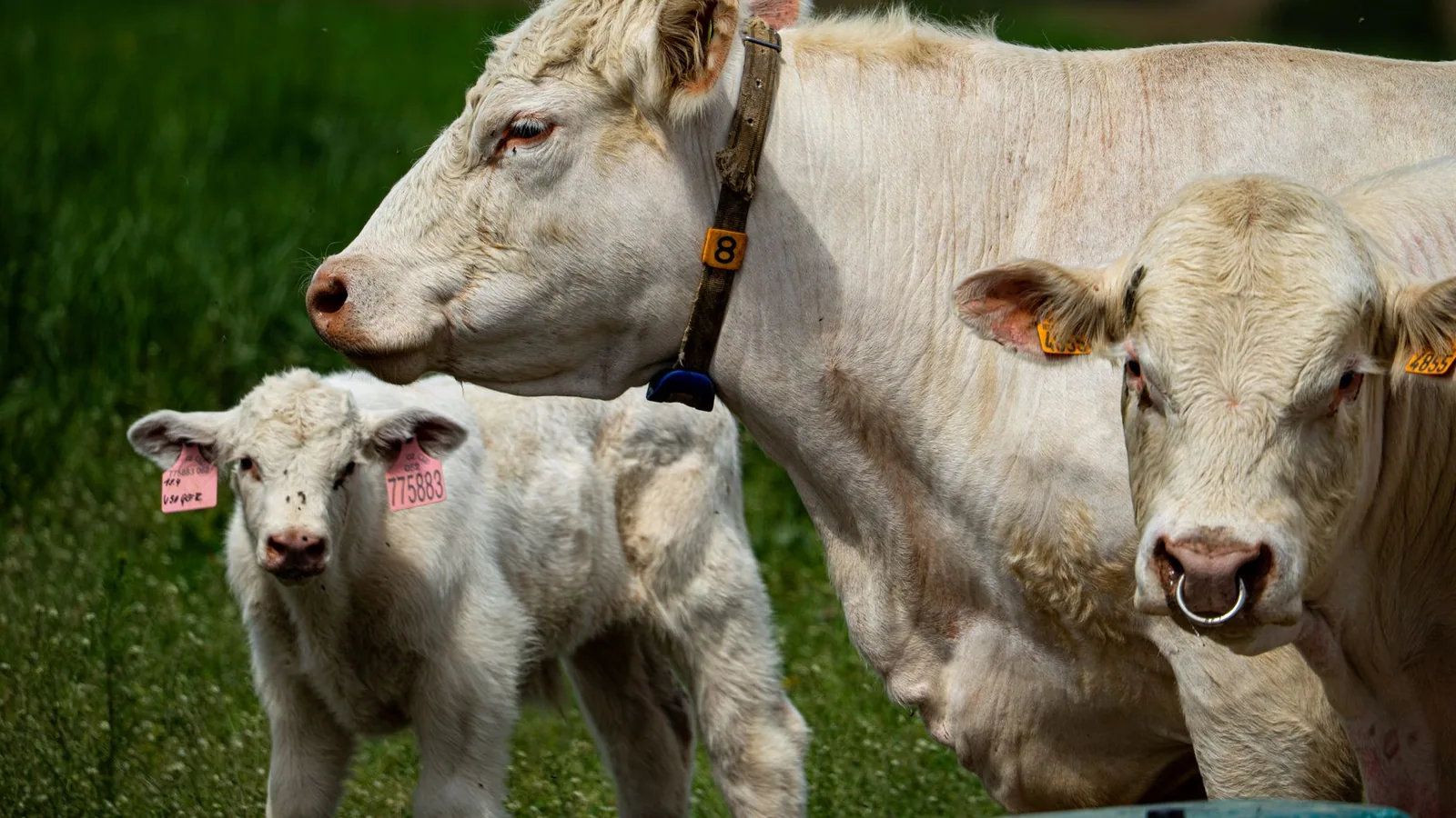
The host range of CCHFV is exceptionally broad, encompassing domestic ruminants (cattle, sheep, goats, and camels), equids, swine, wild ungulates (deer, wild boar, buffalo, giraffe, and various antelope species), lagomorphs, hedgehogs, and multiple species of birds [1, 3-5, 15, 39]. This remarkable host breadth is a reflection of the virus's ability to replicate efficiently in diverse mammalian and avian tissues, a trait that facilitates its maintenance across many ecological niches. Among domestic livestock, cattle and camels consistently exhibit the highest seroprevalence rates globally, often exceeding 50-90% in endemic regions [25, 26, 27, 22]. For instance, serosurveys in Mauritania have documented anti-CCHFV IgG prevalence of 74.6% in cattle and 90.5% in dromedary camels, while studies in Egypt have reported an extraordinary 99.6% seropositivity among camels [26, 27]. These findings underscore the intense exposure pressure on these species, likely attributable to their prolonged lifespan, extensive grazing habits, and heavy tick burdens [22]. Sheep and goats generally exhibit lower seroprevalence, typically ranging from 5% to 30%, although significant geographic variation exists [25, 28, 29, 30]. The age of the animal is a consistently identified risk factor for seropositivity across all livestock species, with older animals demonstrating significantly higher antibody prevalence due to cumulative lifetime exposure to infected ticks [28, 31, 22, 32]. This age-dependent seroprevalence pattern has been documented in cattle, sheep, goats, and camels across Africa, Asia, and Europe, and is considered a reliable indicator of endemic viral circulation [22].
Wild ungulates play a particularly important role in CCHFV ecology, as they are the primary hosts for adult Hyalomma ticks in many ecosystems [13, 5, 33, 14]. Red deer (Cervus elaphus) have been identified as a key sentinel species in Spain, where they harbor high densities of Hyalomma lusitanicum ticks and exhibit seroprevalence rates exceeding 20% in some regions [13, 33, 14]. Similarly, wild boar (Sus scrofa) have been shown to seroconvert following natural exposure, and their expanding populations across Europe may contribute to the northward spread of CCHFV [33, 34]. The Saharan antelope addax (Addax nasomaculatus) has been identified as a competent host for Hyalomma marginatum, the principal vector in North Africa, and has demonstrated exposure to Aigai virus, a recent taxonomic differentiation of CCHFV [35]. Zoo-housed wildlife, including white rhinoceroses (Ceratotherium simum) and dromedary camels, have also been found to harbor anti-CCHFV antibodies, indicating that captive animals in urban and peri-urban settings may serve as sentinels for viral circulation [36]. The detection of CCHFV antibodies in a white rhinoceros in a Spanish zoo, confirmed by virus neutralization testing, represents the first documented evidence of CCHFV exposure in this species and highlights the potential for viral introduction into novel environments through animal translocations [36].
Avian Involvement and the Role of Migratory Birds
The role of birds in CCHFV ecology is a subject of ongoing investigation. While birds are generally considered refractory to clinical disease, they are competent hosts for immature Hyalomma ticks (larvae and nymphs) and may facilitate the long-distance dispersal of infected ticks through migratory flyways [5, 17]. This mechanism is believed to be responsible for the introduction of CCHFV strains from West Africa into southwestern Europe, as evidenced by the close phylogenetic relationship between CCHFV sequences detected in Hyalomma lusitanicum ticks in Spain and those circulating in Mauritania and Senegal [5]. The detection of CCHFV RNA in Rhipicephalus evertsi evertsi and Rhipicephalus guilhoni ticks collected from vegetation in Senegal further supports the notion that multiple tick species, including those that parasitize birds, may contribute to viral maintenance and spread [37]. However, serological surveys in domestic chickens in Kosovo have failed to detect anti-CCHFV antibodies, suggesting that gallinaceous birds may not be significant amplifying hosts [38]. The precise contribution of avian species to CCHFV transmission dynamics remains an important gap in knowledge that warrants further investigation.
Implications for Surveillance and One Health
The asymptomatic nature of CCHFV infection in animals has profound implications for public health surveillance and disease control. Livestock and wild animals function as silent sentinels, and their serological status provides the earliest indication of viral circulation in a given geographic area [18, 23, 39, 30]. The WHO, in collaboration with the WOAH and the FAO, has advocated for the integration of animal health surveillance into CCHFV monitoring programs under the One Health framework, recognizing that human risk is inextricably linked to the ecological dynamics of the virus in animal and tick populations [25, 23, 17]. Serosurveys in livestock have proven instrumental in identifying previously unrecognized endemic foci, as demonstrated by the first detection of CCHFV antibodies in cattle in Croatia, where 28.3% of sheep and 4.3% of horses were seropositive, confirming silent viral circulation in a region previously considered free of the virus [39]. Similarly, the detection of seropositive livestock in the Vushtrri region of Kosovo, an area with no prior history of human CCHF cases, provided evidence of viral spread through animal movement and underscored the need for integrated surveillance [23].
The development of species-independent serological assays, such as the double-antigen ELISA and the recently described split-NanoLuc mix-and-read assays, has greatly facilitated large-scale screening of multiple animal species, enabling researchers to generate robust epidemiological data across diverse ecological settings [40, 9, 41, 11]. These tools, combined with molecular detection methods for viral RNA in ticks and animal blood, form the backbone of contemporary CCHFV surveillance efforts [42, 14, 15]. The identification of risk factors associated with CCHFV exposure in animals, including age, sex, breed, tick infestation, grazing practices, and environmental variables such as normalized difference vegetation index (NDVI) and vapor pressure deficit, provides actionable insights for targeted interventions [25, 43, 31, 27]. For example, the application of acaricides to livestock, particularly in high-risk age groups and during peak tick activity seasons, has been recommended as a cost-effective measure to reduce viral amplification and subsequent spillover to humans [25, 43]. The complex interplay between animal hosts, tick vectors, environmental conditions, and human behavior necessitates a multidisciplinary, One Health approach to effectively mitigate the expanding threat of CCHFV across its geographic range.
Molecular Pathogenesis of CCHFV in Asymptomatic Animal Reservoirs
The remarkable dichotomy between the severe, often fatal hemorrhagic disease elicited by Crimean-Congo hemorrhagic fever virus (CCHFV) in humans and its clinically silent, yet virologically productive, infection in a broad spectrum of animal hosts represents one of the most compelling and unresolved questions in nairovirus biology. This differential pathogenicity is not a matter of host range restriction; rather, it is a profound divergence in disease outcome that underpins the entire ecology of CCHFV as a zoonotic agent. The molecular mechanisms governing this asymptomatic state in reservoir species, including livestock (cattle, sheep, goats, camels) and wild ungulates (e.g., red deer, giraffes, buffalo), are multifactorial, involving viral factors, host cellular permissiveness, innate immune evasion, and the architecture of the host immune response. Understanding these mechanisms is not merely an academic exercise; it is paramount for predicting spillover risk, identifying potential sentinel species, and rationally designing countermeasures, including vaccines that might mimic the protective, disease-sparing immunity observed in animals [4, 1, 16].
Immunodominance of the Nucleocapsid Protein and Humoral Response Dynamics
The serological hallmark of CCHFV infection in asymptomatic animals is the robust and sustained production of antibodies targeting the viral nucleoprotein (NP). The NP is the most abundant viral protein and the primary antigen recognized during serosurveillance across multiple species [9, 11]. The development of species-independent antibody detection assays, such as the double-antigen ELISA and the recently described split-NanoLuc (NanoBiT) mix-and-read assays, has confirmed that anti-NP antibodies are a consistent and reliable marker of past or current infection across diverse taxa, from cattle and camels to wild boar and white rhinoceroses [36, 25, 40]. Critically, the immune response in animals is not solely directed at NP. Multiplex assays have demonstrated that seropositive ruminants frequently harbor antibodies against glycoproteins GP38 and GNe, indicating that natural infection elicits a broader humoral response than often appreciated [9]. However, a key finding in porcine models is that antibody development does not universally correlate with the presence of neutralizing antibodies. Studies in wild boar and domestic pigs revealed that while many animals seroconvert as measured by ELISA and indirect immunofluorescence assays (iIFA), only a subset of these samples possess neutralizing capacity against the virus [34]. This suggests that the immunological control of CCHFV in asymptomatic animal hosts may be less dependent on classical neutralizing humoral immunity and more reliant on other effector functions, such as Fc-mediated mechanisms or complement-dependent activity, particularly for GP38-specific antibodies, which have been shown to be protective in murine models despite lacking neutralizing activity [34, 12].
Cellular Determinants of Restrictive Infection: The Bovine Paradigm
A pivotal molecular clue to the asymptomatic phenotype lies in the differential cellular permissiveness between human and animal cells. The most direct evidence comes from comparative in vitro studies using kidney and adrenal gland cells of human and bovine origin. Infection with CCHFV in bovine kidney cells is marked by a highly restrictive pattern, characterized by a limited cell-to-cell spread that results in small, islet-like foci of infected cells. In stark contrast, human kidney cells become uniformly infected, with the virus spreading rapidly across the entire monolayer [21]. This observation is consistent with the epidemiological reality: cattle are a major amplifying host, yet they never develop clinical illness [18, 4, 30].
The molecular basis for this restriction appears to operate at multiple levels. First, viral replication, as measured by intracellular and extracellular viral RNA, is far more pronounced in human cells, supporting a model where the virus encounters a less permissive intracellular environment in bovine cells [21]. This could involve blocks at the level of viral entry, uncoating, transcription, or translation. Second, the mechanism of spread differs. The islet-like growth in bovine cells suggests a reliance on direct cell-to-cell transmission, potentially via the formation of viral synapses or through exploitation of intercellular junctions, a process that may limit the systemic dissemination seen in humans. The Gn cytoplasmic tail of CCHFV, which contains dual zinc finger motifs that bind viral RNA, has been structurally characterized and is known to be critical for viral assembly and budding [7]. Subtle differences in how the Gn tail interacts with host factors in bovine versus human cells could influence the efficiency of particle assembly and release, contributing to the observed restriction.
Early Cellular Targeting and the Interferon Axis
The early events of infection are decisive in determining disease outcome. Using a fluorescent reporter virus (CCHFV/ZsG) in a murine model, studies have identified mononuclear phagocytic cells, including macrophages and dendritic cells, in lymphatic tissues (spleen, lymph nodes) as the primary early targets of CCHFV [44]. This initial targeting of the innate immune system is a critical juncture. In humans, CCHFV is known to subvert the host interferon (IFN) response, leading to uncontrolled viral replication and a "cytokine storm" that contributes to vascular dysfunction and hemorrhage [1, 3]. In contrast, it is hypothesized that in animal reservoirs, the initial infection of antigen-presenting cells triggers a more tempered and effective innate immune response that contains the virus.
Critically, the commonly used immunocompetent animal models for CCHFV are not natural reservoirs; rodents such as mice are not susceptible unless they are deficient in the interferon-α/β receptor (IFNAR-/-) [44]. This fact underscores that a robust and functional type I IFN response is essential for controlling CCHFV replication in most mammals. The natural reservoir species (e.g., cattle, sheep, deer) likely possess innate immune pathways that are either more efficient at sensing the virus or are less effectively antagonized by CCHFV. This could involve differences in the RIG-I/MDA5 sensing pathways or in the activity of interferon-stimulated genes (ISGs). For example, the Mx proteins or protein kinase R (PKR) in livestock may have a more potent antiviral effect against CCHFV than their human orthologs. This divergence in the early innate immune 'set point' likely dictates whether the infection remains a localized, controlled event or becomes a systemic, fulminant disease.
Persistent Infection and Viral Seeding of Tissues
While CCHFV infection in animals is typically considered acute and self-limiting (with viremia lasting days to a week), emerging evidence from non-human primate models suggests a more complex scenario with implications for long-term viral maintenance. In cynomolgus macaques infected with CCHFV, viral RNA was found to persist in the testes of surviving males, indicating that the virus can establish a niche in immunologically privileged sites [45]. Even more strikingly, in animals with concurrent latent mycobacterial infection (tuberculosis), CCHFV was detected within the lung granulomas for an extended period [45]. This suggests that pre-existing inflammatory or granulomatous lesions can serve as sanctuaries for viral persistence. While the relevance of these findings to natural reservoirs like cattle (which frequently have other infections) is unknown, it raises the possibility that persistent infection in tissues could contribute to the long-term maintenance of the virus within a herd, allowing for transmission to ticks during subsequent feeding events even after the acute viremic phase has resolved. This concept of persistent infection in reservoir hosts, far beyond the brief viremic window, would have profound implications for understanding the enzootic cycle and designing effective surveillance strategies.
The molecular pathogenesis of CCHFV in asymptomatic animal reservoirs is therefore a story of host-specific viral restriction. It involves a combination of a less permissive cellular environment, a more effective and controlled innate immune response, and an antibody response that, while robust, may rely on non-neutralizing effector functions. The recent discoveries of potential viral persistence in tissues further complicate the picture, suggesting that the "asymptomatic carrier" state may be more dynamic and long-lasting than previously assumed. Deciphering the precise host factors that confer this resistance to disease remains a high priority for the development of next-generation therapeutics and vaccines for human use.
Seroepidemiology of CCHFV in Domestic Livestock (Cattle, Sheep, Goats, Buffalo)
The seroepidemiology of Crimean-Congo hemorrhagic fever virus (CCHFV) in domestic livestock constitutes the cornerstone of our understanding of the virus’s ecological maintenance, spatial distribution, and zoonotic risk. Livestock, principally cattle, sheep, goats, and buffalo, serve as the primary amplifying hosts for CCHFV in the peridomestic environment, and their serological profiles provide a robust, cost-effective sentinel system for mapping viral circulation. Unlike humans, these animals do not develop clinical disease following infection, yet they mount a durable antibody response that can be detected months to years post-exposure [18, 4]. This characteristic makes serosurveillance in livestock an indispensable tool for public health authorities, including the World Health Organization (WHO) and the World Organisation for Animal Health (WOAH), which recognize CCHF as a priority zoonosis requiring integrated One Health surveillance.
Species-Specific Seroprevalence Patterns: A Global Perspective
The seroprevalence of CCHFV in livestock varies dramatically across geographic regions, husbandry systems, and species, reflecting the complex interplay between vector ecology, host susceptibility, and environmental drivers. A landmark global review by Spengler et al. (2016) synthesized over five decades of seroepidemiological data, demonstrating that cattle and camels consistently exhibit the highest seroprevalence rates, often exceeding 50% in endemic foci, while small ruminants (sheep and goats) typically show lower but still substantial exposure [18]. This pattern has been corroborated by numerous contemporary studies. In Isiolo County, Kenya, Mukhaye et al. (2024) reported anti-CCHFV IgG seroprevalences of 53.9% in cattle, 11.6% in goats, and 8.6% in sheep, with camels reaching an extraordinary 89.7% [25, 43]. Similarly, in Mauritania, Schulz et al. (2021) found that 69% of cattle and 81% of camels were seropositive, compared to approximately 15% in both sheep and goats [22]. These data underscore a recurring theme: large-bodied ruminants, particularly those with longer lifespans and more extensive tick burdens, accumulate seropositivity over time.
The biological basis for these species differences is multifactorial. Cattle and buffalo are preferential hosts for adult Hyalomma ticks, the principal vectors of CCHFV, due to their large body surface area and prolonged grazing periods [4, 19]. Experimental infection studies have demonstrated that cattle develop a viremia lasting up to 14 days post-infection, providing a substantial window for tick acquisition and subsequent transmission [4]. In contrast, sheep and goats, while susceptible, often exhibit lower viremic titers and shorter duration of infectivity, which may contribute to their comparatively lower seroprevalence in many settings [18, 4]. The role of buffalo, particularly water buffalo (Bubalus bubalis), has been less extensively studied, but available data from India and Egypt indicate seroprevalence rates comparable to or exceeding those in cattle. Mourya et al. (2012) detected CCHFV IgG antibodies in 43% of domestic animals (including buffalo) in a village in Gujarat, India, with one buffalo testing positive for viral RNA by qRT-PCR [42]. In Egypt, Chisholm et al. (2012) identified CCHFV RNA in Hyalomma ticks collected from imported buffalo at an abattoir, confirming that these animals can serve as competent amplifying hosts [24].
Age as the Dominant Risk Factor
Across virtually all livestock species and geographic settings, age emerges as the single most consistent and powerful predictor of CCHFV seropositivity. This relationship is not merely correlative but reflects cumulative exposure over time in an environment where the virus circulates enzootically. In the Turkestan region of Kazakhstan, Myrzakhmetova et al. (2026) reported that adult cattle had 4.06 times higher odds of seropositivity compared to juveniles (43.42% vs. 11.86%), a finding that was highly statistically significant [31]. Similarly, in Uganda, Nyakarahuka et al. (2023) demonstrated that adult and juvenile animals had significantly higher seropositivity than recently born animals, with the effect persisting after adjustment for species, sex, and region [30]. In Burkina Faso, Dahourou et al. (2024) found that seroprevalence in cattle increased progressively with age, with older animals (>5 years) showing rates exceeding 80% [46].
The mechanistic underpinning of this age-dependent pattern is straightforward: older animals have had more opportunities for tick exposure over their lifetimes. However, the relationship is not simply linear. Schulz et al. (2021) in Mauritania made the critical observation that the higher seroprevalence in cattle and camels compared to small ruminants could be largely explained by the fact that cattle and camels were, on average, two to four times older than the sheep and goats sampled [22]. When age was accounted for in multivariate models, the species-specific differences narrowed considerably, suggesting that age is a confounder that must be rigorously controlled in any comparative seroepidemiological study. This finding has profound implications for study design: cross-sectional surveys that do not stratify by age risk producing misleading estimates of species susceptibility and may obscure true geographic or temporal trends in viral circulation.
Geographic Heterogeneity and Environmental Drivers
The seroepidemiology of CCHFV in livestock is characterized by striking geographic heterogeneity, even within relatively small spatial scales. This patchiness reflects the focal distribution of Hyalomma tick populations, which are themselves constrained by climatic and ecological factors. In Kosovo, Zhabari and Xhekaj (2022) documented a seroprevalence of 24.7% in livestock from the Malisheve region, a known endemic focus, compared to only 4.8% in Vushtrri, a region without prior human cases [23]. The detection of seropositive animals in Vushtrri for the first time was interpreted as evidence of viral spread, likely facilitated by the movement of infected livestock. In Croatia, Miletić et al. (2025) reported that almost all seropositive animals originated from coastal and subcoastal areas where Hyalomma ticks are established, with sheep from several farms in Zadar County showing intra-farm seropositivity rates of up to 85.7% [39]. These localized hotspots underscore the importance of farm-level risk factors, including grazing practices, tick control measures, and proximity to wildlife habitats.
Environmental and climatic variables exert a powerful influence on CCHFV seroprevalence in livestock. Mukhaye et al. (2024) in Kenya demonstrated that higher normalized difference vegetation index (NDVI) and higher vapor pressure deficit were significantly associated with CCHFV exposure in livestock, likely because these conditions favor tick survival and questing activity [25, 43]. In the Democratic Republic of the Congo, Lombe et al. (2026) found that high seroprevalence in ruminants correlated with tropical monsoon climate zones, mountain savanna vegetation types, and elevations above 600 meters [6]. These environmental associations are not merely academic; they provide the basis for predictive risk mapping that can guide targeted surveillance and control interventions. The WHO and FAO have emphasized the utility of such spatial epidemiological approaches for anticipating CCHFV emergence in previously unaffected areas, particularly in the context of climate change, which is expected to expand the geographic range of Hyalomma ticks northward into Europe [20, 17].
Temporal Dynamics and the Role of Livestock Movement
Longitudinal serosurveillance data, while less common than cross-sectional surveys, reveal that CCHFV circulation in livestock populations is dynamic and can change rapidly over time. In Spain, where CCHFV was first detected in ticks in 2010 [5], subsequent surveillance has demonstrated sustained viral circulation in livestock and wild ungulates, with evidence of multiple genotypes (I, III, and IV) circulating in Hyalomma lusitanicum ticks collected from cattle and red deer [13, 14]. The detection of seropositive livestock in the Basque Country, an area north of the traditionally recognized vector range, suggests that the virus may be more widely distributed than previously appreciated, possibly due to the transport of infested animals or the northward expansion of tick populations [33].
The movement of livestock, whether through trade, transhumance, or seasonal migration, is a critical driver of CCHFV dissemination. In Kosovo, the first detection of seropositive livestock in the Vushtrri region was attributed to the movement of infected animals from endemic areas [23]. In Egypt, CCHFV RNA was detected in Hyalomma ticks collected from camels imported from Sudan and Somalia at an abattoir, demonstrating that international livestock trade can introduce novel viral variants into new regions [24]. Similarly, in India, retrospective analysis of samples from Gujarat and Rajasthan revealed that CCHFV had been circulating in livestock before the 2011 nosocomial outbreak in Ahmadabad, with phylogenetic analysis linking the Indian strains to the Asian/Middle Eastern lineage IV [42]. These findings highlight the inadequacy of static, geographically bounded surveillance systems and argue for a more dynamic, network-based approach that tracks livestock movements and their associated tick populations.
Implications for Human Risk and Public Health Surveillance
The seroprevalence of CCHFV in livestock is not merely an ecological curiosity; it is a direct and quantifiable predictor of human infection risk. Multiple studies have demonstrated that human seropositivity is significantly associated with household-level livestock seropositivity. In Kenya, Mukhaye et al. (2024) found that belonging to a household with a seropositive herd was a key determinant of human CCHFV exposure (P = 0.041) [25, 43]. In Turkey, Gunes et al. (2009) reported that employment in animal husbandry and history of tick removal from animals were significant risk factors for human seropositivity [47]. These associations are biologically plausible: viremic livestock serve as a source of infection for both ticks and humans, particularly during slaughter, when direct contact with blood and tissues can lead to transmission [4, 3]. The WHO has classified CCHF as a priority disease for research and development, and the WOAH includes CCHFV in its list of notifiable terrestrial animal diseases, underscoring the global recognition of livestock as the critical bridge between the enzootic cycle and human disease.
Given the asymptomatic nature of CCHFV infection in livestock, serological surveillance is the most practical and informative approach for monitoring viral circulation. The development of multi-species, double-antigen ELISA assays, such as the ID Screen® CCHF test, has revolutionized the field by enabling high-throughput, species-independent screening [40, 9]. These assays detect antibodies against the nucleoprotein (NP), which is highly conserved across CCHFV strains, and have demonstrated excellent sensitivity and specificity in cattle, sheep, goats, and camels [11]. The recent development of multiplex assays capable of simultaneously detecting antibodies against NP, glycoprotein Gc, and GP38 provides additional granularity, allowing researchers to differentiate between natural infection and potential future vaccine-induced immunity [9]. Such tools are essential for implementing the One Health surveillance frameworks advocated by the WHO, FAO, and WOAH, which call for integrated human, animal, and environmental monitoring to predict and prevent CCHFV spillover.
The seroepidemiology of CCHFV in domestic livestock reveals a complex, spatially heterogeneous, and temporally dynamic pattern of viral circulation that is shaped by host species, age, environmental conditions, and livestock movement. Cattle and camels consistently exhibit the highest seroprevalence rates, while small ruminants show lower but still epidemiologically significant exposure. Age is the dominant individual-level risk factor, reflecting cumulative tick exposure over time. Geographic heterogeneity is pronounced, with localized hotspots driven by vector ecology and farm-level practices. The strong association between livestock seropositivity and human infection risk underscores the critical importance of livestock surveillance as an early warning system for public health. As CCHFV continues to expand its geographic range in Europe, Africa, and Asia, robust, age-stratified, and spatially explicit serosurveillance in livestock will remain an indispensable component of global efforts to control this emerging zoonotic threat.
Seroepidemiology of CCHFV in Wild Animal Species and Ecological Reservoirs
The seroepidemiology of Crimean-Congo hemorrhagic fever virus (CCHFV) in wild animal populations represents a critical, yet historically understudied, dimension of the virus's complex ecology. Unlike domestic livestock, which have been extensively surveyed due to their direct interface with human populations and economic importance, wild animals occupy a more nuanced role as both sentinel hosts and potential true ecological reservoirs. The World Health Organization (WHO) has classified CCHFV as a priority pathogen, and understanding its sylvatic cycle is paramount for predicting emergence events and implementing effective One Health surveillance strategies. The available serological evidence, drawn from over five decades of research across four continents, reveals a pattern of widespread, often asymptomatic, infection across a remarkable diversity of mammalian taxa, with seroprevalence rates that frequently exceed those observed in domestic livestock in the same geographic regions [18, 4]. This section provides an exhaustive analysis of the seroepidemiological data pertaining to wild animal species, examining the biological mechanisms underlying their role in viral maintenance, the spatial and temporal patterns of exposure, and the critical knowledge gaps that remain.
The Role of Wild Ungulates as Sentinel Hosts and Amplifiers
Wild ungulates, particularly members of the families Cervidae (deer) and Bovidae (antelope, buffalo, and giraffe), have emerged as the most consistently seropositive wild animal group across diverse ecosystems. The red deer (Cervus elaphus) has been identified as a keystone species in the maintenance of CCHFV in southwestern Europe, particularly in Spain. In the initial detection of CCHFV in ticks from the Iberian Peninsula, Hyalomma lusitanicum ticks collected from red deer in Cáceres province yielded viral RNA with 98% genetic similarity to strains circulating in West Africa, suggesting a potential introduction via migratory birds and subsequent establishment in a competent ungulate-tick cycle [5]. Subsequent large-scale surveillance in Spain confirmed that red deer are the host most frequently yielding CCHFV-positive ticks, with the virus detected in 135 of 4,556 tick pools across multiple regions, predominantly in H. lusitanicum [13]. This association is not coincidental; red deer are the primary hosts for adult H. lusitanicum ticks, and their wide home ranges, high population densities, and longevity facilitate both the maintenance of tick populations and the spatial dissemination of the virus. A retrospective study spanning 2017 and 2020-2024 in western Spain further reinforced this finding, detecting CCHFV exclusively in H. lusitanicum ticks with an overall infection rate of 1.54%, with the majority of positive pools originating from wild ungulates, particularly red deer [14]. The sustained circulation of the virus in this region over multiple years, with the detection of both genotype III and other lineages, underscores the role of red deer as a stable reservoir host that supports enzootic transmission.
Beyond Europe, wild ungulates in Africa exhibit remarkably high seroprevalence rates. In a comprehensive seroepidemiological study conducted at the human-livestock-wildlife interface in Isiolo County, Kenya, wild animals (n=87) demonstrated an overall CCHFV seroprevalence of 41.0% (95% CI: 29.1-49.4%), with giraffes exhibiting the highest mean seroprevalence at 75.0% (95% CI: 63.2-79.3%), followed by buffaloes [25, 43]. These figures are striking, particularly when compared to the seroprevalence in sympatric livestock species such as goats (11.6%) and sheep (8.6%), although they are comparable to the exceptionally high seroprevalence observed in camels (89.7%) in the same study area [25]. The high seroprevalence in giraffes and buffaloes likely reflects their long lifespans, their role as preferred hosts for Hyalomma ticks, and their extensive grazing behavior that brings them into frequent contact with tick-infested vegetation. Interestingly, the same study found that age, sex, and species of wild animals were not significant predictors of seropositivity in a mixed-effects logistic regression model (P-values of 0.891, 0.401, and 0.664, respectively), suggesting that exposure risk is more uniformly distributed among wild ungulates compared to livestock, where management practices and age structure play a more pronounced role [25, 43]. This finding implies that in wild populations, ecological factors such as habitat use and tick density may be more important determinants of exposure than individual host characteristics.
In the northern Iberian Peninsula, where the primary vector Hyalomma spp. is not considered permanently established, serological evidence in wild ungulates has revealed cryptic circulation of CCHFV. A study in the Basque Country detected anti-CCHFV antibodies in 2.5% of 1,190 wild boar, 22.2% of 36 red deer, and 2.8% of 36 roe deer sampled between 2014 and 2019 [33]. The highest seroprevalence was found in the southwestern province of Araba (8.6%), and the detection of exposed animals as early as 2014 suggests that viral circulation beyond the traditionally recognized endemic zones of southwestern Spain may be more widespread than previously appreciated [33]. The presence of antibodies in wild boar, a species with high fecundity and extensive ranging behavior, is particularly noteworthy. Wild boar are known to host a variety of tick species, including Dermacentor and Rhipicephalus spp., and their seropositivity may indicate either spillover from infected Hyalomma ticks transported by migratory birds or the potential involvement of alternative tick vectors in viral maintenance [33]. This finding has significant implications for risk assessment in northern Europe, as it suggests that CCHFV can circulate in areas where the classical vector is not abundant, potentially via less efficient but locally abundant tick species.
Carnivores, Omnivores, and the Expanding Host Range
The seroepidemiological evidence for CCHFV infection in carnivores and omnivores is more limited but nonetheless revealing. In Croatia, a multispecies surveillance approach that included companion animals detected anti-CCHFV antibodies in 4.3% of horses and 0.5% of cats, while no antibodies were found in dogs [39]. The detection of a seropositive cat is particularly intriguing, as felids are not typically considered important hosts for Hyalomma ticks. This finding may reflect incidental exposure through predation on infected small mammals or birds, or through contact with contaminated environments. The absence of seropositivity in dogs, despite their close association with humans and livestock, suggests that canids may be less susceptible to infection or that their exposure risk is lower due to differences in tick infestation patterns or behavior [39]. The seropositivity in horses, while lower than that observed in ruminants, is consistent with their role as hosts for Hyalomma ticks and their potential to serve as sentinels for viral circulation in peri-urban and rural environments.
In Spain, a unique study of zoo-housed wildlife provided the first global assessment of CCHFV exposure in captive animals. Among 956 mammals representing 173 species and 38 families across 19 zoos and wildlife rescue centers, anti-CCHFV antibodies were detected in two white rhinoceroses (Ceratotherium simum) and one dromedary camel (Camelus dromedarius), yielding an overall seroprevalence of 0.3% (95% CI: 0.0-0.7) [36]. Virus neutralization testing confirmed the presence of specific neutralizing antibodies in one white rhinoceros, providing unequivocal evidence of past infection. The low seroprevalence in zoo animals, compared to wild populations, likely reflects the controlled environments of zoos, where tick exposure is minimized through regular acaricide treatments and habitat management. However, the detection of seropositive animals in a single zoo in central Spain highlights the potential for localized transmission hotspots even in managed settings, and underscores the importance of including captive wildlife in surveillance programs, particularly in high-risk areas [36].
The Saharan antelope addax (Addax nasomaculatus) has been identified as a significant host for Hyalomma marginatum, the primary vector of CCHFV in North Africa. In a study of natural-living addax in Morocco, 100% of H. marginatum tick pools collected from the animals were positive for Aigai virus (AIGV), a recent taxonomic differentiation of CCHFV [35]. This finding, combined with the detection of other tick-borne pathogens such as Babesia ovis, Anaplasma spp., and Rickettsia spp., demonstrates that the addax is a competent host for maintaining tick populations and exposing them to a diverse array of pathogens. The conservation status of the addax, a critically endangered species, adds an additional layer of complexity, as interventions such as anti-tick vaccines may be necessary both for preserving the species and for mitigating the risk of CCHFV spillover to humans and livestock in shared habitats [35].
Avian Involvement and the Role of Migratory Birds
The role of birds in the ecology of CCHFV has been a subject of considerable debate. While birds are generally considered refractory to CCHFV infection and do not develop detectable viremia, they play a critical indirect role as hosts for immature Hyalomma ticks. Migratory birds, in particular, are implicated in the long-distance dispersal of infected ticks, potentially introducing the virus into new geographic regions. The detection of a CCHFV strain in Spain with 98% genetic similarity to West African isolates, coupled with the lack of similarity to Eastern European strains, strongly supports the hypothesis of introduction via migratory birds traveling along the western coast of Africa [5]. This mechanism is further supported by the observation that Hyalomma marginatum nymphs are commonly found on passerine birds during spring migration, and that these ticks can remain attached for several days, allowing for transport over thousands of kilometers.
Direct serological evidence of CCHFV infection in birds is scarce, but not entirely absent. In Kosovo, a study testing 401 animal sera, including 8 chickens, detected specific antibodies in all mammalian species tested but not in chickens [38]. This finding is consistent with the general understanding that birds do not mount a detectable antibody response to CCHFV, likely due to fundamental differences in their immune system or the inability of the virus to replicate efficiently in avian cells. However, the absence of serological evidence does not preclude the possibility that birds could serve as mechanical vectors, carrying the virus on their feathers or skin without becoming infected themselves. The role of ostriches and other large flightless birds, which are sometimes infested with Hyalomma ticks, remains unexplored and warrants further investigation.
Biological Mechanisms of Asymptomatic Infection in Wild Animals
The consistent observation that wild animals, like domestic livestock, do not develop clinical disease following CCHFV infection is a fundamental aspect of the virus's ecology. This phenomenon is not due to a lack of viral replication; experimental infection studies have demonstrated that wild and domestic animals develop a transient viremia lasting up to 14 days, with viral loads sufficient to infect feeding ticks [4]. The mechanisms underlying this species-specific resistance to disease are not fully understood, but in vitro studies comparing human and bovine kidney cells have provided important insights. CCHFV infection in bovine kidney cells is characterized by a restricted, cell-to-cell spread pattern, with infected cells found in isolated islet-like areas, whereas human kidney cells support robust viral replication leading to 100% infection of the monolayer within three days post-infection [21]. This differential permissiveness suggests that bovine cells, and by extension the cells of other ungulates, possess intrinsic antiviral mechanisms that limit viral spread and cytopathology. The identification of these mechanisms could have profound implications for understanding CCHFV pathogenesis and for developing therapeutic interventions.
The lack of clinical signs in wild animals has significant epidemiological consequences. Asymptomatic, viremic animals continue to forage, migrate, and interact with ticks, facilitating the maintenance and dissemination of the virus without the behavioral changes that would typically accompany illness. This silent transmission cycle makes it exceedingly difficult to detect viral circulation without active serological surveillance. The long lifespan of many wild ungulates, such as giraffes and buffaloes, allows for repeated exposure and the maintenance of high antibody titers, which can persist for years. This longevity of antibody responses is a key reason why seroprevalence in wild animals is often higher than in livestock, where animals are typically slaughtered at younger ages [22]. The age-stratified seroprevalence data from livestock studies, which consistently show higher seropositivity in older animals, are directly applicable to wild populations, where the age structure is often skewed towards older individuals due to lower predation and hunting pressure in protected areas [31, 22].
Spatial and Temporal Patterns in Wild Animal Seroprevalence
The seroepidemiology of CCHFV in wild animals exhibits pronounced spatial heterogeneity, reflecting the distribution of competent tick vectors, suitable habitat, and host density. In Spain, the highest seroprevalence in wild ungulates is concentrated in the southwestern regions, particularly in the autonomous community of Extremadura, where H. lusitanicum is abundant and red deer populations are dense [13, 14]. The detection of CCHFV in ticks from red deer in this region over multiple years, with an infection rate of 1.54% in H. lusitanicum, indicates stable, enzootic transmission [14]. In contrast, seroprevalence in wild ungulates in the Basque Country, located over 500 km to the north, is an order of magnitude lower (2.5%), suggesting that viral circulation in this region is sporadic and likely dependent on the annual influx of infected ticks carried by migratory birds [33]. This spatial gradient highlights the importance of vector distribution as the primary driver of CCHFV circulation in wildlife.
In Africa, the spatial patterns are influenced by a complex interplay of climatic factors, vegetation, and land use. In Kenya, the high seroprevalence in wild ungulates (41.0%) was observed in Isiolo County, a semi-arid region characterized by a human-livestock-wildlife interface [25, 43]. The study found that environmental factors such as the Normalized Difference Vegetation Index (NDVI) and vapor pressure deficit were significantly associated with CCHFV exposure in livestock, and it is reasonable to assume that similar factors influence exposure risk in wild animals [25, 43]. Areas with higher NDVI, indicating greener vegetation, likely support higher tick densities and provide more favorable conditions for tick survival and host-seeking behavior. The high seroprevalence in giraffes and buffaloes in this region may reflect their preference for riparian habitats and wooded savannas, where tick abundance is highest.
Temporal patterns in wild animal seroprevalence are less well-documented due to the logistical challenges of longitudinal sampling. However, the available data suggest that seroprevalence can fluctuate seasonally, in concert with tick activity. In Senegal, a study of sentinel sheep in Agnam found that the risk of CCHFV infection was significantly higher during the dry season, which corresponds to the peak activity period for adult Hyalomma ticks [37]. This seasonal pattern is likely to be similar in wild ungulates, which are exposed to the highest tick challenge during the dry season when animals congregate around limited water sources. The persistence of antibodies in wild animals means that seroprevalence surveys conducted at any time of year will detect past infections, but the detection of active viral RNA is rare and typically confined to
Role of Hyalomma Ticks as Biological Vectors and Viral Maintenance Hosts
The perpetuation of Crimean-Congo hemorrhagic fever virus (CCHFV) in nature is inextricably linked to ticks of the genus Hyalomma, which function not merely as mechanical vectors but as genuine biological vectors and long-term viral maintenance hosts. This fundamental ecological relationship underpins the entire epidemiology of CCHF, as recognized by the World Health Organization (WHO) which classifies CCHFV as a high-priority pathogen precisely because of its tick-borne transmission cycle and potential for widespread dissemination. The Hyalomma tick-virus interface represents a sophisticated co-evolutionary system wherein the virus is perpetuated through transstadial and transovarial transmission pathways, enabling viral persistence even in the absence of vertebrate reservoirs for extended periods [4, 19]. Understanding the biological mechanisms, vector competence, and maintenance dynamics of Hyalomma ticks is therefore essential for comprehending CCHFV ecology and for designing effective surveillance and control strategies.
Biological Vector Mechanisms and Viral Maintenance
The concept of Hyalomma ticks as biological vectors implies that CCHFV undergoes replication within the tick tissues, not merely mechanical transfer on mouthparts. Experimental evidence demonstrates that once a Hyalomma tick acquires CCHFV through feeding on a viremic vertebrate host, the virus disseminates within the tick's hemolymph and internal organs, including the salivary glands, reproductive tissues, and midgut epithelium [4, 19]. This systemic infection allows for subsequent transmission to naive vertebrate hosts during later blood meals. Critically, Hyalomma ticks exhibit transstadial transmission, meaning that the virus persists through the molting process from one life stage to the next, from larva to nymph, and from nymph to adult [4, 19]. This biological feature ensures that once a tick becomes infected, it remains infectious for the remainder of its life cycle, which can span several years depending on environmental conditions. Transovarial transmission has been documented in Hyalomma species, whereby infected female ticks pass the virus to their offspring through the eggs [19, 3]. This vertical transmission mechanism is particularly significant because it provides a mechanism for viral persistence within tick populations independently of vertebrate reservoirs, allowing CCHFV to overwinter or persist during periods when vertebrate hosts are scarce or non-viremic.
A comprehensive systematic review and meta-analysis of CCHFV infection in ticks revealed that among 31 tick species found to harbor the virus, 15 species have been confirmed as proven vectors through rigorous experimental and field evidence, while an additional 16 species are considered potential vectors pending further confirmation [19]. The same analysis identified that there are no significant differences in CCHFV infection rates between tick species, between male and female ticks, or across different continents, suggesting a broad and relatively uniform vector competence across the Hyalomma genus [19]. This finding underscores that Hyalomma ticks universally serve as both reservoirs and vectors, with a low but epidemiologically significant burden of CCHFV in natural enzootic cycles being sufficient to maintain viral circulation and occasionally spill over into human populations [19]. The World Organisation for Animal Health (WOAH) recognizes this tick-borne transmission as the primary mechanism for CCHFV maintenance in livestock systems, emphasizing the need for integrated vector management.
Key Hyalomma Vector Species and Geographic Distribution
The genus Hyalomma comprises multiple species that serve as primary vectors in different geographic regions, each with distinct ecological preferences and host associations. In Europe, particularly in the Iberian Peninsula, Hyalomma lusitanicum has emerged as the dominant vector species responsible for maintaining CCHFV in wildlife and livestock. Extensive surveillance in Spain has consistently detected CCHFV in H. lusitanicum ticks, with a large multidisciplinary study analyzing 12,584 ticks from 4,556 pools across numerous regions finding CCHFV in 135 pools, most commonly in H. lusitanicum [13]. This tick species was associated with the highest prevalence of viral detection, and genetic analysis revealed the circulation of CCHFV genotypes I, III, and IV in Spanish tick populations, indicating multiple introductions or long-term co-circulation of diverse viral lineages [13]. Further supporting the prominent role of H. lusitanicum, retrospective surveillance in southwestern Cáceres province over multiple years (2017 and 2020-2024) found that CCHFV was exclusively detected in H. lusitanicum ticks, with an overall infection rate of 1.54% (95% CI: 1.14-2.03), and most positive pools originated from ticks collected from wild ungulates, particularly red deer (Cervus elaphus) [14]. This sustained detection over multiple years confirms the establishment of enzootic circulation in this region and highlights the importance of H. lusitanicum as the primary vector maintaining the virus in wild ungulate populations.
In Africa, Hyalomma rufipes has been identified as a key vector species. Following the first symptomatic human case of CCHF in southeastern Senegal in 2023, entomological investigations revealed that H. rufipes was the first tick species found infected in this region, with a minimum field infection rate of 13.3% [48]. This finding is consistent with the broader distribution of H. rufipes across West Africa, where it commonly infests cattle and other livestock. In Senegal, tick infestation rates were significantly higher in cattle (8.2%) compared to sheep (1.2%), while goats were not infested, indicating species-specific host preferences that influence viral transmission dynamics [48]. In the Middle East and Central Asia, Hyalomma anatolicum serves as the principal vector, alongside Hyalomma marginatum and Hyalomma dromedarii. In Iran, a comprehensive spatial analysis of tick distribution over three decades (1979-2018) identified H. marginatum and H. anatolicum as the most important CCHFV vectors, reported in over 38.7% of provinces and classified as invasive species [49]. These two species accounted for the highest levels of CCHFV infection, with detection in 18 of 31 provinces [49]. Similarly, in India, Hyalomma anatolicum anatolicum ticks collected from livestock in Gujarat were found positive for CCHFV by PCR and yielded viral isolates, confirming their role in the enzootic cycle [42]. The detection of CCHFV RNA in H. anatolicum ticks in northeastern Iran further supported this species' vectorial capacity, with an infection rate of 15% in tested specimens [50]. In Armenia, molecular investigations during 2022-2024 detected CCHFV RNA in 96 pools of ticks collected from cattle across eight species, with positive samples confined to the Syunik and Tavush regions, and phylogenetic analysis revealed the presence of both Europe 1 (Subgroup V) and Europe 3 (Subgroup VII) lineages [15]. In Egypt, H. excavatum and H. dromedarii were found carrying CCHFV RNA in ticks collected from camels imported from Sudan and Somalia at an abattoir in Giza, with sequence analysis revealing a previously unrecorded viral variant [24]. The detection of CCHFV in H. dromedarii is particularly notable given the extensive camel trade networks in North Africa and the Middle East, which can facilitate long-distance dispersal of infected ticks [24, 35].
Tick-Host-Virus Transmission Dynamics
The transmission cycle of CCHFV involving Hyalomma ticks is complex and involves a diverse array of vertebrate hosts that serve as blood meal sources for different tick life stages. Larval and nymphal Hyalomma ticks predominantly feed on small mammals, hares, and ground-feeding birds, while adult ticks preferentially feed on large ungulates including cattle, sheep, goats, camels, and wild ruminants such as red deer [4, 5]. This bipartite feeding pattern is crucial for maintaining the enzootic cycle, as it connects different ecological compartments and ensures that the virus can circulate among both wildlife and domestic animal populations. The red deer (Cervus elaphus) has been identified as the host that most frequently yields CCHFV-positive ticks in Spain, and this wild ungulate plays a central role in the maintenance of the virus in natural ecosystems [13, 14]. Similarly, the Saharan antelope addax (Addax nasomaculatus) has been documented as an important host species for adult Hyalomma marginatum in Morocco, with ticks collected from these animals testing positive for Aigai virus (AIGV), a recent taxonomic differentiation of CCHFV, with 100% prevalence in tested tick pools [35]. This finding expands the known host range for CCHFV and highlights the potential role of threatened wildlife species in viral maintenance.
The ability of Hyalomma ticks to remain infected with CCHFV for extended periods, coupled with their long lifespan and capacity to survive in diverse environmental conditions, makes them exceptionally effective viral maintenance hosts. The virus can persist in unfed ticks for months to years, and infected ticks can transmit the virus to susceptible vertebrates during blood meals that may occur weeks or even months after the initial infection [4, 19]. This temporal separation between acquisition and transmission is a hallmark of biological vector systems and contributes to the epidemiological unpredictability of CCHF outbreaks. The role of migratory birds in transporting immature Hyalomma ticks across long distances has been implicated in the introduction of CCHFV into new geographic areas. The detection of CCHFV in H. lusitanicum ticks in Spain in 2010, with a strain showing 98% genetic similarity to sequences from Mauritania and Senegal, suggests that migratory birds traveling along the West African flyway could introduce virus-infected ticks into southwestern Europe [5]. This mechanism of long-distance dispersal explains the emergence of CCHFV in areas where the vector tick species are established but where the virus was previously absent [20, 5].
Co-infections and Ecological Complexity
Hyalomma ticks are also vectors for other pathogens of veterinary and medical importance, and co-infections with CCHFV and other tick-borne agents can influence transmission dynamics and host immunity. In ticks collected from addax in Morocco, H. marginatum pools were found to harbor not only AIGV (CCHFV) but also Babesia ovis (75% of pools), Anaplasma spp. (75%), Rickettsia spp. (50%), and Theileria spp. (25%) [35]. Similarly, Hyalomma anatolicum is the principal vector for Theileria annulata, T. equi, and T. lestoquardi in animals, in addition to CCHFV in humans, highlighting the multi-pathogen vectorial capacity of this tick species [51]. The development of anti-tick vaccines targeting multiple epitopes of H. anatolicum has shown promise in reducing tick infestation and potentially disrupting the transmission of both CCHFV and Theileria parasites, with vaccine efficacy reaching 93.3-96.9% against larval stages and 86.4-89.9% against adult ticks in rabbit models [51]. Such integrated approaches, recommended by the FAO for sustainable tick management, could simultaneously reduce the burden of multiple tick-borne diseases.
Environmental and climatic factors profoundly influence Hyalomma tick distribution, abundance, and viral infection rates, thereby shaping the geographic risk of CCHFV transmission. In Kenya, environmental factors such as normalized difference vegetation index (NDVI) and vapor pressure deficit were significantly associated with CCHFV exposure in livestock, with areas of higher NDVI and higher vapor pressure deficit showing increased seroprevalence [25, 43]. Spatial analysis in Kazakhstan demonstrated that seroprevalence ranged from 10% to 76.6% across districts, with communal grazing practices identified as a significant risk factor [31]. In the Democratic Republic of the Congo, high seroprevalences in cattle (94.6% in Tanganyika and 90.7% in Lualaba) were associated with specific climate conditions (tropical monsoon) and vegetation types (mountain savanna), and higher elevation (>600 m) [6, 52]. The expanding geographic range of Hyalomma ticks due to climate change and anthropogenic factors is a major concern for public health authorities, including the European Centre for Disease Prevention and Control (ECDC), which monitors the northward expansion of H. marginatum into previously unaffected regions of Europe [20, 17]. The detection of CCHFV antibodies in livestock in Croatia, with intra-farm seropositivity rates up to 85.7% in sheep from Zadar County, confirms localized virus circulation likely influenced by vector distribution and farm-level practices [39]. In Hungary, the first serological evidence of CCHFV in healthy blood donors (0.37% seroprevalence) coincided with the first detection of H. marginatum in the country in 2011, illustrating the direct link between vector establishment and viral emergence [53].
Diagnostic Detection and Isolation of CCHFV in Animal and Tick Samples
The accurate and timely detection of Crimean-Congo hemorrhagic fever virus (CCHFV) in animal and tick samples is foundational to understanding its ecology, mapping endemic foci, and implementing effective One Health surveillance strategies. Unlike human infections, which may present with severe hemorrhagic manifestations, CCHFV infection in vertebrate hosts is invariably subclinical, characterized by a transient, low-magnitude viremia lasting only a few days to a week, followed by seroconversion [4, 21]. This asymptomatic carrier state in livestock and wildlife, coupled with the prolonged infectivity of the tick vector, creates a unique diagnostic challenge. Consequently, a multi-pronged diagnostic approach, integrating molecular detection of viral RNA, virus isolation in permissive cell lines, and serological screening for anti-CCHFV antibodies, is essential for a comprehensive understanding of viral circulation. Each modality offers distinct advantages and limitations, and their application must be carefully tailored to the specific epidemiological question, the sample matrix, and the temporal stage of infection.
Molecular Detection of Viral RNA
Reverse transcription quantitative polymerase chain reaction (RT-qPCR) and conventional reverse transcription PCR (RT-PCR) represent the most sensitive and specific methods for the direct detection of CCHFV in animal blood and tick homogenates, particularly during the acute phase of viremia [42, 13]. These assays typically target the highly conserved small (S) genomic segment encoding the nucleoprotein (NP), ensuring broad reactivity across diverse viral lineages [42, 5]. The utility of these methods for active surveillance is well-documented. For instance, during the investigation of a nosocomial outbreak in Ahmadabad, India, qRT-PCR successfully identified CCHFV RNA in livestock (cattle, buffalo, and goat) and in Hyalomma anatolicum anatolicum ticks, confirming active viral circulation at the rural village residence of the index case [42]. Similarly, a large-scale study in Spain involving over 12,000 ticks collected from animals and vegetation utilized molecular methods to detect CCHFV in 135 pools, revealing the widespread distribution of genotypes I, III, and IV, predominantly in Hyalomma lusitanicum [13]. This study underscores the power of molecular screening in ticks, which act as both vectors and reservoirs, providing a sentinel signal for virus presence even in the absence of reported human cases.
However, the ephemeral nature of viremia in animals imposes a critical limitation. In serosurveillance studies conducted in Croatia, for example, despite detecting anti-CCHFV antibodies in 7.4% of farm animals (including sheep with seroprevalence rates up to 85.7% on individual farms), no viral RNA could be detected by qRT-PCR [39]. This absence of RNA is consistent with the transient viremia typical of CCHFV infection in animals, where viral clearance occurs rapidly after seroconversion [4, 39]. Therefore, a negative molecular result in an animal serum sample does not rule out prior infection; it merely indicates that the animal is not currently viremic. The strict temporal window for RNA detection necessitates sampling animals very shortly after infection, which is logistically challenging in field settings.
The choice of molecular target and the design of primers are also critical. Ticks can harbor multiple CCHFV genotypes simultaneously, and some studies have demonstrated that using a single PCR method may underestimate viral diversity. A retrospective surveillance study in western Spain (2017-2024) employed two distinct PCR methods to analyze over 3,000 ticks and successfully identified the circulation of genotype III and, to a lesser extent, genotype I, reinforcing the key role of H. lusitanicum [14]. The use of degenerate primers or multiplex assays that can detect multiple lineages is therefore recommended for comprehensive molecular surveillance. The detection of RNA in ticks collected from vegetation or from migrating birds has provided evidence for the introduction of CCHFV into previously non-endemic areas, such as southwestern Europe, where a strain of CCHFV detected in H. lusitanicum showed 98% genetic similarity to viruses circulating in West Africa, suggesting introduction via migratory birds or livestock trade [5].
Virus Isolation in Cell Culture and Animal Models
Virus isolation remains the definitive gold standard for confirming the presence of infectious CCHFV and is essential for detailed virological characterization, including antigenic analysis, virulence studies, and the development of medical countermeasures. Isolation is typically performed in biosafety level 4 (BSL-4) containment facilities, reflecting the high pathogenicity of the virus to humans [42, 4]. The most commonly used cell line for primary isolation is Vero E-6 (African green monkey kidney cells), which are highly permissive to CCHFV infection. In the Indian outbreak investigation, CCHFV was successfully isolated from human blood samples (cases C and E) by inoculating Vero E-6 cells, with cytopathic effect and viral antigen confirmed by immunofluorescence [42]. Additionally, the virus was isolated via intracerebral inoculation of suckling Swiss albino mice, a classical but more labor-intensive method [42].
The biology of CCHFV replication in cells of different species offers insights into the mechanisms of asymptomatic infection in animals. Comparative in vitro studies have shown that in primary bovine kidney cells, CCHFV replication is largely restricted, and viral spread occurs primarily through cell-to-cell contact, forming isolated foci of infection. In contrast, in human kidney cells, the virus replicates to high titers and rapidly spreads throughout the monolayer, causing a productive lytic infection [21]. This differential permissiveness may explain the lack of clinical disease in cattle and other livestock, compared to the severe pathogenesis observed in humans. The isolation of virus from animal samples, while challenging due to short viremic windows, is critical for obtaining contemporary viral strains for phylogenetic and functional analyses. For example, isolates from livestock and ticks in India were sequenced and found to belong to the Asian/Middle East lineage IV (similar to a strain from Tajikistan), linking the outbreak to a broader geographic circulation network [42].
Antigen Detection Assays
Detection of viral antigens, particularly the nucleoprotein, provides an alternative to molecular methods for diagnosing active infection, and it can be performed using simpler, more cost-effective platforms such as antigen-capture ELISA. This approach is especially valuable in resource-limited settings where qRT-PCR may not be feasible. In a study in the Zhambyl region of Kazakhstan, an antigen-capture ELISA was used in parallel with RT-qPCR to screen tick pools for CCHFV. Positive tick pools were identified by both methods, and the antigen test proved useful for confirming infection in ticks collected from sheep and cattle [54]. Similarly, in Oman, an antigen-capture ELISA was employed on tick homogenates, identifying 9 positive samples out of 174, with results corroborated by RT-PCR in a subset of samples [55].
More recently, the development of sensitive electrochemical immunosensors has opened new avenues for rapid, point-of-care antigen detection. One such platform, based on carbon nanofibers modified with copper (II) phosphate nanoflowers, functionalized with anti-CCHFV antibodies, demonstrated a limit of detection of 3.30 PFU/mL in buffer and performed reliably in spiked human serum samples [56]. While this technology is still under development for field use, it holds promise for deployment in abattoirs or remote pastoralist communities for near-real-time screening of livestock, enabling rapid identification of viremic animals that pose a risk to slaughterhouse workers and butchers.
Serological Surveillance: Antibody Detection
Given the narrow window for direct pathogen detection, serological surveillance for anti-CCHFV IgG and IgM antibodies is the cornerstone of epidemiological studies in animal populations. These assays detect past exposure and indicate the prevalence and geographic distribution of the virus [18, 25]. The development of robust, species-independent serological tools has been a major priority for the WHO and the World Organisation for Animal Health (WOAH), given the diversity of livestock and wildlife hosts involved in the CCHFV cycle.
The most widely adopted format is the double-antigen (DA) ELISA, which uses recombinant CCHFV nucleoprotein as the capture and detection antigen. This format is advantageous because it is multispecies, detecting total IgG irrespective of the host species’ isotype, and it avoids the need for species-specific secondary antibodies [40, 30, 11]. The ID Screen® CCHF Double Antigen Multi-species ELISA has been extensively validated and used in large-scale surveillance studies. For example, in a comprehensive study at the human/livestock/wildlife interface in Isiolo County, Kenya, this DA ELISA revealed an overall anti-CCHFV IgG seroprevalence of 53.9% in cattle, 89.7% in camels, and 41.0% in wild animals (with giraffes showing a remarkable 75.0% seroprevalence) [25]. This high exposure level in camels, in particular, has been corroborated by studies in other regions, such as Egypt, where a staggering 99.61% of 513 sampled dromedary camels were seropositive, indicating near-universal exposure in that population [26].
However, reliance on NP alone may not capture the full immune response. Research has shown that antibodies to the viral glycoproteins, particularly GP38, develop in many infected animals, and their detection can enhance the specificity and retrospective precision of serosurveys. A novel split-NanoLuc (NanoBiT) mix-and-read assay has been developed to detect anti-NP and anti-GP38 antibodies simultaneously, providing results in approximately 30 minutes without the need for washing steps. This assay demonstrated 100% concordance between anti-GP38 and anti-NP antibody detection in human convalescent sera and showed 95.2% sensitivity and 98.9-99.7% specificity, comparable to the gold-standard commercial ELISA [40]. Encouragingly, this assay is species- and isotype-agnostic, making it ideal for animal surveillance. Similarly, a multiplex suspension microarray assay has been developed for ruminants, allowing simultaneous detection of antibodies against NP, the GN ectodomain (GNe), and GP38. In a field study, 40% of naturally infected animals that were NP-positive also showed antibodies against GNe or GP38, confirming the complexity and heterogeneity of the humoral response to CCHFV [9]. This multiplex capability is crucial for DIVA (differentiating infected from vaccinated animals) strategies as vaccines advance toward licensure.
Confirmatory testing is a critical step, given the potential for cross-reactivity with other orthonairoviruses, particularly those within the Nairobi sheep disease genogroup [40, 34]. The virus neutralization test (VNT) remains the gold standard for confirmation, as it specifically detects functional, neutralizing antibodies. In a serosurvey of zoo animals in Spain, ELISA-positive samples from a white rhinoceros and a dromedary camel were subjected to VNT, which confirmed the presence of specific neutralizing antibodies in the rhinoceros, providing the first evidence of CCHFV exposure in captive wildlife [36]. However, VNT is technically demanding, requires BSL-4 facilities, and may not detect non-neutralizing antibodies, which are common in some species. For example, a study in swine found that only a subset of sera that were reactive in both commercial and in-house ELISAs and indirect immunofluorescence assays (iIFA) actually contained neutralizing antibodies, highlighting that the immune response to CCHFV in swine may not be strongly associated with neutralizing activity [34]. This observation underscores the need for a multi-test algorithm when establishing seroprevalence in novel host species.
Interpretation and Integration of Diagnostic Data
The integration of molecular, antigenic, and serological data is essential for a complete epidemiological picture. The transient nature of viremia means that the presence of antibodies is a more reliable indicator of past infection in a population, while RNA detection, though rarer, provides definitive proof of recent active viral circulation and allows for genetic characterization. Studies that combine these approaches are particularly powerful. For instance, in a longitudinal study in Senegal, sentinel sheep were monitored over a year; 38.24% showed anti-CCHFV IgG, but only a small number of ticks (Rhipicephalus evertsi evertsi and Rhipicephalus guilhoni) were found to contain viral RNA [37]. This pattern indicates that while the virus is endemic and animals are regularly exposed, the force of infection may be moderate, and active transmission to ticks occurs at discrete intervals.
WHO and WOAH have highlighted the need for integrated surveillance systems that link human, animal, and vector data. The detection of CCHFV RNA in ticks, even at low levels, is a powerful early warning system. In Spain, the detection of CCHFV in 1.54% of H. lusitanicum ticks over multiple years confirmed the sustained circulation of the virus in an endemic focus [14]. In Armenia, systematic molecular screening of 3,158 ticks from livestock across 10 regions identified viral RNA in 96 pools (from Hyalomma and 7 other species) exclusively in the Syunik and Tavush regions, providing the first evidence of viral maintenance in the country since the 1970s [15]. The phylogenetic placement of these sequences into Europe 1 (Subgroup V) and Europe 3 (Subgroup VII) lineages has critical implications for understanding regional viral dispersal [15].
A comprehensive diagnostic algorithm for animal and tick samples should thus incorporate initial high-throughput screening by multispecies NP-based ELISA for serology, followed by confirmatory VNT or GP38-based assays on positives. For active surveillance, RT-qPCR should be performed on whole blood from livestock and on pooled tick homogenates, with positive samples subjected to virus isolation and Sanger or next-generation sequencing for genetic characterization. The recent advances in rapid, multiplex, and portable technologies, from the 30-minute NanobiT assay to electrochemical sensors, promise to decentralize CCHFV diagnostics, empowering field veterinarians and public health authorities to detect viral activity at the animal-human interface with unprecedented speed and accuracy, a critical step toward mitigating zoonotic spillover.
Transmission Dynamics and Nosocomial Spillover from Infected Livestock to Humans
The transmission of Crimean-Congo Hemorrhagic Fever Virus (CCHFV) from infected livestock to humans represents a critical juncture in the pathogen’s ecology, bridging a silent, asymptomatic enzootic cycle with a severe, often fatal human disease. Unlike the vector-borne transmission that dominates the natural cycle, spillover from livestock to humans frequently occurs through direct percutaneous or mucosal exposure to viremic blood and tissues, a route that carries profound implications for occupational health and healthcare infrastructure. Understanding the precise mechanisms, epidemiological drivers, and risk factors for this spillover is essential for designing effective public health interventions, as recognized by the World Health Organization (WHO), which lists CCHF as a priority disease requiring urgent research and development.
The Asymptomatic Viremic Bridge: Livestock as Amplifying Hosts
The fundamental prerequisite for livestock-to-human transmission is the transient, high-titer viremia that characterizes CCHFV infection in domestic ruminants. Experimental infection studies have definitively established that cattle, sheep, goats, and camels develop a viremia lasting up to 14 days, with viral loads sufficient to infect naïve ticks and, critically, to pose a risk to humans handling the animal [4, 22]. This viremic phase is entirely subclinical; livestock do not exhibit fever, hemorrhagic signs, or any overt indicators of illness [18, 4, 3]. This asymptomatic carriage is the cornerstone of the virus’s success as a zoonotic pathogen. It allows infected animals to be moved, traded, and slaughtered without any suspicion of infection, creating a mobile and invisible reservoir of infectious virus. The role of livestock as amplifying hosts is not merely passive; they serve as a biological bridge, concentrating the virus from the tick vector and presenting it in high concentrations to humans during the slaughter process [4, 2]. The World Organisation for Animal Health (WOAH) emphasizes that this silent circulation in livestock is a primary indicator of CCHFV endemicity and a direct precursor to human cases.
The magnitude of this risk is underscored by seroprevalence studies across endemic regions. In Kenya, a study at the human-livestock-wildlife interface found that the presence of a seropositive herd was the single most significant determinant of human exposure at the household level [25, 43]. This finding directly links the force of infection in livestock to the risk of spillover in humans. Similarly, in Mauritania, a One Health retrospective study revealed that 90.5% of dromedaries and 74.6% of cattle were seropositive for CCHFV, indicating a massive viral burden in the animal population that directly threatens pastoralist communities [27]. The high seroprevalence in camels, often exceeding 80-99% in some African and Middle Eastern studies, positions them as particularly potent reservoirs due to their longevity, close contact with humans, and the cultural and economic importance of their milk, meat, and blood [26, 22, 57]. The viremic load in these animals is not a theoretical risk; molecular detection of CCHFV RNA has been confirmed in cattle, goats, and camels during active surveillance, proving that these animals are not just serological sentinels but are actively shedding virus at the time of sampling [42, 25].
The Slaughterhouse and Abattoir Nexus: A High-Risk Occupational Interface
The most well-documented and epidemiologically significant route of livestock-to-human spillover occurs during the slaughter and butchering of viremic animals. The process of exsanguination, evisceration, and carcass dressing creates an aerosol of blood and exposes workers to high concentrations of virus through cuts, abrasions, and mucous membranes [4, 3]. This occupational hazard is so pronounced that butchers, slaughterhouse workers, and veterinarians are consistently identified as high-risk populations in seroprevalence studies globally [16, 47, 58]. In Turkey, a high-risk population study found that CCHFV seroprevalence was significantly associated with a history of tick removal from animals and employment in animal husbandry or farming, but the risk is amplified for those handling the carcass [47]. In Cameroon, a study of animal workers, including slaughterers and butchers, confirmed a 4.6% seroprevalence of CCHFV, providing direct evidence of occupational exposure in a region where clinical cases are underreported [58].
The risk is not confined to formal slaughterhouses. In many endemic regions, particularly in rural Africa, Asia, and the Middle East, home slaughter and the consumption of fresh blood are common cultural practices. The handling of raw meat and blood from an animal that was viremic at the time of death presents a direct and immediate infection risk. The 2011 nosocomial outbreak in Ahmadabad, India, provides a stark illustration of this pathway. The index case (Case A) was a rural resident whose likely source of infection was contact with CCHFV-infected Hyalomma ticks and livestock at his village residence [42]. The subsequent infection of medical professionals (Cases C and E) and the husband of the index case (Case D) highlights how a single spillover event from livestock can cascade into a secondary nosocomial crisis [42]. This outbreak underscores the critical need for surveillance at the livestock level to prevent the initial human infection.
Nosocomial Spillover: The Amplification of Risk in Healthcare Settings
Once a human case is established following livestock contact, the potential for secondary, nosocomial transmission becomes a grave concern. CCHFV is highly infectious in the healthcare setting, with human-to-human transmission occurring through direct contact with blood, body fluids, and tissues of infected patients [42, 20, 3]. The initial symptoms of CCHF, fever, myalgia, headache, are non-specific, meaning patients often present to primary care or emergency departments before a diagnosis is suspected [20, 2]. This diagnostic window is the period of highest risk for healthcare workers (HCWs), who may not be using appropriate barrier precautions. The case fatality rate for nosocomial CCHF is notoriously high, often exceeding 30%, as seen in the Spanish cases and the Indian outbreak [42, 20, 17].
The dynamics of nosocomial spillover are driven by the high viral load in human blood during the acute phase of illness. Needlestick injuries, splashes to mucous membranes, and contact with contaminated surfaces or linens are well-documented routes of transmission [3]. The 2011 Ahmadabad outbreak is a textbook example: two medical professionals (a doctor and a nurse) who cared for the index patient became infected, and the husband, who likely provided care at home, also succumbed [42]. This pattern of infection, from patient to HCW to family member, demonstrates the explosive potential of CCHFV in a hospital environment. The WHO has issued strict guidelines for the management of suspected CCHF cases, including isolation, strict barrier nursing, and the use of personal protective equipment (PPE), but these measures are often difficult to implement in resource-limited settings where the disease is most prevalent. The emergence of CCHF in Spain, where 20 autochthonous human cases including six fatalities have been reported since 2013, highlights that even in developed healthcare systems, nosocomial transmission remains a tangible threat [36, 20, 17].
Environmental and Behavioral Drivers of Spillover Risk
The probability of a spillover event is not random; it is modulated by a complex interplay of environmental, animal-level, and human behavioral factors. Environmental conditions that favor tick survival and reproduction, such as high normalized difference vegetation index (NDVI) and vapor pressure deficit, have been shown to significantly increase CCHFV exposure in livestock [25, 43]. This, in turn, elevates the probability that any given animal entering the human food chain will be viremic. The movement of livestock, both for trade and transhumance, is a powerful driver of viral spread. The detection of CCHFV in ticks from imported camels in Egypt, originating from Sudan and Somalia, demonstrates how animal trade can introduce novel viral strains into new geographic areas, creating new spillover risks [24]. Similarly, the spread of CCHFV from the historically endemic Malisheve region to the previously non-endemic Vushtrri region in Kosovo was attributed to the movement of infected animals, underscoring the role of livestock trade in expanding the geographic footprint of the virus [23].
Human behavior is the final determinant of spillover. Male gender and age over 30 years are consistently identified as risk factors for CCHFV seropositivity in humans, reflecting the occupational exposure of men who are primarily responsible for animal husbandry, slaughter, and tick removal [25, 43, 47]. In pastoralist communities, the close physical proximity of humans and livestock, often within the same dwelling, creates a high-risk interface. The practice of applying acaricides, while effective at reducing tick burdens, does not eliminate the risk of exposure to blood during slaughter [25]. The lack of awareness about the asymptomatic nature of CCHFV in animals means that farmers and butchers may not perceive a healthy-looking animal as a potential source of a lethal virus. This knowledge gap is a critical barrier to prevention. The Food and Agriculture Organization (FAO) and WOAH advocate for integrated tick control programs and public health education campaigns targeting at-risk occupational groups to reduce the incidence of spillover. Ultimately, the transmission of CCHFV from livestock to humans is a preventable tragedy, but it requires a concerted, One Health approach that bridges veterinary surveillance, occupational safety, and healthcare preparedness.
References
[1] Ozdarendeli A. Crimean, Congo Hemorrhagic Fever Virus: Progress in Vaccine Development. Diagnostics. 2023. DOI: https://doi.org/10.3390/diagnostics13162708
[2] Dai S, Dèng F, Wáng H, Ning Y. Crimean-Congo Hemorrhagic Fever Virus: Current Advances and Future Prospects of Antiviral Strategies. Viruses. 2021. DOI: https://doi.org/10.3390/v13071195
[3] Karanam SK, Nagvishnu K, Uppala P, Edhi S, Varri SR. Crimean-Congo hemorrhagic fever: Pathogenesis, transmission and public health challenges. World Journal of Virology. 2025. DOI: https://doi.org/10.5501/wjv.v14.i1.100003
[4] Spengler J, Estrada-Peña A, Garrison A, Schmaljohn C, Spiropoulou C, Bergeron É, et al.. A chronological review of experimental infection studies of the role of wild animals and livestock in the maintenance and transmission of Crimean-Congo hemorrhagic fever virus. Antiviral Research. 2016. DOI: https://doi.org/10.1016/j.antiviral.2016.09.013
[5] Estrada-Peña A, Palomar A, Santibáñez P, Sánchez N, Habela M, Portillo A, et al.. Crimean-Congo Hemorrhagic Fever Virus in Ticks, Southwestern Europe, 2010. Emerging Infectious Diseases. 2012. DOI: https://doi.org/10.3201/eid1801.111040
[6] Lombe B, Munyeku-Bazitama Y, Kashitu-Mujinga G, Mukadi PK, Mampasi H, Tshilenge CG, et al.. Seroprevalence of Crimean-Congo Hemorrhagic Fever Virus Infection in Humans and Domestic Ruminants, Democratic Republic of the Congo. Emerging Infectious Diseases. 2026. DOI: https://doi.org/10.3201/eid3204.250969
[7] Estrada DF, Guzman RND. Structural Characterization of the Crimean-Congo Hemorrhagic Fever Virus Gn Tail Provides Insight into Virus Assembly*. Journal of Biological Chemistry. 2011. DOI: https://doi.org/10.1074/jbc.M110.216515
[8] Garrison A, Moresco V, Zeng X, Cline CR, Ward MD, Ricks K, et al.. Nucleocapsid protein-specific monoclonal antibodies protect mice against Crimean-Congo hemorrhagic fever virus. Nature Communications. 2024. DOI: https://doi.org/10.1038/s41467-024-46110-4
[9] Hoste ACR, Djadjovski I, Jiménez-Clavero MÁ, Rueda P, Barr J, Sastre P. Multiplex Assay for Simultaneous Detection of Antibodies against Crimean-Congo Hemorrhagic Fever Virus Nucleocapsid Protein and Glycoproteins in Ruminants. Microbiology spectrum. 2023. DOI: https://doi.org/10.1128/spectrum.02600-22
[10] Pongombo BL, Saito T, Miyamoto H, Mori-Kajihara A, Kajihara M, Saijo M, et al.. Mapping of Antibody Epitopes on the Crimean-Congo Hemorrhagic Fever Virus Nucleoprotein. Viruses. 2022. DOI: https://doi.org/10.3390/v14030544
[11] Shrivastava N, Shrivastava A, Ninawe SM, Sharma S, Kumar JS, Alam SI, et al.. Development of Multispecies Recombinant Nucleoprotein-Based Indirect ELISA for High-Throughput Screening of Crimean-Congo Hemorrhagic Fever Virus-Specific Antibodies. Frontiers in Microbiology. 2019. DOI: https://doi.org/10.3389/fmicb.2019.01822
[12] Chapman N, Borisevich V, Kose N, Myers L, Priest S, Bergeron É, et al.. Human antibody targeting Crimean-Congo hemorrhagic fever virus glycoprotein 38 protects mice against heterologous virus challenge. Journal of Clinical Investigation. 2026. DOI: https://doi.org/10.1172/JCI191440
[13] Sánchez-Seco M, Sierra MJ, Estrada-Peña A, Valcárcel F, Molina R, Arellano ERd, et al.. Widespread Detection of Multiple Strains of Crimean-Congo Hemorrhagic Fever Virus in Ticks, Spain. Emerging Infectious Diseases. 2021. DOI: https://doi.org/10.3201/eid2802.211308
[14] Gonzales JL, Bernard C, Negredo A, S´anchez-Mora P, Habela M, Peso Td, et al.. Detection and genetic characterization of Crimean-Congo hemorrhagic fever virus in ticks from western Spain (2017, 2020-2024). Frontiers in Veterinary Science. 2026. DOI: https://doi.org/10.3389/fvets.2026.1789622
[15] Manucharyan A, Melik-Andreasyan G, Atoyan S, Danielyan R. Molecular Confirmation and Sequencing of Crimean-Congo Hemorrhagic Fever Virus in Ticks in Armenia (2022-2024). Vector Borne and Zoonotic Diseases. 2026. DOI: https://doi.org/10.1177/15303667261449543
[16] Tipih T, Burt F. Crimean, Congo Hemorrhagic Fever Virus: Advances in Vaccine Development. BioResearch Open Access. 2020. DOI: https://doi.org/10.1089/biores.2019.0057
[17] Norman FF, Arce OA, Díaz‐Menéndez M, Belhassen-García M, González-Sanz M. Changes in the epidemiology of Crimean-Congo Hemorrhagic Fever: impact of travel and a OneHealth approach in the European region.. Travel Medicine and Infectious Disease. 2025. DOI: https://doi.org/10.1016/j.tmaid.2025.102806
[18] Spengler J, Bergeron É, Rollin P. Seroepidemiological Studies of Crimean-Congo Hemorrhagic Fever Virus in Domestic and Wild Animals. PLoS Neglected Tropical Diseases. 2016. DOI: https://doi.org/10.1371/journal.pntd.0004210
[19] Nasirian H. Ticks infected with Crimean-Congo hemorrhagic fever virus (CCHFV): A decision approach systematic review and meta-analysis regarding their role as vectors.. Travel Medicine and Infectious Disease. 2022. DOI: https://doi.org/10.1016/j.tmaid.2022.102309
[20] Eslava M, Carlos S, Reina G. Crimean-Congo Hemorrhagic Fever Virus: An Emerging Threat in Europe with a Focus on Epidemiology in Spain. Pathogens. 2024. DOI: https://doi.org/10.3390/pathogens13090770
[21] Földes K, Farzani TA, Ergünay K, Ozkul A. Differential Growth Characteristics of Crimean-Congo Hemorrhagic Fever Virus in Kidney Cells of Human and Bovine Origin. Viruses. 2020. DOI: https://doi.org/10.3390/v12060685
[22] Schulz A, Barry Y, Stoek F, Ba A, Schulz J, Haki M, et al.. Crimean-Congo hemorrhagic fever virus antibody prevalence in Mauritanian livestock (cattle, goats, sheep and camels) is stratified by the animal’s age. PLoS Neglected Tropical Diseases. 2021. DOI: https://doi.org/10.1371/journal.pntd.0009228
[23] Zhabari Z, Xhekaj B. Serological data suggest the spread of Crimean-Congo hemorrhagic fever virus in domestic animals in Kosovo - a short communication. Veterinarski arhiv (Tisak). 2022. DOI: https://doi.org/10.24099/vet.arhiv.1217
[24] Chisholm KE, Dueger E, Fahmy N, Samaha H, Zayed A, Abdel-Dayem M, et al.. Crimean-Congo Hemorrhagic Fever Virus in Ticks from Imported Livestock, Egypt. Emerging Infectious Diseases. 2012. DOI: https://doi.org/10.3201/eid1801.111071
[25] Mukhaye E, Akoko J, Nyamota R, Mwatondo A, Muturi M, Nthiwa D, et al.. Exposure patterns and the risk factors of Crimean Congo hemorrhagic fever virus amongst humans, livestock and selected wild animals at the human/livestock/wildlife interface in Isiolo County, upper eastern Kenya. PLoS Neglected Tropical Diseases. 2024. DOI: https://doi.org/10.1371/journal.pntd.0012083
[26] Elmahallawy E, El-Zayat M, Frías M, Albalawi WO, Eissa SA, El-khadragy MF, et al.. Serosurvey of Crimean-Congo Hemorrhagic Fever Virus in dromedary camels (Camelus dromedarius) in Egypt. Frontiers in Veterinary Science. 2026. DOI: https://doi.org/10.3389/fvets.2026.1792421
[27] Ghassem AE, Apolloni A, Vial L, Bouvier R, Bernard C, Khayar MS, et al.. Risk factors associated with Crimean-Congo hemorrhagic fever virus circulation among human, livestock and ticks in Mauritania through a one health retrospective study. BMC Infectious Diseases. 2023. DOI: https://doi.org/10.1186/s12879-023-08779-8
[28] Raheemi H, Afsheen Z, Abbas H, Rizwan H, Sargison N, Hamdard E, et al.. Serosurveillance of Crimean-Congo hemorrhagic fever virus antibodies in livestock as a reservoir for human infection in Afghanistan. One Health. 2025. DOI: https://doi.org/10.1016/j.onehlt.2025.101065
[29] Gahn MCB, Diouf G, Cissé N, Ciss M, Bordier M, Ndiaye M, et al.. Large-Scale Serological Survey of Crimean-Congo Hemorrhagic Fever Virus and Rift Valley Fever Virus in Small Ruminants in Senegal. Pathogens. 2024. DOI: https://doi.org/10.3390/pathogens13080689
[30] Nyakarahuka L, Kyondo J, Telford C, Whitesell A, Tumusiime A, Mulei S, et al.. Seroepidemiological investigation of Crimean Congo hemorrhagic fever virus in livestock in Uganda, 2017. PLoS ONE. 2023. DOI: https://doi.org/10.1371/journal.pone.0288587
[31] Myrzakhmetova B, Kutumbetov L, Tlenchiyeva T, Tussipova A, Zhapparova G, Bissenbayeva K, et al.. Seroprevalence of Antibodies of Crimean-Congo Hemorrhagic Fever Virus in Cattle in the Turkestan Region of Kazakhstan. Iranian Journal of Veterinary Medicine. 2026. DOI: https://doi.org/10.32598/ijvm.20.2.1005804
[32] Dzikwi-Emennaa A, Meseko CA, Emennaa P, Adeyinka A, Adamu A, Adegboye O. Detection of Crimean-Congo Hemorrhagic Fever Virus Antibodies in Cattle in Plateau State, Nigeria. Viruses. 2022. DOI: https://doi.org/10.3390/v14122618
[33] Cevidanes A, Barandika J, Aduriz G, Hurtado A, García-Pérez A, Barral M. Exposure to Crimean-Congo Hemorrhagic Fever Virus in Wild Ungulates in the Basque Country, Northern Iberian Peninsula. Transboundary and Emerging Diseases. 2024. DOI: https://doi.org/10.1155/tbed/8553577
[34] Bost C, Castro-Scholten S, Sadeghi B, Cano‐Terriza D, Frías M, Jiménez‐Ruiz S, et al.. Approaching the complexity of Crimean-Congo hemorrhagic fever virus serology: a study in swine.. Journal of Virological Methods. 2024. DOI: https://doi.org/10.1016/j.jviromet.2024.114915
[35] Rodríguez O, Fuente Gdl, Mera IGFd, Vaz-Rodrigues R, Gortázar C, Fuente Jdl. The Saharan antelope addax (Addax nasomaculatus) as a host for Hyalomma marginatum, tick vector of Crimean-Congo hemorrhagic fever virus.. Ticks and Tick-borne Diseases. 2022. DOI: https://doi.org/10.1016/j.ttbdis.2022.102034
[36] Beato-Benítez A, Gonzálvez M, Fischer K, Martínez-Nevado E, Gutjahr B, Navarro-López R, et al.. Serosurveillance of Crimean-Congo hemorrhagic fever virus in zoo animals, Spain, 2007-2024.. Veterinary Microbiology. 2025. DOI: https://doi.org/10.1016/j.vetmic.2025.110787
[37] Mhamadi M, Badji A, Dieng I, Gaye A, Ndiaye EH, Ndiaye M, et al.. Crimean, Congo Hemorrhagic Fever Virus Survey in Humans, Ticks, and Livestock in Agnam (Northeastern Senegal) from February 2021 to March 2022. Tropical Medicine and Infectious Disease. 2022. DOI: https://doi.org/10.3390/tropicalmed7100324
[38] Fajs L, Humolli I, Saksida A, Knap N, Jelovšek M, Korva M, et al.. Prevalence of Crimean-Congo Hemorrhagic Fever Virus in Healthy Population, Livestock and Ticks in Kosovo. PLoS ONE. 2014. DOI: https://doi.org/10.1371/journal.pone.0110982
[39] Miletić GM, Čorić I, Kovač S, Škrinjarić A, Taslaman MK, Božiković M, et al.. First Serological Evidence of Crimean-Congo Hemorrhagic Fever Virus Infections in Croatia: A Multispecies Surveillance Approach Emphasising the Role of Sentinel Hosts. Viruses. 2025. DOI: https://doi.org/10.3390/v17101335
[40] Karaaslan E, Chiang C, Kurutaş GÖ, Barkay O, Güler NSÇ, Kalkan MY, et al.. Rapid, sensitive, and species-independent detection of Crimean Congo hemorrhagic fever virus nucleoprotein and GP38 antibodies. EBioMedicine. 2025. DOI: https://doi.org/10.1016/j.ebiom.2025.105857
[41] Belij-Rammerstorfer S, Limon G, Maze E, Hannant K, Hughes E, Tchakarova SR, et al.. Development of anti-Crimean-Congo hemorrhagic fever virus Gc and NP-specific ELISA for detection of antibodies in domestic animal sera. Frontiers in Veterinary Science. 2022. DOI: https://doi.org/10.3389/fvets.2022.913046
[42] Mourya D, Yadav P, Shete A, Gurav Y, Raut C, Jadi R, et al.. Detection, Isolation and Confirmation of Crimean-Congo Hemorrhagic Fever Virus in Human, Ticks and Animals in Ahmadabad, India, 2010-2011. PLoS Neglected Tropical Diseases. 2012. DOI: https://doi.org/10.1371/journal.pntd.0001653
[43] Mukhaye E, Akoko J, Nyamota R, Mwatondo A, Muturi M, Nthiwa D, et al.. Exposure patterns and the risk factors of zoonotic Crimean Congo hemorrhagic fever virus amongst pastoralists, livestock and selected wild animals species at the human/livestock/wildlife interface in Isiolo County, upper eastern Kenya. medRxiv. 2024. DOI: https://doi.org/10.1101/2024.03.20.24304573
[44] Welch SR, Ritter J, McElroy A, Harmon JR, Coleman-McCray J, Scholte F, et al.. Fluorescent Crimean-Congo hemorrhagic fever virus illuminates tissue tropism patterns and identifies early mononuclear phagocytic cell targets in Ifnar-/- mice. PLoS Pathogens. 2019. DOI: https://doi.org/10.1371/journal.ppat.1008183
[45] Smith DR, Shoemaker CJ, Zeng X, Garrison A, Golden JW, Schellhase C, et al.. Persistent Crimean-Congo hemorrhagic fever virus infection in the testes and within granulomas of non-human primates with latent tuberculosis. PLoS Pathogens. 2019. DOI: https://doi.org/10.1371/journal.ppat.1008050
[46] Dahourou LD, Savadogo M, Dera M, Abga L, Ouoba B, Tapsoba RSA, et al.. Detection of Crimean-Congo hemorrhagic fever virus antibodies in cattle in Kenedougou and Mouhoun provinces in Burkina Faso. Open Veterinary Journal. 2024. DOI: https://doi.org/10.5455/OVJ.2024.v14.i8.19
[47] Gunes T, Engin A, Poyraz O, Elaldı N, Kaya S, Dokmetas I, et al.. Crimean-Congo Hemorrhagic Fever Virus in High-Risk Population, Turkey. Emerging Infectious Diseases. 2009. DOI: https://doi.org/10.3201/eid1503.080687
[48] Sadio B, Ngom D, Sow B, Diagne C, Gaye A, Khoule A, et al.. Re-emergence of Crimean Congo hemorrhagic fever virus in Kedougou, south-eastern Senegal in 2023: Epidemiological and zoological investigations of the first symptomatic human case. One Health. 2025. DOI: https://doi.org/10.1016/j.onehlt.2025.101040
[49] Hanafi-Bojd A, Jafari S, Telmadarraiy Z, Abbasi-Ghahramanloo A, Moradi-Asl E. Spatial Distribution of Ticks (Arachniada: Argasidae and Ixodidae) and Their Infection Rate to Crimean-Congo Hemorrhagic Fever Virus in Iran. Journal of Arthropod-Borne Diseases. 2021. DOI: https://doi.org/10.18502/jad.v15i1.6485
[50] Saghafipour A, Mousazadeh-Mojarrad A, Arzamani N, Telmadarraiy Z, Rajabzadeh R, Arzamani K. Molecular and seroepidemiological survey on Crimean-Congo Hemorrhagic Fever Virus in Northeast of Iran. Medical Journal of The Islamic Republic of Iran. 2019. DOI: https://doi.org/10.34171/mjiri.33.41
[51] Contreras M, nimo SAJó, Nandi A, Manisha, Solanki V, Tiwari V, et al.. Protective Efficacy of Multiple Epitope-Based Vaccine against Hyalomma anatolicum, Vector of Theileria annulata and Crimean, Congo Hemorrhagic Fever Virus. Vaccines. 2023. DOI: https://doi.org/10.3390/vaccines11040881
[52] Halbrook M, Pongombo BL, Merritt S, Vakaniaki E, Lubunda DK, Tshingula C, et al.. Serologic evidence of crimean-congo hemorrhagic fever virus exposure among livestock and farmers in the Democratic Republic of the Congo. medRxiv. 2025. DOI: https://doi.org/10.1371/journal.pntd.0013648
[53] Magyar N, Kis Z, Barabás É, Nagy A, Henczkó J, Damjanova I, et al.. New geographical area on the map of Crimean-Congo hemorrhagic fever virus: First serological evidence in the Hungarian population.. Ticks and Tick-borne Diseases. 2020. DOI: https://doi.org/10.1016/j.ttbdis.2020.101555
[54] Bryant-Genevier J, Bumburidi Y, Kazazian L, Seffren V, Head J, Berezovskiy D, et al.. Prevalence of Crimean-Congo Hemorrhagic Fever Virus among Livestock and Ticks in Zhambyl Region, Kazakhstan, 2017.. American Journal of Tropical Medicine and Hygiene. 2022. DOI: https://doi.org/10.4269/ajtmh.21-1092
[55] Jvmah A, Adedapo A, Hussain M, AL-Maklady S, ALmaewaly MG, Rajamony S, et al.. Cross-sectional survey of Crimean-Congo hemorrhagic fever virus in the sultanate of Oman. Journal of Veterinary Medicine and Animal Health. 2016. DOI: https://doi.org/10.5897/JVMAH2016.0472
[56] Rahmati Z, Raoof J. Electrochemical immunosensor for sensitive detection of Crimean-Congo hemorrhagic fever virus based on a flexible electrode of carbon nanofibers modified with copper (II) phosphate nanoflowers.. Biosensors & bioelectronics. 2025. DOI: https://doi.org/10.1016/j.bios.2025.118291
[57] Champour M, Mohammadi G, Chinikar S, Razmi G, Shah-Hosseini N, Khakifirouz S, et al.. Seroepidemiology of Crimean-Congo hemorrhagic fever virus in one-humped camels (Camelus dromedarius) population in northeast of Iran.. Journal of Vector Borne Diseases. 2014. DOI: https://doi.org/10.4103/0972-9062.130163
[58] Machuetum GL, Tchatchueng-Mbougua JB, Munyeku-Bazitama Y, Mizuma K, Essima G, Mounchili L, et al.. Detection of Crimean-Congo hemorrhagic fever virus and Rift Valley fever virus antibodies in animal workers in Cameroon. Frontiers in Veterinary Science. 2025. DOI: https://doi.org/10.3389/fvets.2025.1646715
Disclaimer: This article is for educational and informational purposes only. It is not intended to substitute for professional veterinary advice, diagnosis, treatment, or regulatory guidance. Always consult a licensed veterinarian or qualified specialist regarding animal health, disease diagnosis, and therapeutic decisions.