
Rabies Lyssavirus
Overview and Taxonomy of Rabies Lyssavirus
Introduction and Nomenclature
The etiological agent of rabies, one of the most feared and ancient zoonotic diseases known to humanity, is a neurotropic RNA virus classified within the genus Lyssavirus, family Rhabdoviridae, order Mononegavirales [1-3, 26, 32, 36, 50, 54, 55]. The term “rabies” is derived from the Latin word for “madness,” while the genus name Lyssavirus originates from the Greek lyssa, meaning “violence” or “fury,” a sign of the violent neurological symptoms characteristic of the disease [1, 2]. The canonical and type species of this genus is Rabies lyssavirus (RABV), historically and commonly referred to as rabies virus, which serves as the prototypical model for understanding the virology, pathogenesis, and molecular biology of the entire genus [3, 4, 5, 6, 7, 8]. The virus is the causative agent of an acute, progressive, and invariably fatal encephalomyelitis that affects all warm-blooded vertebrates, representing the highest case-fatality rate of any conventional infectious disease [9, 1, 10, 11, 12, 5, 13].
Virion Structure and Genomic Organization
The RABV virion exhibits the classic bullet-shaped morphology characteristic of the Rhabdoviridae family, measuring approximately 75 nm in diameter and 180 nm in length [12, 8]. The viral particle is composed of a lipid envelope derived from the host cell plasma membrane, which encloses a helically symmetric ribonucleoprotein (RNP) core. The RNP core consists of the single-stranded, negative-sense RNA genome tightly encapsidated by the nucleoprotein (N). Associated with this RNP complex are the RNA-dependent RNA polymerase (L, or large protein) and its essential cofactor, the phosphoprotein (P) [10, 14, 8, 15]. The matrix protein (M) forms a bridge between the RNP core and the cytoplasmic tail of the glycoprotein (G) trimeric spikes that protrude from the viral envelope [10, 14, 8]. The G protein is the sole surface antigen and the primary target for virus-neutralizing antibodies, making it the critical immunogen for all currently licensed rabies vaccines [9, 16, 14, 17, 6, 18, 19].
The RABV genome is an unsegmented, negative-sense RNA molecule of approximately 12 kilobases (kb) in length [20, 21, 8]. It encodes five structural proteins in the conserved order: 3'-N-P-M-G-L-5', flanked by a short, non-coding leader sequence at the 3' terminus and a trailer sequence at the 5' terminus [10, 22, 12, 8]. The leader RNA is crucial for transcription and replication, and its high degree of conservation across the Lyssavirus genus has been exploited for pan-lyssavirus diagnostic assays, such as the LN34 real-time RT-PCR [23, 24, 25, 22]. The N protein is the most abundant and highly conserved protein, encapsidating the genomic RNA to protect it from cellular nucleases and serving as a scaffold for the replication complex [10, 14, 26, 24, 27]. The P protein is a multifunctional protein essential for viral replication and pathogenesis, playing a key role in polymerase processivity, antagonism of the host interferon system, and, as recently discovered, possessing intrinsic RNA duplex-unwinding activities, functioning as an RNA helicase and chaperone, a first for the Rhabdoviridae family [10, 3, 14]. The M protein orchestrates viral assembly and budding, and is also a potent virulence factor involved in the modulation of host cell processes including the inhibition of NLRP3 inflammasome assembly, the induction of autophagy, and the degradation of neuronal microtubules through the reprogramming of mitochondrial metabolism [6, 33-35]. The L protein is the large multifunctional RNA-dependent RNA polymerase responsible for both transcription of viral mRNAs and replication of the full-length genome [10, 14, 22]. Finally, the G protein is a trimeric class I fusion protein that mediates virus attachment to host cell receptors and subsequent membrane fusion, a process critical for neuroinvasion [16, 14, 6, 28].
Species Demarcation and the Expanding Lyssavirus Genus
Historically, the Lyssavirus genus was considered to contain only a single species, Rabies lyssavirus. However, the application of advanced molecular phylogenetic techniques and enhanced global surveillance efforts have dramatically expanded our understanding of the viral diversity within this genus, leading to the official recognition of 17 or more distinct species by the International Committee on Taxonomy of Viruses (ICTV) [29, 12, 5, 18]. These viral species are distributed across the globe, with many being associated with specific chiropteran (bat) hosts, while others circulate primarily among terrestrial carnivores [12, 5, 15]. The current known members include, but are not limited to: Rabies lyssavirus (RABV), Lagos bat virus (LBV), Mokola virus (MOKV), Duvenhage virus (DUVV), European bat lyssavirus type 1 (EBLV-1), European bat lyssavirus type 2 (EBLV-2), Australian bat lyssavirus (ABLV), Irkut virus (IRKV), Khujand virus (KHUV), West Caucasian bat virus (WCBV), Shimoni bat virus (SHIBV), Bokeloh bat lyssavirus (BBLV), Lleida bat lyssavirus (LLEBV), Gannoruwa bat lyssavirus (GBLV), Taiwan bat lyssavirus (TWBLV), and the recently discovered Kotalahti bat lyssavirus (KBLV) and Phala bat lyssavirus (PBLV) [30, 17, 31, 12, 32, 18]. The discovery of novel lyssaviruses, such as PBLV in South Africa and TWBLV in Taiwan, underscores the importance of continued surveillance, particularly in bat populations, which represent the primordial ecological reservoir for all known lyssaviruses [30, 17, 29, 33, 15].
Phylogenetic Groupings and Implications for Cross-Protection
The genetic diversity within the Lyssavirus genus is substantial, with amino acid sequence homologies ranging from as low as 50% to over 90% depending on the protein being compared [26, 15]. Based on phylogenetic analyses, antigenic cross-reactivity, and the level of cross-protection afforded by existing rabies vaccines, the genus is broadly categorized into at least four phylogroups (I, II, III, and a putative IV) [34, 35, 18]. This classification is of immense practical and public health significance, as current human and veterinary rabies vaccines, which are based on RABV (a phylogroup I member), provide robust protective immunity only against other phylogroup I viruses, such as EBLV-1, EBLV-2, ABLV, DUVV, and IRKV [17, 31, 25, 34, 36, 5, 37, 18]. In contrast, vaccines offer little to no protection against members of phylogroups II (e.g., LBV, MOKV) or III (e.g., WCBV, LLEBV), highlighting a critical gap in global rabies preparedness [17, 34, 35, 32, 5, 18]. This antigenic divergence is driven by variability in the G protein, particularly within antigenic sites responsible for neutralizing antibody recognition [17, 34, 6, 18]. The development of a truly pan-lyssavirus vaccine, capable of eliciting broad protection across all phylogroups, remains a high-priority research goal, with promising strategies including recombinant vectors expressing chimeric glycoproteins or cocktails of antigens representing diverse phylogroups [34, 35, 18].
Host Range, Reservoirs, and Global Distribution
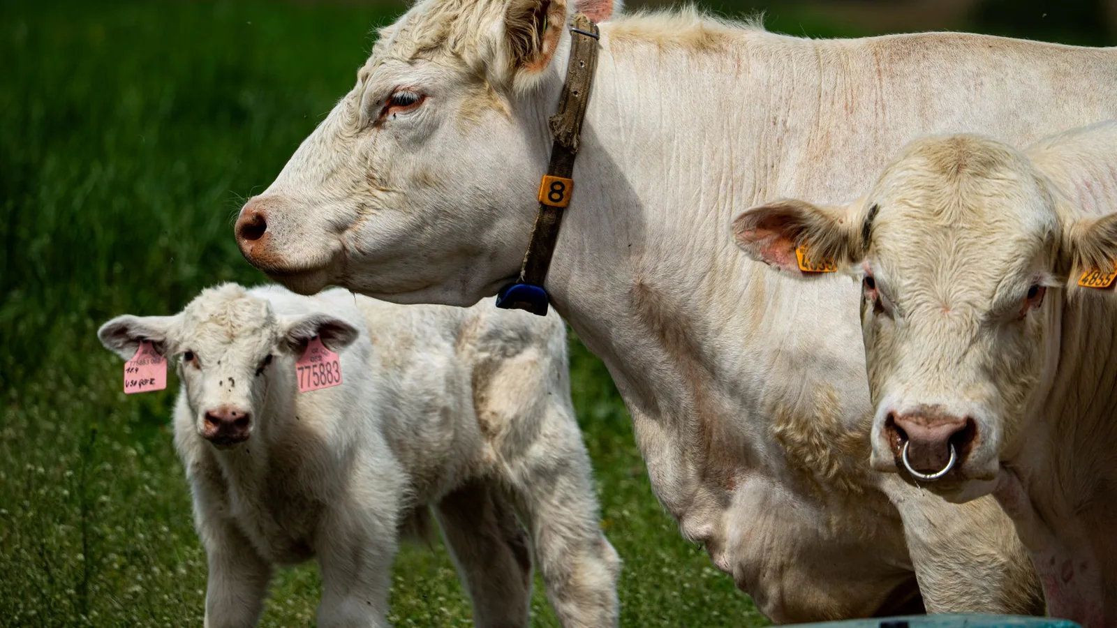
RABV has a remarkably broad host range, capable of infecting essentially all mammalian species, but its perpetuation in nature is maintained by specific reservoir hosts that vary geographically [9, 1, 11, 5, 38]. On a global scale, the domestic dog (Canis lupus familiaris) is the principal reservoir and vector for human rabies, responsible for over 99% of all human transmission events, predominantly across Africa and Asia [1, 38, 39, 21, 37, 13]. In the developed world and specific ecological niches, a diverse array of wildlife reservoirs sustain the virus. In North America, distinct RABV variants circulate in raccoons (Procyon lotor), striped skunks (Mephitis mephitis), red and arctic foxes (Vulpes vulpes, Vulpes lagopus), and numerous bat species [40, 41, 15, 42]. In Europe, the red fox is the primary terrestrial reservoir, alongside the serotine bat (Eptesicus serotinus) for EBLV-1 [27, 43, 44, 45]. The epizootiology of rabies in South Africa is particularly complex, with canid and mongoose biotypes of RABV circulating in species such as the black-backed jackal (Canis mesomelas), bat-eared fox (Otocyon megalotis), yellow mongoose (Cynictis penicillata), and the aardwolf (Proteles cristatus), with evidence of ongoing host-switching events [46, 47]. Similarly, in South America, bats of the genus Myotis are considered ancestral spreaders of the majority of RABV diversity on the continent, with frequent cross-species transmission to other mammals [15]. The World Health Organization (WHO), the World Organisation for Animal Health (WOAH), and the Food and Agriculture Organization of the United Nations (FAO) have jointly prioritized the global elimination of dog-mediated human rabies by 2030, a goal whose success hinges on a deep understanding of the taxonomic diversity, reservoir ecology, and transmission dynamics of Rabies lyssavirus and its related lyssaviruses [5, 39, 13].
The Molecular Basis for Diagnostic and Therapeutic Strategies
The taxonomic and genetic characterization of lyssaviruses directly informs both diagnostic approaches and therapeutic development. The high conservation of the N gene and the leader RNA region across the genus has enabled the design of highly sensitive pan-lyssavirus real-time RT-PCR assays, such as the LN34 assay, which has demonstrated exceptional performance in detecting all known members of the genus, including divergent viruses like WCBV and LLEBV, and is recommended by the WHO and WOAH for confirmatory diagnosis [13, 22-25, 44]. This assay has revolutionized rabies diagnostics by providing a robust, high-throughput alternative to the gold-standard direct fluorescent antibody test (DFAT), particularly for degraded, formalin-fixed, or low-positive samples [48, 23, 49, 24, 25, 41, 50]. Detailed knowledge of the G protein's structure and the genomic sequences of different lyssaviruses is essential for the design of novel biologics. For instance, the identification of highly conserved epitopes on the RABV-G protein, such as those recognized by broad-spectrum neutralizing monoclonal antibodies (mAbs) like NM57 and SOJB, which lock the glycoprotein in a pre-fusion state, offers a promising avenue for next-generation immunotherapeutics [6]. Similarly, the functional characterization of the P protein’s RNA unwinding activity provides a new target for antiviral drug development, as demonstrated by the efficacy of peptides designed to inhibit its oligomerization domain [3]. The molecular taxonomy of Rabies lyssavirus is thus not merely an academic exercise but the foundational science underpinning the global fight against this unrelenting pathogen.
Molecular Pathogenesis of Rabies Lyssavirus
Introduction: A Paradigm of Neurotropic Invasion and Immune Subversion
The molecular pathogenesis of Rabies lyssavirus (RABV) represents a sophisticated paradigm of neurotropism, immune evasion, and cellular reprogramming that culminates in a nearly invariably fatal encephalomyelitis. Unlike many acute cytolytic viruses, RABV has evolved a strategy centered on preserving host cell integrity to facilitate its centripetal spread through the peripheral nervous system, followed by massive centrifugal dissemination and profound neuronal dysfunction without triggering rapid cell death. This host-pathogen dynamic is orchestrated by a compact genome encoding five structural proteins, the nucleoprotein (N), phosphoprotein (P), matrix protein (M), glycoprotein (G), and RNA-dependent RNA polymerase (L), each possessing multifunctional domains that engage in a complex interplay with host cellular machinery. The disease's distinctive neuropathogenesis stems from the virus's ability to hijack axonal transport mechanisms, suppress innate immune signaling, manipulate autophagy and apoptosis, and ultimately induce neuronal cytoskeletal degradation. Understanding these molecular mechanisms at a granular level is essential for developing targeted therapeutics, designing pan-lyssavirus vaccines, and informing global rabies control strategies endorsed by the WHO, WOAH, and FAO.
The Glycoprotein (G): Orchestrating Entry, Tropism, and Neuroinvasiveness
The viral glycoprotein (G) is the master regulator of host cell entry, neurotropism, and the primary target of neutralizing antibodies. RABV-G is a class III viral fusion protein that arranges as trimers on the viral envelope and mediates receptor recognition and pH-dependent membrane fusion. The interaction between G and host receptors dictates the virus's marked neurotropism. While the nicotinic acetylcholine receptor (nAChR) at the neuromuscular junction was historically considered the primary receptor, subsequent research has identified additional molecules, including the neural cell adhesion molecule (NCAM) and the p75 neurotrophin receptor (p75NTR), which facilitate entry into multiple cell types, including neurons and keratinocytes.
Recent structural studies have provided atomic-level resolution of the molecular basis for antibody-mediated neutralization. The crystal structures of neutralizing monoclonal antibodies NM57 and SOJB in complex with RABV-G reveal that these antibodies recognize overlapping epitopes spanning the pleckstrin-homology domain (PHD) and the junction between PHD and the fusion domain (FD) [6]. Critically, two residues at positions 226 and 231 within the PHD serve as key determinants for antibody recognition, and these antibodies function by locking the PHD/FD local structure in a pre-fusion-like state, thereby inhibiting the structural transitions essential for membrane fusion [6]. This structural insight has profound implications for designing broad-spectrum therapeutics. The NM57 and SOJB antibodies neutralize a majority of phylogroup I lyssaviruses, including divergent members like European bat lyssavirus 1 (EBLV-1) and Duvenhage virus (DUVV), highlighting conserved structural vulnerabilities in the glycoprotein that can be exploited for pan-lyssavirus antibody cocktails.
The antigenic diversity of G determines both vaccine efficacy and the risk of immune escape. RABV and related lyssaviruses are categorized into phylogroups based on cross-neutralization profiles, with current vaccines providing robust protection only against phylogroup I. Distant lyssaviruses, such as Mokola virus (MOKV) and Lagos bat virus (LBV) in phylogroup II, and West Caucasian bat virus (WCBV) in phylogroup III, escape vaccine-induced immunity entirely. To address this limitation, innovative approaches using epitope chimerization have been developed. By replacing a single antigenic site of the RABV glycoprotein with the corresponding site from MOKV (creating RABVG-cAS1), researchers have generated recombinant vaccine candidates that induce high antibody titers against both RABVG and MOKVG, protecting mice against challenge with RABV, Irkut virus (IRKV), and MOKV [18]. This molecular engineering strategy paves the way for a truly pan-lyssavirus vaccine capable of protecting against the entire spectrum of lyssavirus diversity.
Beyond immune recognition, G also influences pathogenesis through its impact on viral spread. Studies using recombinant viruses expressing heterologous glycoproteins have demonstrated that the M and G proteins together determine neuroinvasiveness. For example, recombinant viruses where the matrix and glycoprotein of LBV replaced those of an attenuated RABV strain (SPBN-LBVM-LBVG) proved lethal to mice following intramuscular inoculation, whereas viruses expressing only LBV G were not [35]. This indicates that the molecular compatibility between M and G is critical for efficient viral assembly, budding, and dissemination through the nervous system, a finding with direct implications for reverse genetics-based vaccine development.
The Phosphoprotein (P): A Multifunctional Hub for Replication, RNA Processing, and Immune Evasion
The phosphoprotein (P) is a remarkably multifunctional molecule that serves both as an essential cofactor for viral RNA synthesis and as a potent antagonist of the host interferon (IFN) response. The P protein exists in multiple isoforms expressed from alternative translation start sites, each contributing distinct functions to the viral lifecycle. Recent discoveries have fundamentally expanded our understanding of P's molecular capabilities. Most strikingly, the phosphoprotein of RABV possesses intrinsic RNA duplex-unwinding activities, a paradigm-shifting finding for the Rhabdoviridae family, which previously lacked any known RNA helicase or chaperone [3]. RABV-P exhibits two distinct unwinding mechanisms: a nucleoside triphosphate (NTP)-dependent helicase-like activity and an NTP-independent duplex-unwinding and annealing activity. The C-terminal domain of P is required for both activities, whereas the central oligomerization domain (COD) is specifically essential for NTP-dependent unwinding [3]. This RNA chaperone function is likely crucial for resolving secondary structures in the negative-sense genomic RNA during replication and transcription, and for facilitating the correct folding of newly synthesized viral RNAs. The discovery that targeting the COD with designed peptides (P90 and P110) effectively inhibits RABV replication in cells opens a novel avenue for antiviral drug development against this recalcitrant pathogen [3].
Simultaneously, the P protein is a master regulator of the host antiviral response. It suppresses interferon induction and signaling through multiple mechanisms, including sequestering STAT proteins in the cytoplasm, inhibiting IRF-3 phosphorylation, and blocking the formation of the IFN-stimulated gene factor 3 (ISGF3) complex. The intrinsically disordered regions (IDPRs) within P, as elucidated by disorder-based bioinformatics, are central to these multifunctional interactions [10]. The high degree of intrinsic disorder in P, and to a lesser extent in N, allows these proteins to adopt multiple conformations and interact with a diverse array of host partners. This structural plasticity contributes to the formation of Negri bodies, the cytoplasmic inclusion bodies that are pathognomonic for rabies. These membraneless organelles, formed through liquid-liquid phase separation driven by N and P interactions, concentrate viral replication machinery while simultaneously sequestering host innate immune sensors, effectively creating a protected "viral factory" shielded from cellular detection [10].
The Matrix Protein (M): A Nexus of Egress, Innate Immune Suppression, and Neuronal Damage
The matrix protein (M) is increasingly recognized as a central orchestrator of viral egress, innate immune evasion, and neuronal pathology. The M protein mediates virus budding by interacting with the host endosomal sorting complexes required for transport (ESCRT) pathway. A critical molecular mechanism involves the PPxY late-domain motif within RABV-M, which recruits the E3 ubiquitin-protein ligase NEDD4 [51]. This interaction is particularly remarkable because NEDD4 then is a bridge to recruit MAP1LC3 (LC3) via its LC3-interacting region (LIR), thereby hijacking the host autophagy machinery for viral benefit. RABV-M not only promotes autophagosome accumulation by preventing autophagosome-lysosome fusion but also uses these accumulated autophagosomes as platforms for efficient viral budding, with NEDD4 knockdown significantly reducing M-induced autophagosome accumulation and extracellular virus-like particle production [51]. This PPxY-NEDD4-autophagy axis is shared among M proteins across nearly all lyssavirus species, including Australian bat lyssavirus (ABLV) and European bat lyssaviruses (EBLV-1 and EBLV-2), indicating a conserved evolutionary strategy for exploiting the autophagy system to enhance replication [51].
Concurrently, the M protein subverts innate immunity by directly inhibiting the NLRP3 inflammasome, a critical component of the host inflammatory response. RABV-M blocks both the priming step and the activation step of NLRP3 inflammasome assembly by competing with NEK7 for binding to NLRP3, thereby restricting downstream apoptosis-associated speck-like protein containing a CARD (ASC) oligomerization [52]. The serine residue at position 158 of M is a critical determinant of this inhibitory activity; a single G158S mutation in a lab-attenuated RABV decreases interleukin-1β production in bone-marrow-derived dendritic cells, facilitating virus invasion into the brain and elevating pathogenicity in mice [52]. This mechanism reveals how RABV dampens the inflammatory milieu at the peripheral site of inoculation, allowing the virus to reach the central nervous system (CNS) without triggering a robust antiviral response.
Perhaps most devastatingly, the M protein directly contributes to the neurological dysfunction that defines clinical rabies. RABV-M induces microtubule degradation by reprogramming mitochondrial metabolism [53]. The M protein binds to the mitochondrial adenine nucleotide translocator Slc25a4 (ANT1), disrupting ATP/ADP exchange and reducing cellular ATP levels. This energy crisis leads to decreased NAD+ production in the cytosol, which in turn triggers calcium release from the endoplasmic reticulum and mitochondria into the cytoplasm. The resulting calcium influx activates calpains, calcium-dependent cysteine proteases, which then degrade α-tubulin, a core component of the neuronal cytoskeleton [53]. The 57th amino acid of M is the critical molecular switch determining this activity; a mutation at this residue significantly reduces α-tubulin degradation in vitro and axonal degeneration in vivo [53]. This mechanism explains the axonal self-destruction observed in RABV-infected neurons and provides a direct molecular link between viral protein expression and the neurodegenerative changes underlying clinical paralysis and behavioral alterations.
The Nucleoprotein (N) and RNA-Dependent RNA Polymerase (L): Genome Encapsidation and Replication Fidelity
The N protein encapsidates the viral genomic RNA to form the nucleocapsid, which serves as the template for transcription and replication by the L polymerase. The N protein's intrinsic disorder, as identified through bioinformatic analyses of the Pasteur Vaccins strain, facilitates its participation in liquid-liquid phase separation and Negri body formation, processes that are essential for efficient viral RNA synthesis [10]. The L protein, the largest of the RABV proteins, is a multifunctional polymerase with RNA-dependent [RNA polymerase](/knowledge/bioinformatics/rna-polymerase-structure-transcription-mechanisms 2), capping, and polyadenylation activities. Both N and L harbor multiple immunodominant epitopes that have been identified through comprehensive in silico prediction [14]. While the glycoprotein contains the majority of B-cell and T-cell epitopes, the N and L proteins also contain highly conserved peptides that elicit cellular immune responses, making them attractive targets for inclusion in next-generation recombinant vaccines and diagnostic tools. Phylogenetic analyses of RABV isolates have identified specific marker mutations in the N gene, such as V56I in the Eurasian group, L/V95W in the Central group, and D101N/S/T and N/G106D, that correlate with geographic distribution and host species, providing molecular epidemiological markers for tracking viral spread [26].
Host Restriction Factors and the Antiviral Battle
The host cell mounts a multifaceted antiviral response that RABV must overcome to establish productive infection. The long non-coding RNA (lncRNA) EDAL (EZH2 degradation-associated lncRNA) exerts potent antiviral activity by degrading EZH2, thereby reducing trimethylation of histone H3 at lysine 27 (H3K27me3) and promoting transcription of the antiviral gene Pcp4l1 [11]. EDAL restricts RABV replication in a highly cell-specific and infection route-dependent manner, it is effective in primary granule neurons following intranasal infection but not in cortical neurons, astrocytes, or after intramuscular inoculation [11]. This suggests that the antiviral efficacy of host factors varies dramatically across different neuronal subtypes, potentially explaining the vulnerability of certain brain regions to RABV pathology.
Similarly, receptor transporter protein 4 (RTP4) is upregulated in response to RABV infection and suppresses viral replication by binding directly to viral genomic RNA (vRNA) through its zinc finger domain (ZFD) [54]. The cysteine residue at position 95 within the 3CxxC motif of RTP4 is essential for this antiviral function [54]. Recombinant virus expressing RTP4 shows significantly reduced pathogenicity in mice, highlighting RTP4 as a promising target for therapeutic intervention. In another host-virus interaction, the tetraspanin CD9 facilitates RABV infection, and its ligand IL-16 exerts anti-RABV effects by binding to CD9 on the surface of viral particles [55]. These findings suggest that host factors involved in membrane organization and extracellular vesicle biology play critical roles in modulating RABV entry and spread.
Autophagy and Apoptosis: A Delicate Balance for Viral Persistence
RABV must carefully balance the induction of autophagy (which can be co-opted for viral replication) and apoptosis (which prematurely terminates viral production). The virus induces both processes, but the timing and molecular regulation determine the outcome of infection. Early in infection, RABV-M-induced autophagy provides membranes and energy for viral replication, while simultaneously blocking autophagosome-lysosome fusion to prevent degradation of viral components [51, 8]. As infection progresses, the virus can induce apoptosis through mitochondrial pathways, but this is delayed sufficiently to allow viral replication and assembly. The interplay between these processes is crucial for neuropathogenesis; too rapid apoptosis would halt viral spread and expose the infection to immune surveillance, while uncontrolled autophagy would deplete cellular resources. This delicate balance is achieved through the multifunctional nature of M and other viral proteins that simultaneously engage multiple host signaling pathways.
Bat-Variant Adaptations and Host-Switching
The molecular pathogenesis of RABV is further shaped by adaptation to specific reservoir hosts. Bat-borne RABV variants exhibit distinct molecular signatures that enable maintenance in chiropteran populations. In the Americas, Myotis bats have been identified as potential ancestral spreaders of much of bat RABV diversity, with cross-species transmission influenced by host genetic similarity and geographic overlap [15]. The big brown bat (Eptesicus fuscus) variant Ef-W1 has demonstrated the capacity for spillover and host shift into mesocarnivores, as documented in repeated outbreaks in Flagstaff, Arizona. Whole-genome sequencing revealed that the 2021-2023 epizootic in striped skunks originated from a distinct Ef-W1 lineage compared to earlier 2001-2009 outbreaks [40]. Naturally infected skunks showed greater viral RNA loads in the brain compared to skunks infected with a South-Central skunk RABV variant, suggesting that this bat-adapted variant may have unique pathogenic properties that enhance its ability to replicate in mesocarn
Epidemiological Dynamics and Global Burden of Rabies Lyssavirus
Rabies Lyssavirus (RABV) remains one of the most formidable zoonotic pathogens known to medical and veterinary science, exacting a staggering toll on human and animal populations across the globe. The epidemiological landscape of this neurotropic virus is characterized by complex transmission networks spanning domestic animals, wildlife, and humans, with profound disparities in burden that reflect socioeconomic inequities, ecological heterogeneity, and the variable capacity of public health systems to implement effective control measures. Understanding the nuanced dynamics of RABV circulation, the factors driving its persistence, and the true magnitude of its global impact is essential for informing evidence-based interventions and progressing toward the ambitious goal of eliminating dog-mediated human rabies by 2030.
Global Mortality and Morbidity Estimates
The World Health Organization (WHO) and the World Organisation for Animal Health (WOAH) have long recognized rabies as a neglected tropical disease that disproportionately affects the most vulnerable populations in low- and middle-income countries. Current estimates indicate that rabies causes approximately 59,000 human deaths annually worldwide, with a loss of 3.7 million disability-adjusted life years (DALYs) and associated economic losses reaching 8.6 billion USD per year [39, 13, 56]. These figures, while staggering, almost certainly represent a substantial underestimation of the true burden, as surveillance systems in many endemic regions are severely limited, and cases, particularly in rural areas with limited access to healthcare, frequently go undiagnosed and unreported [5, 56]. The case-fatality rate of rabies approaches 100% once clinical symptoms manifest, making it the infectious disease with the highest case fatality rate known to humanity [9, 10, 5]. This near-certain lethality, combined with the absence of any effective antiviral therapy once the virus reaches the central nervous system, underscores the critical importance of prevention through vaccination and timely post-exposure prophylaxis (PEP) [10, 57, 58].
The geographic distribution of human rabies deaths is profoundly uneven. Asia and Africa bear the overwhelming brunt of the burden, accounting for more than 95% of all human fatalities [39, 13, 56, 59]. Within Asia, India consistently reports the highest number of human rabies deaths globally, followed by other densely populated nations with large stray dog populations and limited vaccination coverage [1, 57]. In Africa, the disease remains hyperendemic across much of the continent, with countries such as Tanzania, Nigeria, South Africa, and Malawi reporting substantial numbers of cases [39, 46, 21, 60]. The burden in Latin America has declined significantly over recent decades due to sustained mass dog vaccination campaigns, although focal outbreaks persist, particularly in remote Amazonian regions where bat-mediated transmission plays an increasingly recognized role [2, 61]. Europe, North America, and much of the developed world have successfully eliminated terrestrial rabies through aggressive wildlife vaccination and strict import regulations, though sporadic cases from bat-associated lyssaviruses and imported infections continue to pose a low but persistent risk [44, 62, 63, 64, 65].
Canine Rabies: The Primary Engine of Human Disease
Domestic dogs (Canis lupus familiaris) are unequivocally the most important reservoir and vector of RABV, responsible for over 99% of human rabies cases worldwide [1, 38, 39, 37]. The dynamics of canine rabies transmission are shaped by a complex interplay of ecological, behavioral, and anthropogenic factors. In endemic regions, large populations of free-roaming, unvaccinated, and often ownerless dogs sustain continuous viral circulation, creating a persistent threat to human populations [39, 60]. The virus is maintained within dog populations through direct transmission via saliva-laden bites, with the incubation period typically ranging from weeks to months, allowing infected animals to travel considerable distances and expose numerous contacts before developing clinical signs [38, 66]. Puppies less than three months of age, often excluded from routine vaccination campaigns due to age restrictions, can harbor and transmit the virus, representing a particularly challenging demographic for control programs [60].
The epidemiology of canine rabies exhibits pronounced spatial and temporal heterogeneity. In many endemic settings, incidence peaks during specific seasons, often correlating with dog breeding cycles, increased human-animal interactions during agricultural seasons, or the arrival of new susceptible cohorts of young dogs [38, 43, 67]. Urban and peri-urban environments, characterized by high human and dog densities, facilitate rapid viral propagation, while rural areas with dispersed populations may experience more sporadic spillover events from wildlife reservoirs [67, 68]. The recent war in Ukraine has dramatically illustrated how disruptions to veterinary services and vaccination campaigns can precipitate a resurgence of canine rabies, with cases increasing fivefold in some regions and new foci emerging in previously controlled areas [38, 67, 69, 70]. Similarly, the illegal importation of rabid puppies from endemic countries into rabies-free regions, such as the documented case in Germany in 2021, highlights the ongoing risk of viral reintroduction and the challenges of maintaining freedom from disease in a globally connected world [64, 71].
The "Zero by 30" initiative, a collaborative global framework led by the WHO, WOAH, the Food and Agriculture Organization (FAO), and the Global Alliance for Rabies Control (GARC), aims to eliminate human deaths from dog-mediated rabies by 2030 [13, 56]. Achieving this ambitious goal requires sustained mass vaccination of at least 70% of the dog population in endemic areas, combined with improved access to PEP, enhanced surveillance, and community engagement [39, 13, 61]. However, numerous barriers persist, including inadequate funding, vaccine shortages, logistical challenges in reaching remote populations, and cultural misconceptions about vaccination, as documented in studies from Tanzania and Indonesia [39, 72, 68]. The economic constraints faced by low-income countries are particularly acute, with the cost of dog vaccination programs and PEP placing a substantial burden on already strained healthcare systems [39, 56].
Wildlife Reservoirs and Sylvatic Cycles
While dogs are the primary source of human infection, RABV and other lyssaviruses are maintained in a diverse array of wildlife reservoirs across different continents, creating complex sylvatic cycles that complicate control efforts and pose ongoing spillover risks [5, 46, 43, 15].
In Africa, the epidemiological picture is particularly intricate. South Africa harbors multiple canid RABV biotypes maintained in distinct wildlife hosts, including the black-backed jackal (Canis mesomelas), bat-eared fox (Otocyon megalotis), and yellow mongoose (Cynictis penicillata) [46, 47]. The aardwolf (Proteles cristatus), an insectivorous member of the hyena family, has recently emerged as a significant reservoir in the Northern Cape Province, with evidence suggesting a host shift from other canid species [47]. Foxes (Vulpes vulpes) are the dominant wildlife reservoir in much of Europe and Russia, where they sustain the sylvatic cycle and periodically spill over into domestic animals and humans [43, 67, 70]. The red fox is responsible for the majority of rabies cases in the Russian Federation, where the disease remains enzootic across vast swathes of the East European Plain, and oral rabies vaccination (ORV) programs have been deployed to reduce incidence [43].
In North America, distinct RABV variants circulate in specific wildlife hosts. The raccoon (Procyon lotor) rabies variant (RRVV) is actively managed through large-scale ORV campaigns in the eastern United States [42]. Spillover events from raccoons into other species, including skunks, foxes, and domestic animals, are common, and density-dependent management strategies are critical for controlling spread [40, 42]. The striped skunk (Mephitis mephitis) is a major reservoir for distinct RABV variants in the central United States, and recent emergence of a big brown bat (Eptesicus fuscus) variant (Ef-W1) in skunks in Flagstaff, Arizona, illustrates the potential for host shifts and the establishment of new transmission cycles [40]. Similarly, gray foxes and Arctic foxes maintain unique viral lineages in different regions of the continent [5].
The role of mesocarnivores as both reservoirs and spillover hosts adds considerable complexity to rabies epidemiology. Cats, while not typically serving as long-term reservoirs, are important vectors that can transmit RABV to humans, particularly in settings where they interact with rabid wildlife [37]. The recent confirmation of European bat lyssavirus type 1 (EBLV-1) infection in a domestic cat in the Netherlands, following contact with a dead bat, underscores the risk of spillover from wildlife into domestic animals and the subsequent exposure of humans [63]. Indeed, 46.8% of rabies cases in Ukraine during 2019-2024 involved domestic carnivores (dogs and cats), highlighting the interconnectedness of wildlife and domestic cycles [67].
Bat-Associated Lyssaviruses: A Growing Concern
Bats represent the ancestral reservoir of the lyssavirus genus, and diverse bat species across all continents (except Antarctica) harbor a remarkable array of RABV variants and other lyssavirus species [12, 5, 15]. In the Americas, bats are the only known wildlife reservoir of RABV, and insectivorous, frugivorous, and hematophagous (vampire) bat species all contribute to viral circulation [37, 15]. Vampire bats, which feed on the blood of livestock and occasionally humans, are particularly important vectors in Latin America, where they cause substantial economic losses in cattle and sporadic human fatalities [16, 61]. The genus Myotis has been proposed as a potential ancestral spreader of much of the RABV diversity in the Americas, with cross-species transmission events shaped by host genetic relatedness and geographic overlap [15].
Beyond the Americas, a growing number of novel lyssaviruses have been identified in bat populations worldwide, raising concerns about vaccine efficacy and the potential for emergence. European bat lyssaviruses type 1 (EBLV-1) and type 2 (EBLV-2) are endemic in serotine bats (Eptesicus serotinus) and other species across Europe, causing sporadic spillover infections in humans and domestic animals [27, 44, 45, 63]. EBLV-1 was detected for the first time in the United Kingdom in 2018, likely introduced from Brittany, France, and has since been identified in 34 cases, with evidence of sustained circulation [44]. West Caucasian bat virus (WCBV) and Lleida bat lyssavirus (LLEBV), both highly divergent from RABV and classified in phylogroups outside the range of current vaccine protection, have been detected in the Schreiber's bent-winged bat (Miniopterus schreibersii) across Europe, with seroprevalence reaching up to 70% in some populations [32]. The lack of cross-protection afforded by conventional rabies vaccines against these divergent lyssaviruses represents a significant gap in public health preparedness [17, 31, 34, 32, 18].
Other notable bat-associated lyssaviruses include Lagos bat virus (LBV), endemic in Africa and currently comprising four lineages, which has been implicated in several human exposures but not yet confirmed human fatalities [29, 35]. Mokola virus (MOKV), another African lyssavirus, has been isolated from shrews and a single rodent, and serological evidence suggests bushveld gerbils (Gerbilliscus leucogaster) may serve as a reservoir in South Africa [33]. Duvenhage virus, also African, has caused fatal human infections following bat bites [12]. Australian bat lyssavirus (ABLV), enzootic in both fruit bats (Pteropus spp.) and insectivorous bats (Saccolaimus flaviventris), has been responsible for three human fatalities in Australia, demonstrating that lyssaviruses are a global threat not confined to developing nations [12, 73, 74]. Taiwan bat lyssavirus (TWBLV), first isolated from Japanese house bats in 2018, and Phala bat lyssavirus (PBLV) from South Africa, further expand the known diversity of these pathogens [30, 17, 31]. The Kotalahti bat lyssavirus (KBLV) from Finland was initially detected solely through RNA detection, and subsequent studies have demonstrated that current rabies vaccines provide cross-protection against this virus, offering some reassurance, but the potential for future emergence of vaccine-resistant variants remains [31]. The vast majority of bat sampling efforts globally still yield negative results, as exemplified by long-term surveillance in Israel, where zero out of 294 bats tested over 27 years were positive for RABV [62]. Yet even in such settings, the absence of detection does not confirm absence of risk, and sustained surveillance is crucial [62, 75].
Diagnostic Surveillance and Molecular Epidemiology
Accurate and timely diagnosis is the cornerstone of effective rabies surveillance and control. For decades, the direct fluorescent antibody test (DFA/FAT) on brain tissue has been the gold standard for post-mortem diagnosis, but this method requires high-quality reagents, skilled technicians, and well-preserved specimens [48, 76, 25]. The advent of molecular diagnostics, particularly real-time reverse transcription-polymerase chain reaction (RT-qPCR), has revolutionized rabies detection, offering enhanced sensitivity, specificity, and the ability to test degraded or formalin-fixed samples [12, 13, 22-24]. The LN34 pan-lyssavirus real-time RT-PCR assay, which targets the highly conserved leader region and nucleoprotein gene, has emerged as a transformative tool for global surveillance [22]. Multi-site evaluations have demonstrated that the LN34 assay achieves diagnostic sensitivity of 99.90% and specificity of 99.68% compared to DFA, and can detect viral RNA in fresh, frozen, archived, deteriorated, and formalin-fixed brain tissue [25]. The assay can identify early infections that DFA might miss, as demonstrated by the detection of rabies virus in two raccoons in Pennsylvania where antigen levels were low and confined to caudal brain structures, suggesting early viral invasion [23, 49]. The LN34 assay has also proven valuable for testing roadkill specimens, enabling enhanced surveillance in areas where sample collection is otherwise impractical [41], and has been successfully applied to formalin-fixed human brain tissue for both diagnosis and genetic characterization [24]. The development of a multiplexed version (LN34M) further streamlines workflow, reduces costs, and improves quality control [76, 50].
Molecular epidemiological techniques have provided profound insights into the transmission dynamics and evolutionary history of RABV. Phylogenetic analyses of nucleoprotein (N) and glycoprotein (G) gene sequences have revealed distinct viral lineages associated with specific hosts and geographic regions, facilitating the tracking of viral spread and identification of cross-species transmission events [16, 26, 27, 46, 44]. In South Africa, for example, phylogenetic analysis of 1,374 sequences from the cytoplasmic domain of the glycoprotein and the G-L intergenic region has elucidated the interconnected transmission networks involving black-backed jackals, bat-eared foxes, yellow mongooses, and domestic dogs, highlighting the challenges of rabies elimination in settings with multiple wildlife reservoirs [46]. Similarly, studies in the Russian Federation have identified four marker mutations in the nucleoprotein that distinguish Eurasian and Central Asian viral groups, and phylogenetic analysis confirmed that a human case in the country was due to EBLV-1a, underscoring the value of genotyping for understanding hidden transmission mechanisms [26]. Bayesian phylodynamic analyses have estimated that EBLV-1 emerged in Europe only approximately 175 years ago, providing a temporal framework for understanding its current distribution [27]. The application of metagenomic next-generation sequencing (mNGS) has further enhanced diagnostic capabilities, enabling the detection of rabies virus in cerebrospinal fluid from cases with atypical presentations, such as paralytic rabies mimicking stroke [77].
The Role of Host Immunity and Viral Evasion
The epidemiological dynamics of RABV are profoundly influenced by the interplay between host immune responses and viral immune evasion strategies. The virus has evolved multiple mechanisms to subvert host defenses, facilitating its spread to the central nervous system before the adaptive immune response can be mounted [10, 4, 52]. The matrix protein (M protein) plays a critical role in suppressing NLRP3 inflammasome activation, thereby inhibiting the production of pro-inflammatory cytokines and limiting the recruitment of immune cells to the site of infection [52]. By binding to NLRP3 and competing with NEK7, M protein prevents the downstream oligomerization of ASC and the subsequent activation of caspase-1, effectively dampening the innate immune response [52]. The phosphoprotein (P protein) exhibits RNA duplex-unwinding activities that are essential for viral replication, and its intrinsically disordered regions facilitate the formation of Negri bodies and contribute to immune evasion [10, 3]. The nucleoprotein (N protein) also contains disordered regions that may aid in subverting host antiviral responses [10].
At the same time, the host mounts a diverse array of antiviral responses. Long non-coding RNAs such as EDAL (EZH2 degradation-associated lncRNA) have been shown to restrict RABV replication in a cell-specific and infection route-dependent manner by degrading EZH2 and promoting the transcription of the antiviral gene Pcp4l1 [11, 78]. Similarly, receptor transporter protein 4 (RTP4) binds viral genomic RNA and suppresses genome amplification, playing a crucial role in host defense [54]. Interleukin-16 (IL-16) exerts anti-RABV effects through binding to CD9 on the surface of viral particles, and the tetraspanin CD9 facilitates viral entry, making it a potential therapeutic target [55]. The induction of autophagy and apoptosis during RABV infection represents a double-edged sword: while autophagy can help clear viral components, the virus has evolved to hijack the autophagic pathway for efficient egress, using the PPxY motif of M protein to recruit NEDD4 and prevent autophagosome-lysosome fusion [51, 8]. The M protein also disrupts mitochondrial metabolism by binding to Slc25a4, reducing NAD+ production and triggering calcium influx that activates calpains, leading to the degradation of neuronal microtubules and contributing to the neurodegenerative changes observed in rabies [53].
The antibody response, particularly neutralizing antibodies (nAbs) against the viral glycoprotein (G protein), is a critical correlate of protection [17, 6, 79]. The WHO and WOAH have established a threshold of 0.5 IU/mL as indicative of adequate seroconversion following vaccination [17, 36, 79]. However, studies have shown that protection against more divergent lyssaviruses within phylogroup I may require titers 10-fold or higher than this standard, and current rabies vaccines provide no protection against phylogroup II or III lyssaviruses [17, 31, 34, 35, 18]. The development of pan-lyssavirus vaccines,
Diagnostic Approaches for Rabies Lyssavirus Infection
The diagnosis of rabies lyssavirus (RABV) infection, whether in animals or humans, remains a critical pillar of public health and veterinary practice, directly influencing the administration of life-saving post-exposure prophylaxis (PEP) and informing epidemiological surveillance. Given the nearly 100% case-fatality rate once clinical signs manifest, and the rapidity with which intervention must occur, diagnostic methods must be both exquisitely sensitive and specific, and ideally, capable of delivering results with minimal delay [80, 1, 10]. The World Health Organization (WHO) and the World Organisation for Animal Health (WOAH) have established standards that define the gold-standard approaches, yet the landscape of rabies diagnostics is evolving rapidly, driven by the need for higher throughput, objective interpretation, and the ability to detect divergent lyssaviruses, including those not readily neutralized by current vaccines [17, 5, 37, 59].
The Established Gold Standard: Direct Fluorescent Antibody Test (DFA)
For decades, the direct fluorescent antibody (DFA) test, performed on fresh or frozen brain tissue, has been the cornerstone of post-mortem rabies diagnosis across the globe [48, 25]. The test relies on the direct detection of viral nucleoprotein (N) antigen in impression smears or tissue sections using fluorescein-isothiocyanate (FITC)-conjugated anti-rabies antibodies. Its sensitivity and specificity, when performed by skilled personnel with high-quality conjugates, have been well documented, and it remains the primary method recommended by WOAH for routine animal diagnosis [22, 5]. However, the DFA test suffers from several inherent limitations that have spurred the development of alternative and complementary platforms. Subjective interpretation by microscopists can lead to variability, particularly in cases with low antigen burden, such as early infections or degraded specimens [48, 23, 49]. The requirement for a fluorescence microscope and a skilled technician limits its application in resource-limited settings, where rabies is often most prevalent [25, 22]. The DFA test cannot reliably differentiate between individual lyssavirus species or provide genotyping information, which is essential for molecular epidemiology [26, 24].
The Ascendancy of Molecular Diagnostics: Real-Time RT-PCR and the LN34 Pan-Lyssavirus Assay
Reverse transcription polymerase chain reaction (RT-PCR), and in particular real-time RT-PCR, has emerged as a transformative tool for rabies diagnosis. These assays offer superior sensitivity, the ability to detect viral RNA in degraded or formalin-fixed tissues, and an objective, quantitative readout that reduces operator dependency [48, 81, 25]. The most significant recent advance in this arena is the LN34 pan-lyssavirus real-time RT-PCR assay, developed and validated by the United States Centers for Disease Control and Prevention (CDC) and collaborating laboratories. This assay targets the highly conserved non-coding leader region and the 5’ end of the nucleoprotein (N) gene, employing degenerate primers and a locked-nucleotide-modified TaqMan probe to achieve broad-spectrum detection across the entire Lyssavirus genus [22, 50]. The LN34 assay has been subjected to extensive multi-site evaluation, demonstrating diagnostic sensitivity of 99.90% and specificity of 99.68% when compared to the DFA test across thousands of samples from Africa, the Americas, Asia, and Europe [25]. Crucially, it detected rabies RNA in all DFA-positive samples in that study and resolved 80 samples that were inconclusive or untestable by DFA [25].
The enhanced sensitivity of the LN34 assay has proven especially valuable for identifying early or low-level infections that evade standard antigen detection. In Pennsylvania, routine testing of 6,855 animal samples identified two cases that were initially DFA-negative but subsequently confirmed as positive by LN34 RT-PCR [23, 49]. In both cases, a raccoon and a juvenile raccoon, viral antigen and RNA were present at very low levels and were more abundant in the brain stem and rostral spinal cord than in the cerebellum or cortex, a pattern suggestive of early infection before extensive dissemination to cortical structures [23, 49]. Such findings underscore the power of molecular diagnostics to reduce the number of false-negative DFA results, which could otherwise lead to missed opportunities for PEP administration. The LN34 assay has been successfully applied to formalin-fixed, paraffin-embedded human brain tissue, where conventional PCR fails due to nucleic acid fragmentation. In a case from the Dominican Republic, the LN34 assay amplified a 165 bp fragment from fixed tissue, enabling both diagnosis and rapid genetic typing that linked the virus to a circulating RABV strain in Haiti [24]. Similarly, the LN34 assay has been optimized into a multiplexed format (LN34M) that combines the pan-lyssavirus and internal control reactions into a single well, reducing costs and simplifying setup while maintaining excellent performance [76, 50].
Other real-time RT-PCR approaches have also been validated. A pan-lyssavirus SYBR green-based assay, developed by the Animal and Plant Health Agency (APHA) in the UK, uses dissociation curve analysis to differentiate lyssavirus species and can detect even the most divergent members of the genus, such as West Caucasian bat virus and Lleida bat lyssavirus [81]. The LN34 assay, however, has become the de facto standard for many reference laboratories due to its widespread validation, ease of implementation, and compatibility with automated extraction platforms [48, 50].
Serological Methods: Quantifying Neutralizing Antibodies and Detecting Past Exposure
Serological testing plays a dual role in rabies diagnostics: evaluating vaccine-induced immunity and detecting natural exposure in surveillance studies. The gold standard for quantification of rabies virus-neutralizing antibodies (rVNA) is the rapid fluorescent focus inhibition test (RFFIT) [79]. The RFFIT involves mixing serial dilutions of serum with a fixed dose of live challenge virus standard (CVS), infecting susceptible cells, and then counting fluorescent foci. Despite its reliability, the RFFIT is labor-intensive, subjective, and requires highly trained personnel and expensive fluorescence microscopy, limiting its throughput [79]. To address these drawbacks, a novel in-cell ELISA-based neutralization test (icNT) has been developed. This assay replaces microscopic counting with a colorimetric readout that can be automatically quantified using a standard microplate reader. Validation against 200 sera with known RFFIT titers showed excellent sensitivity and specificity, with very good intra- and inter-assay precision [79]. The icNT can thus accelerate the processing of large numbers of samples for routine vaccination monitoring or for screening antiviral compounds [79].
For laboratories lacking cell culture facilities, competitive ELISA (cELISA) test systems provide an alternative. A recently developed cELISA for dogs demonstrated high correlation with the fluorescent antibody virus neutralization (FAVN) test (R² = 0.988), a diagnostic sensitivity of 94.9%, and specificity of 83.1%, with a lower detection threshold of less than 0.02 IU/mL [82]. This test shows no cross-reactivity with antibodies against common canine viruses, making it a practical tool for evaluating mass vaccination campaigns [82]. In human diagnostics, serology is primarily used to confirm the adequacy of PEP and to investigate rare cases of convalescence. For instance, detection of rabies antibodies in cerebrospinal fluid (CSF) is a key laboratory criterion for confirming active lyssavirus encephalitis, particularly in survivors of clinical rabies [57].
Serosurveys are also essential for understanding the ecology of lyssaviruses in wildlife. For example, in a study of bat populations in Belgium, 32% of blood samples contained neutralizing antibodies against European bat lyssavirus 1 (EBLV-1), providing evidence of widespread past exposure even when virus itself was not detected in saliva samples [45]. Similarly, a micro-neutralization test identified Mokola virus-neutralizing antibodies in 87.80% of Bushveld gerbils in South Africa, suggesting a potential reservoir species [33]. Such serological data are invaluable for risk assessment and guiding surveillance efforts.
Emerging Technologies: Metagenomic Next-Generation Sequencing (mNGS) and Proteomics
The diagnostic armamentarium for rabies continues to expand, with metagenomic next-generation sequencing (mNGS) emerging as a powerful tool for ante-mortem diagnosis, especially in challenging cases. Traditional PCR-based ante-mortem detection of RABV in CSF, saliva, or skin biopsies has limited sensitivity. mNGS offers an unbiased, hypothesis-free approach capable of capturing the entire pathogen genome from low-titer specimens. In a reported case of paralytic rabies with a stroke-like onset, mNGS of CSF detected nine unique rabies virus sequences sharing >99% nucleotide homology with a known RABV strain, confirming the diagnosis within a short window before the patient’s death [77]. This technology shortens the diagnostic time and increases sensitivity compared to conventional PCR or DFA, particularly when serological or virological tests are negative [77]. Although still restricted to specialized laboratories, the falling cost of sequencing and the development of portable platforms may make mNGS more accessible in the future.
Proteomic profiling represents another frontier. A recent study analyzed brain tissue, CSF, and serum from rabies-infected and uninfected dogs using mass spectrometry. It identified 54 significantly differentially abundant proteins in brain, 299 in CSF, and 280 in serum, with pathways related to neurodegeneration and spinocerebellar ataxia being enriched [7]. Although still in the early stages, such biomarker discovery could lead to the development of point-of-care tests that detect infection before the onset of clinical signs, a critical capability for both human and animal health [7].
Special Diagnostic Scenarios: Poor-Quality Samples, Roadkill, and Formalin-Fixed Tissues
One of the greatest strengths of molecular methods is their ability to extract diagnostic information from specimens that are unsuitable for DFA. Roadkill, for example, is an important component of enhanced rabies surveillance (ERS) but often yields autolyzed or contaminated tissues. The LN34 real-time RT-PCR assay successfully identified rabies virus RNA in 2.7% of 299 roadkill specimens collected across ten U.S. states, findings that would have been missed by antigen detection alone [41]. Similarly, the assay has been proven effective on archived, deteriorated, and formalin-fixed brain tissues, where the short amplicon length (165 bp) overcomes the fragmentation caused by formalin fixation [24, 25].
Contextual Challenges and the Need for Integrated Diagnostics
The choice of diagnostic approach must be tailored to the epidemiological context. In regions where multiple lyssavirus species circulate, such as Africa and Europe, pan-lyssavirus assays like LN34 are indispensable because they can detect divergent viruses, including Lagos bat virus, Mokola virus, and EBLV-1 [29, 81, 27, 45]. Cross-protection from RABV-based vaccines is not guaranteed against distantly related lyssaviruses, making accurate identification of the infecting species crucial for risk management [17, 31, 18]. In contrast, in the Americas, where RABV is the exclusive lyssavirus, diagnostic panels may focus on RABV-specific detection, though the LN34 assay still provides superior sensitivity and standardization [22]. The ongoing threat of rabies re-introduction into disease-free areas, through illegal pet importation [64], transboundary spread of wildlife variants [40, 46], or disruption of vaccination campaigns due to conflict [70], demands robust, portable, and rapid diagnostic capacity at national borders and in field settings.
The diagnostic landscape for rabies lyssavirus infection has moved decisively beyond the limitations of the DFA test. Real-time RT-PCR, particularly the LN34 pan-lyssavirus assay, has become the new benchmark for post-mortem diagnosis, offering enhanced sensitivity, objective readouts, and the ability to genotype the virus. Serological methods, including the icNT and cELISA, provide high-throughput solutions for monitoring vaccine-induced immunity. Emerging technologies such as mNGS hold promise for ante-mortem diagnosis, while proteomics may eventually yield novel biomarkers for early detection. The integration of these tools, guided by internationally accepted standards from WHO and WOAH, is essential for achieving the global goal of zero human deaths from dog-mediated rabies by 2030 and for understanding the broader ecology of lyssaviruses in wildlife reservoirs [5, 37, 13].
Structural and Functional Virology of Rabies Lyssavirus Proteins
The Rabies lyssavirus (RABV) virion, a prototypical member of the Rhabdoviridae family, exhibits a characteristic bullet-shaped morphology approximately 180 nm in length and 75 nm in diameter. This architecture is the product of a remarkably efficient genetic economy, as the virus encodes only five structural proteins from its single-stranded, negative-sense RNA genome of approximately 12 kilobases. These proteins, the nucleoprotein (N), phosphoprotein (P), matrix protein (M), glycoprotein (G), and the large RNA-dependent [RNA polymerase](/knowledge/bioinformatics/rna-polymerase-structure-transcription-mechanisms 2) (L), orchestrate a sophisticated life cycle that enables the virus to evade host immunity, invade the peripheral nervous system, and cause a nearly invariably fatal encephalitis [1, 10, 4]. The functional interplay between these proteins, particularly their roles in transcription, replication, assembly, and immune evasion, is central to understanding the pathogenicity of RABV and represents a critical frontier for the development of novel antiviral strategies [80, 3].
The Glycoprotein (G): The Master Key for Entry and the Prime Immunological Target
The RABV glycoprotein (G) is the most extensively studied of the five viral proteins, owing to its dual role in mediating host cell entry and serving as the primary target for the host neutralizing antibody response [14, 6]. The G protein forms trimeric spikes that project from the viral envelope, giving the virion its characteristic surface appearance. Its function is not merely structural; it is the molecular determinant of viral tropism, neuroinvasiveness, and pathogenicity. The G protein mediates attachment to host cell receptors, a process that remains incompletely understood but involves interactions with nicotinic acetylcholine receptors, the neural cell adhesion molecule (NCAM), the low-affinity nerve growth factor receptor (p75NTR), and potentially other molecules [4, 28]. Following receptor binding, the virus is internalized via clathrin-mediated endocytosis. The acidic environment of the endosome triggers a dramatic conformational rearrangement of the G protein, transitioning from its pre-fusion state to a post-fusion state that drives the fusion of the viral envelope with the endosomal membrane, releasing the viral ribonucleocapsid (RNP) complex into the cytoplasm [6].
The antigenic structure of the G protein is of paramount importance for vaccine and therapeutic development. It contains multiple antigenic sites (I, II, III, IV, and minor sites) that are recognized by virus-neutralizing antibodies (VNAs) [6, 18]. The WHO and WOAH recognize a serum neutralizing antibody titre of ≥0.5 IU/mL as a correlate of protection for rabies vaccination [36, 64]. However, the G protein is also the primary driver of antigenic diversity among lyssaviruses. The genus Lyssavirus is divided into phylogroups based on the genetic and antigenic relatedness of their G proteins. Current rabies vaccines, which are based on RABV (a phylogroup I member), provide robust protection against other phylogroup I lyssaviruses (e.g., Duvenhage virus, European bat lyssavirus types 1 and 2, Australian bat lyssavirus) but fail to protect against more divergent phylogroup II (e.g., Lagos bat virus, Mokola virus) or phylogroup III (e.g., West Caucasian bat virus) viruses [17, 31, 34, 35, 18]. This antigenic gap is a major driver for the development of next-generation pan-lyssavirus vaccines. Strategies such as epitope chimerization, where antigenic sites from divergent lyssaviruses (e.g., Mokola virus) are grafted onto the RABV G backbone, have shown promise in eliciting broad cross-neutralizing antibody responses in animal models [18]. The structural characterization of broadly neutralizing monoclonal antibodies (mAbs) like NM57 and SOJB, which lock the G protein in a pre-fusion state and block the essential structural transition between the pleckstrin-homology domain (PHD) and the fusion domain (FD), provides a mechanistic blueprint for the rational design of universal vaccines and immunotherapeutics [6]. The critical role of the G protein is further underscored by the fact that recombinant viruses expressing heterologous G proteins, such as the cSN-TWBLV and cSN-KBLV constructs, are viable and can be used to assess cross-protection conferred by existing vaccines against emerging lyssaviruses [17, 31].
The Nucleoprotein (N) and Phosphoprotein (P): The Replication and Immune Evasion Machinery
The N and P proteins are the core components of the viral RNP complex, which serves as the template for both transcription and replication. The N protein encapsidates the viral genomic RNA, forming a helical nucleocapsid that protects the RNA from cellular nucleases and provides the necessary structural scaffold for the viral polymerase [10, 14]. The N protein is highly conserved among lyssaviruses, with amino acid sequence homology reaching 78-93% across the genus, making it an excellent target for pan-lyssavirus diagnostic assays such as the LN34 real-time RT-PCR, which targets the leader RNA and the N gene [26, 48, 76, 25, 22, 50].
The P protein is a multifunctional adaptor protein that is indispensable for viral replication and a master regulator of the host antiviral response. It acts as a non-catalytic cofactor for the L polymerase, tethering it to the N-RNA template to facilitate processive RNA synthesis [10, 3]. Recent groundbreaking research has revealed that the RABV P protein possesses intrinsic RNA duplex-unwinding activities, a function previously unknown for any rhabdovirus-encoded protein. This study demonstrated that the C-terminal domain of P is required for both an NTP-dependent helicase-like activity and an NTP-independent duplex-unwinding and annealing activity, with the central oligomerization domain (COD) being essential for the former [3]. This RNA chaperone-like activity is likely crucial for resolving secondary structures in the viral RNA genome during replication and transcription, highlighting a novel and potentially druggable target. Indeed, peptides designed to target the COD (P90 and P110) were shown to effectively inhibit RABV replication in cell culture [3].
Beyond its role in viral RNA synthesis, the P protein is a potent antagonist of the host innate immune system, particularly the interferon (IFN) response. The P protein achieves this through multiple mechanisms, including the inhibition of interferon regulatory factor 3 (IRF-3) phosphorylation and the sequestration of signal transducer and activator of transcription (STAT) proteins in the cytoplasm, thereby blocking IFN-stimulated gene (ISG) expression [10, 4]. The high level of intrinsic disorder predicted in the P protein, as revealed by disorder-based bioinformatics analyses, is hypothesized to facilitate these promiscuous protein-protein interactions, allowing it to effectively subvert multiple host signaling pathways [10]. This intrinsic disorder is also thought to contribute to the formation of Negri bodies, the characteristic intracytoplasmic inclusion bodies in infected neurons, which are sites of viral replication and may serve to concentrate viral components while shielding them from cellular sensors [10].
The Matrix Protein (M): The Architect of Budding and a Key Player in Pathogenesis
The matrix protein (M) is a small, multifunctional protein that lies beneath the viral envelope, bridging the RNP core with the cytoplasmic tail of the G protein. Its primary function is to orchestrate viral assembly and budding. The M protein drives the condensation of the RNP into a tightly coiled helical structure, a process essential for the formation of infectious virions. It then recruits the host ESCRT (Endosomal Sorting Complexes Required for Transport) machinery to facilitate membrane scission and viral egress. This is achieved through a conserved PPxY (Proline-Proline-x-Tyrosine) late domain motif in the M protein, which interacts with the E3 ubiquitin-protein ligase NEDD4 [51]. This interaction is a critical step in the budding process, as NEDD4 ubiquitinates the M protein, which in turn recruits the ESCRT machinery. Remarkably, RABV-M hijacks this pathway not only for budding but also to subvert the host autophagy system. The M protein, via its PPxY motif and NEDD4, recruits the autophagy protein LC3, leading to the accumulation of autophagosomes while simultaneously blocking their fusion with lysosomes. This creates a protected niche that supports viral replication and facilitates efficient egress of viral particles [51].
The M protein is also a central player in RABV-induced pathogenesis and neuronal dysfunction. It has been demonstrated that the M protein can inhibit the activation of the NLRP3 inflammasome, a critical component of the innate immune response. Specifically, the M protein competes with the host kinase NEK7 for binding to NLRP3, thereby blocking the downstream oligomerization of the adaptor protein ASC and the subsequent production of the pro-inflammatory cytokine IL-1β. This immune evasion strategy is critical for the virus to establish infection in the periphery and invade the central nervous system (CNS) without being cleared by a robust inflammatory response. A single serine residue at position 158 of the M protein was identified as critical for this inhibitory function; a G158S mutation in a lab-attenuated RABV strain led to increased IL-1β production and reduced pathogenicity in mice [52].
The M protein directly contributes to the degenerative changes observed in RABV-infected neurons. Infection with RABV, or overexpression of its M protein, disrupts mitochondrial metabolism by binding to the adenine nucleotide translocator Slc25a4. This interaction reduces cellular ATP and NAD+ levels, leading to an influx of Ca²⁺ from the endoplasmic reticulum and mitochondria into the cytoplasm. This calcium surge activates calpains, Ca²⁺-dependent proteases that degrade α-tubulin, a core component of the neuronal cytoskeleton [53]. This mechanism provides a molecular explanation for the axonal degeneration and neuronal dysfunction that are hallmarks of rabies encephalitis. The 57th amino acid of the M protein was identified as a key determinant of this microtubule-degrading activity, with a mutation at this site reducing α-tubulin degradation in vitro and axonal degeneration in vivo [53].
The Large Polymerase (L): The Viral Replicative Engine
The L protein is the largest of the RABV proteins and functions as the viral RNA-dependent RNA polymerase (RdRp). It is a multifunctional enzyme responsible for both the transcription of viral mRNAs and the replication of the full-length genomic RNA. The L protein possesses all the enzymatic activities required for RNA synthesis, including the RdRp catalytic domain, a capping enzyme domain, and a poly(A) polymerase domain [10, 14]. The L protein does not function in isolation; it requires the P protein as a processivity factor to engage with the N-RNA template. The interaction between the L and P proteins is a critical regulatory point in the viral life cycle, and targeting this interface is a promising antiviral strategy, as demonstrated by the inhibitory peptides targeting the P protein's COD [3]. The L gene is also a target for molecular diagnostics and phylogenetic analyses, as it contains conserved regions that can be used for pan-lyssavirus detection [14, 22].
Protein-Protein Interactions and the Viral Life Cycle
The five structural proteins of RABV do not function as isolated entities but rather as an integrated molecular machine. The life cycle begins with the G protein mediating entry. Once inside the cell, the RNP complex (N-RNA-P-L) is released into the cytoplasm, where primary transcription occurs. The L-P polymerase complex transcribes the negative-sense genome into five monocistronic mRNAs. As the infection progresses, the polymerase switches from transcription to replication, producing a full-length positive-sense antigenome, which then is a template for the synthesis of progeny negative-sense genomes. The newly synthesized genomes are immediately encapsidated by the N protein. The M protein then orchestrates the assembly of the RNP and its transport to the plasma membrane, where it interacts with the cytoplasmic domain of the G protein to drive budding [4, 12, 51, 53]. The intricate network of interactions between these proteins, such as the N-P interaction for replication, the M-N interaction for RNP condensation, and the M-G interaction for budding, represents a vast landscape of potential targets for antiviral intervention. In-silico screening studies have identified several promising compounds, such as the fungal metabolite Chaetoglobosin C and the phytochemical Gallocatechin, which show high binding affinity to these critical viral proteins, though their efficacy requires further validation in vitro and in vivo [80, 9]. The continued elucidation of the structural and functional virology of these five proteins is not merely an academic exercise; it is the essential foundation for the rational design of novel therapeutics, improved vaccines, and more sensitive diagnostics to combat this ancient and devastating disease.
Antiviral Strategies and In Silico Screening of Natural Compounds Against Rabies Lyssavirus
The development of effective antiviral therapies against Rabies lyssavirus (RABV) remains one of the most formidable challenges in infectious disease research. Despite over a century of vaccine availability, rabies continues to exact a devastating toll, with an estimated 59,000 human deaths annually, predominantly in Asia and Africa [1, 5, 13]. The case fatality rate of rabies approaches 100% once clinical symptoms manifest, and currently, there exists no specific antiviral drug approved for the treatment of established rabies encephalitis [10, 56, 57]. The World Health Organization (WHO) and the World Organisation for Animal Health (WOAH) have prioritized the global elimination of dog-mediated human rabies by 2030, yet the absence of effective therapeutics beyond post-exposure prophylaxis (PEP) represents a critical gap in the armamentarium against this lethal pathogen [13, 56]. The Milwaukee protocol and other experimental therapeutic regimens have yielded inconsistent and largely disappointing results, with survivors often suffering severe neurological sequelae [12, 57]. This therapeutic void has catalyzed intensive research into novel antiviral strategies, with particular emphasis on the in silico screening of natural compounds, a approach that leverages computational power to identify potential inhibitors of viral replication from vast chemical libraries derived from fungal, plant, and other natural sources [80, 9].
The Rationale for In Silico Screening in Rabies Antiviral Discovery
The molecular pathogenesis of rabies offers multiple druggable targets, each representing a potential Achilles' heel in the viral life cycle. The RABV genome encodes five structural proteins: the glycoprotein (G), nucleoprotein (N), phosphoprotein (P), matrix protein (M), and the RNA-dependent RNA polymerase (L) [10, 14]. Each of these proteins performs essential functions that can theoretically be disrupted by small molecule inhibitors. The glycoprotein mediates viral attachment and entry into host cells, making it a prime target for entry inhibitors and neutralizing antibodies [14, 6, 59]. The L protein, as the viral polymerase, is essential for genome replication and transcription, and its conserved catalytic domains represent attractive targets for nucleoside analogue inhibitors [80, 14]. The phosphoprotein is a non-catalytic cofactor for the polymerase and, crucially, has recently been demonstrated to possess intrinsic RNA duplex-unwinding activities, both NTP-dependent helicase-like activity and NTP-independent RNA chaperone functions, that are essential for viral replication [3]. The matrix protein orchestrates viral assembly and budding, modulates host gene expression, and, as recent research has revealed, plays a multifaceted role in immune evasion by inhibiting NLRP3 inflammasome assembly through direct binding to NLRP3 [52] and by hijacking the host autophagy machinery via recruitment of NEDD4 through its PPxY motif to facilitate efficient viral egress [51]. The matrix protein also induces neuronal microtubule degradation by reprogramming mitochondrial metabolism, a process linked to the neuropathogenesis of rabies [53].
Traditional antiviral drug discovery is resource-intensive and time-consuming. In silico screening, encompassing molecular docking, molecular dynamics simulations, and pharmacokinetic/toxicity prediction (ADMET), offers a rapid and cost-effective alternative for identifying lead compounds from extensive natural product libraries [80, 9]. This computational approach has gained particular traction in the search for anti-rabies compounds due to the urgent need for novel therapeutics and the rich, underexplored biodiversity of natural product sources, including fungal metabolites and medicinal plant phytochemicals [80, 9].
Fungal Metabolites as a Reservoir of Anti-RABV Compounds
Fungi represent a prolific source of structurally diverse secondary metabolites with a long history of pharmaceutical application, including antiviral activity. A comprehensive in silico study by Meroy et al. (2025) systematically evaluated the interactions between 52 fungal metabolites and four key RABV proteins, RABV-G, RABV-L, RABV-P, and RABV-M, using molecular docking simulations [80]. From this initial library, 18 metabolites were selected for further physicochemical and pharmacokinetic evaluation based on their binding affinities. The study identified several compounds with exceptional promise. Chaetoglobosin A and Cladospirone B demonstrated encouraging interactions with both RABV-G and RABV-L, suggesting potential dual-target activity that could impair both viral entry and genome replication [80]. Most significantly, Chaetoglobosin C exhibited the highest binding affinity among all tested compounds against both glycoprotein and polymerase targets, positioning it as a leading candidate for further development [80].
The mechanistic implications of these findings are profound. The ability of Chaetoglobosin C to bind with high affinity to RABV-L suggests that it may interfere with the catalytic activity of the RNA-dependent RNA polymerase, a function essential for viral replication and one that has no close homologue in mammalian cells, thus offering a favorable therapeutic index. Similarly, binding to RABV-G could block the critical pH-dependent conformational changes required for membrane fusion during viral entry [6]. The ADMET profiling of these fungal metabolites revealed generally favorable pharmacokinetic characteristics; however, the study authors explicitly highlighted two critical limitations: poor blood-brain barrier (BBB) permeability and suboptimal bioavailability [80]. These are particularly salient challenges for rabies therapeutics, as the virus replicates within the central nervous system (CNS), and any effective antiviral must achieve therapeutic concentrations in brain tissue. The BBB represents a formidable obstacle for drug delivery to the CNS, and improving the CNS penetration of these lead compounds will be essential for their translation into viable clinical candidates [80].
Phytochemical Screening from Medicinal Plants: The Case of Boswellia dalzielii
Parallel to the investigation of fungal metabolites, plant-derived natural products have also been subjected to rigorous computational screening for anti-rabies activity. A notable study by Oranu et al. (2025) employed molecular docking and virtual screening to evaluate 149 phytochemical constituents derived from Boswellia dalzielii (Hutch), a medicinal plant used in traditional African medicine, against the RABV glycoprotein (PDB ID: 8B8V) [9]. The study utilized polyethylene glycol (PEG) as a reference ligand, which exhibited a mean binding energy of -3.3 Kcal/mol. Remarkably, the vast majority of the phytoconstituents demonstrated superior binding affinities, with binding energies ranging from -8.6 to -3.8 Kcal/mol, indicating that these natural compounds interact more favorably with the viral target than the reference ligand [9].
Among the ten front-runner compounds identified, gallocatechin displayed the highest binding affinity at -8.6 Kcal/mol, suggesting potent anti-RABV potential [9]. However, gallocatechin failed to satisfy Lipinski’s Rule of Five, a standard set of criteria used to predict the oral bioavailability and drug-likeness of a compound, and also exhibited concerning toxicity profiles. In contrast, luteolin (-7.7 Kcal/mol), epicatechin (-7.6 Kcal/mol), and cianidanol (-7.3 Kcal/mol) not only demonstrated robust binding affinities but also passed both Lipinski’s Rule of Five and toxicity evaluations [9]. These three flavonoids, therefore, represent the most promising leads from this plant source for further preclinical development. The binding of these flavonoids to the RABV glycoprotein likely interferes with the protein's ability to mediate viral attachment to host cell receptors or to undergo the structural rearrangements necessary for membrane fusion. The identification of multiple active compounds from a single plant source also raises the possibility of synergistic effects, a common feature of traditional plant-based remedies, where the combined action of several constituents may yield greater efficacy than any single purified compound [9].
Targeting the Phosphoprotein: A Novel Paradigm in Antiviral Design
A paradigm-shifting discovery has emerged from the characterization of the RNA-unwinding activities of the RABV phosphoprotein (P). Mu et al. (2026) provided the first demonstration that a rhabdovirus-encoded protein possesses both NTP-dependent helicase-like RNA duplex-unwinding activity and NTP-independent duplex-unwinding and annealing activity [3]. The C-terminal domain of RABV-P is essential for these RNA-unwinding functions, while the NTP-dependent helicase-like activity additionally requires the central oligomerization domain (COD) [3]. This discovery is of extraordinary significance for antiviral development because it identifies a previously unrecognized enzymatic activity in the viral life cycle that is essential for replication. The researchers capitalized on this finding by designing two peptides, designated P90 and P110, specifically targeted against the COD. These peptides effectively inhibited RABV replication in cell culture, validating the phosphoprotein as a druggable target and opening an entirely new avenue for therapeutic intervention [3]. The RNA chaperone and helicase activities of P are likely required for resolving secondary structures in the viral RNA genome during transcription and replication, processes that are critical for efficient viral RNA synthesis. Small molecules or peptidomimetics that disrupt these activities could, therefore, function as potent replication inhibitors. This strategy is conceptually analogous to the development of helicase inhibitors against other RNA viruses, such as hepatitis C virus, and holds substantial promise for the discovery of novel anti-rabies agents [3].
Exploiting Host-Virus Interactions for Therapeutic Gain
Beyond direct-acting antivirals that target viral proteins, an alternative and increasingly sophisticated strategy involves targeting host cellular factors that are hijacked by the virus for its replication. The rationale for this approach is that host targets are genetically stable, reducing the likelihood of drug resistance emergence, and that multiple viruses may exploit the same host pathways, offering potential for broad-spectrum antiviral activity.
One of the most compelling host-directed antiviral strategies against RABV involves the exploitation of the matrix protein's dependence on the host E3 ubiquitin-protein ligase NEDD4 for efficient viral egress. Yuan et al. (2024) elegantly demonstrated that RABV-M uses its PPxY motif to recruit NEDD4, which in turn recruits LC3 via its LC3-interacting region (LIR) to hijack autophagosomes for viral budding [51]. Critically, this mechanism is shared by M proteins of most lyssaviruses, suggesting a conserved vulnerability [51]. Pharmacological inhibition of NEDD4 or disruption of the M-NEDD4 interaction could, therefore, impair viral release and reduce the viral burden. The M protein of lyssaviruses independently inhibits NLRP3 inflammasome assembly by competing with NEK7 for binding to NLRP3, thereby suppressing IL-1β production and facilitating viral invasion of the brain [52]. Small molecules that restore NLRP3 inflammasome activation or that enhance IL-1β signaling could potentially counteract this immune evasion strategy, bolstering the host's innate immune response against the invading pathogen.
Another promising host target is the tetraspanin CD9, which has been shown to facilitate RABV infection. Zhang et al. (2025) demonstrated that CD9 significantly enhances RABV replication in cells and that its ligand, IL-16, effectively inhibits RABV replication in vitro and prolongs survival in a mouse model of rabies [55]. The delivery of single-chain antibodies against RABV-G or IL-16 using adeno-associated virus (AAV) vectors carrying exocytotic and membrane-penetrating peptides successfully suppressed RABV replication both in vitro and in vivo, pointing towards a novel gene therapy-based approach for rabies treatment [55]. While still at an early stage, this strategy exemplifies the potential of harnessing host biology to combat viral infection.
The Promise and Pitfalls of Nanobody Therapy
Monoclonal antibodies (mAbs) have revolutionized the treatment of several viral infections, and their application to rabies is an area of active investigation. Current PEP relies on rabies immunoglobulin (RIG) of either equine or human origin, but these products have limitations, including supply shortages, batch-to-batch variability, and the risk of immune interference with vaccination [59, 83]. Recombinant mAbs, particularly "cocktails" of two or more mAbs targeting non-overlapping epitopes, have been proposed by WHO as a safer and more standardized alternative to RIG [59]. The development of mAbs that broadly neutralize phylogroup I lyssaviruses, such as NM57 and SOJB, which function by locking the glycoprotein in a pre-fusion state and blocking the structural transition between the pleckstrin-homology domain and the fusion domain, represents a significant advance [6]. Huaman et al. (2024) demonstrated that peripheral administration of a single human mAb, F11, protected mice from lethal rabies even when the virus was already replicating robustly in the CNS, and this protection was critically dependent on Fc effector function, which recruited CD4+ T cells and myeloid cells to the brain [84]. This finding challenges the dogma that antibodies are ineffective once infection has reached the CNS and suggests that mAb therapy, combined with immune engagement, could be a viable therapeutic strategy even in late-stage disease.
An even more exotic approach involves the use of nanobodies, single-domain antibody fragments derived from camelids. Agostinho et al. (2025) explored the therapeutic potential of anti-RABV recombinant monoclonal nanobodies in a post-exposure mouse model [19]. Although one of the nanobodies was able to reduce viral load in the brain following intracerebral injection, no significant improvement in survival was observed [19]. This underscores the immense challenge of achieving therapeutic concentrations within the CNS and the need for highly potent agents with exceptional brain penetration. The failure of nanobodies as a monotherapy in this model does not diminish their potential utility as part of a multi-pronged therapeutic regimen, but it highlights the formidable biological barriers that must be overcome for any antiviral to be clinically effective against rabies.
RNA-Based Antiviral Strategies: Exploiting Long Non-Coding RNAs
The discovery of host long non-coding RNAs (lncRNAs) with intrinsic antiviral activity has opened a new frontier in rabies therapeutics. Sui et al. (2022) identified the lncRNA EZH2 degradation-associated lncRNA (EDAL), which effectively inhibits RABV infection both in vitro and in vivo by degrading EZH2 and promoting the transcription of the antiviral gene Pcp4l1 [11]. Intriguingly, this restriction was cell-specific (active in primary granule neurons but not in cortical neurons or astrocytes) and infection route-dependent (effective after intranasal but not intramuscular infection) [11]. This cell-specificity likely reflects the differential expression of the molecular machinery required for EDAL function in different neuronal subtypes and highlights the complexity of host-virus interactions within the CNS. A second host factor, receptor transporter protein 4 (RTP4), was also identified as a potent restriction factor against RABV. RTP4 binds directly to viral genomic RNA (vRNA) and suppresses viral genome amplification, with the zinc finger domain (ZFD) and a critical cysteine residue (C95) within the 3CxxC motif being essential for its antiviral function [54]. These findings suggest that therapeutic upregulation of EDAL, RTP4, or other endogenous restriction factors could represent a novel strategy to enhance the intrinsic antiviral state of neurons, making them less permissive to RABV replication [11, 54]. The differential expression of lncRNAs and mRNAs between wild-type and lab-attenuated RABV-infected mouse brains further reveals the complex transcriptional reprogramming that occurs during infection and identifies ion transport and immune-related pathways as key areas of virus-host conflict that could be therapeutically modulated [78].
The Pan-Lyssavirus Vaccine Imperative and Cross-Protective Antivirals
The antigenic diversity of lyssaviruses poses a significant challenge for both vaccine development and antiviral therapy. While current rabies vaccines provide excellent protection against RABV and other phylogroup I lyssaviruses, they confer inadequate protection against more divergent phylogroup II (e.g., Mokola virus, Lagos bat virus) and phylogroup III viruses (e.g., West Caucasian bat virus, Lleida bat lyssavirus) [35, 32, 18]. The development of a pan-lyssavirus vaccine is a high priority, and strategies employing epitope chimerization, where antigenic sites from divergent lyssaviruses are grafted onto an attenuated RABV backbone, have shown promise in inducing broad neutralizing antibody responses against both RABV and MOKV [18]. Similarly, recombinant RABV expressing the Lagos bat virus glycoprotein demonstrated the ability to elicit sera that neutralized both phylogroup I and II lyssaviruses [35]. From an antiviral perspective, the discovery of broadly neutralizing mAbs, such as NM57 and SOJB, which recognize conserved epitopes within the glycoprotein and neutralize the majority of phylogroup I lyssaviruses, provides a foundation for developing pan-lyssavirus antibody-based therapeutics [6]. Continued surveillance for emerging lyssaviruses, such as Taiwan bat lyssavirus, Kotalahti bat lyssavirus, and Phala bat lyssavirus, is essential to ensure that antiviral strategies remain effective against the full spectrum of circulating viral diversity [30, 17, 31].
Vaccination and Immunoprophylaxis for Rabies Lyssavirus
Historical Foundation and Immunological Principles
The cornerstone of rabies prevention rests upon the seminal work of Louis Pasteur and Émile Roux, who developed the first rabies vaccine in 1885 using desiccated nerve tissue from infected rabbits [10]. This foundational achievement established the principle that prophylactic immunization could prevent a disease that, once symptomatic, approaches 100% case fatality [10, 5]. Modern rabies vaccines have evolved substantially from these crude nerve tissue preparations to highly purified, inactivated cell-culture vaccines that induce robust neutralizing antibody (nAb) responses against the rabies lyssavirus (RABV) glycoprotein (G), the primary target of protective immunity [14, 6, 79]. The correlate of protection, recognized by the World Health Organization (WHO) and the World Organisation for Animal Health (WOAH), is a virus-neutralizing antibody (VNA) titre of ≥0.5 IU/mL in serum, as measured by the rapid fluorescent focus inhibition test (RFFIT) or the fluorescent antibody virus neutralization (FAVN) test [36, 79]. This threshold was established empirically and remains the globally accepted standard for assessing vaccine efficacy in both humans and animals [34, 36].
The RABV glycoprotein is a trimeric class I fusion protein that mediates viral attachment to host cell receptors and subsequent membrane fusion. Vaccination elicits antibodies that block these critical entry steps, with neutralizing epitopes concentrated in antigenic sites I, II, III, and the pleckstrin-homology domain (PHD) of the G protein [6]. Recent structural studies have elucidated the molecular basis of neutralization, demonstrating that broad-spectrum monoclonal antibodies such as NM57 and SOJB bind highly overlapping epitopes involving both the PHD and the junction between PHD and the fusion domain, effectively locking the glycoprotein in a pre-fusion conformation and preventing the structural transitions essential for membrane fusion [6]. These insights have profound implications for vaccine design and the development of monoclonal antibody-based immunoprophylaxis [6, 59].
Vaccine Platforms and Immunogenicity
Contemporary rabies vaccines for human use are based on inactivated RABV propagated in cell culture, including purified chick embryo cell vaccine (PCECV), Vero cell vaccine, and human diploid cell vaccine (HDCV) [31, 36]. These vaccines are safe, highly immunogenic, and induce protective antibody responses in virtually all immunocompetent recipients following a complete pre-exposure prophylaxis (PrEP) or post-exposure prophylaxis (PEP) regimen [10, 64]. Veterinary rabies vaccines, which are similarly based on inactivated RABV strains, have been instrumental in controlling canine rabies, which accounts for >99% of human rabies cases globally [1, 38, 39, 37]. However, significant species-specific differences in vaccine responsiveness exist. A large-scale study of companion animals revealed that a significantly higher proportion of dogs (10.7%) than cats (3.5%) failed to achieve the protective titre of ≥0.5 IU/mL following routine vaccination [36]. Factors associated with inadequate seroconversion in dogs included the use of specific vaccine brands, vaccination exceeding six months prior to antibody testing, and large body size (>60 cm), suggesting that vaccine dose or metabolic factors may influence immunogenicity [36]. Conversely, cats generally mount robust and durable antibody responses, although they remain epidemiologically important as vectors of lyssavirus transmission and are frequently undervaccinated in many regions [37].
The duration of protective immunity conferred by rabies vaccination is a subject of ongoing investigation. While the WHO recommends booster intervals of one to three years for humans, studies demonstrate that protective antibody titres can persist for many years following a complete primary series [36]. In veterinary medicine, triennial vaccination is standard for dogs and cats in many jurisdictions, although the immunological memory that confers protection against lethal challenge likely far outlasts measurable serum antibody levels [36, 37]. The licensing of rabies vaccines is typically based on challenge studies in animal models, where survival following intracranial or intramuscular inoculation with virulent RABV serves as the definitive efficacy endpoint [31, 36, 35].
The Challenge of Cross-Protection Against Divergent Lyssaviruses
A critical limitation of current rabies vaccines is that they are based solely on RABV (the prototype lyssavirus within phylogroup I) and do not provide uniform protection against all 17 recognized lyssavirus species [17, 29, 34, 5, 18]. Lyssaviruses are classified into phylogroups based on glycoprotein genetic diversity and antigenic cross-reactivity. Phylogroup I, which includes RABV, European bat lyssavirus types 1 and 2 (EBLV-1, EBLV-2), Australian bat lyssavirus (ABLV), Duvenhage virus (DUVV), and others, is generally covered by current vaccines [17, 34, 36, 18]. However, phylogroups II (including Lagos bat virus [LBV] and Mokola virus [MOKV]) and III (including West Caucasian bat virus [WCBV] and Lleida bat lyssavirus [LLEBV]) are antigenically distinct and are not neutralized by RABV vaccine-induced antibodies [17, 34, 35, 32, 18].
Experimental evidence underscores this vulnerability. Mice vaccinated with a commercial RABV-based veterinary vaccine and subsequently challenged with EBLV-2 showed only 75-80% survival, whereas 100% survived homologous RABV challenge [36]. Similarly, recombinant viruses expressing the glycoprotein of Kotalahti bat lyssavirus (KBLV), a divergent phylogroup I lyssavirus, demonstrated that 92% of vaccinated mice survived challenge, but this required significantly higher neutralizing antibody titres than the standard 0.5 IU/mL threshold [31]. For Taiwan bat lyssavirus (TWBLV), neutralization equivalent to 0.5 IU/mL of WHO standard sera required 2.5 IU/mL of WHO sera, representing a fivefold increase in the protective titre [17]. These data indicate that while cross-neutralization occurs within phylogroup I, the degree of protection is variable and may depend on high antibody levels, which are not uniformly achieved in all vaccinees [17, 34].
The public health implications are substantial. Phylogroup II viruses, particularly LBV and MOKV, are endemic in Africa and have caused documented human exposures, yet no licensed biologics offer protection [29, 33, 18]. WCBV, a phylogroup III lyssavirus identified in European bats, induces 100% lethality in Syrian hamsters following intramuscular inoculation, and current rabies vaccines confer no protection [32]. LLEBV, also in phylogroup III, appears less neuroinvasive but remains a theoretical threat [32]. The seroprevalence of antibodies against WCBV and LLEBV in Miniopterus schreibersii bat populations across eight European countries has reached up to 70%, indicating widespread circulation and potential for human exposure [32]. These findings underscore the urgent need for a pan-lyssavirus vaccine that can protect against all phylogroups [35, 18].
Development of Pan-Lyssavirus Vaccines
To address the antigenic gap, researchers have employed reverse genetics to engineer recombinant vaccines expressing chimeric glycoproteins or incorporating immunogens from divergent lyssaviruses. One promising strategy involves replacing the antigenic site I of the RABV glycoprotein with the corresponding epitope from MOKV, creating a chimera (RABVG-cAS1) that elicits high antibody titres against both RABV and MOKV glycoproteins [18]. In mouse challenge studies, this vaccine protected against RABV, Irkut virus (IRKV), and MOKV, representing a significant improvement over homologous RABV vaccines [18]. Another approach utilized the attenuated RABV strain SPBN as a backbone to express the glycoprotein and matrix protein of LBV [35]. The recombinant virus SPBNGAS-LBVG-GAS, which inserted the LBV glycoprotein between two mutated RABV G genes, induced serum antibodies capable of neutralizing phylogroup I viruses (RABV, DUVV) and phylogroup II viruses (LBV, MOKV) in vaccinated mice [35]. This construct was attenuated and non-lethal following intramuscular inoculation, suggesting its potential as a safe, broadly protective vaccine [35].
The rational design of a pan-lyssavirus vaccine requires consideration of the minimum antigenic repertoire needed to cover the genetic and antigenic diversity of the genus. Based on live virus neutralization assays, a minimum of seven distinct lyssavirus glycoprotein antigens would be required to stimulate a comprehensive protective response, with representative immunogens from phylogroups II and III essential for breadth [34]. This goal is biologically achievable, as demonstrated by the successful generation of recombinant viruses expressing heterologous glycoproteins, but faces regulatory and manufacturing hurdles before clinical deployment [35, 18].
Passive Immunoprophylaxis: Immunoglobulin and Monoclonal Antibodies
Passive immunoprophylaxis is an indispensable component of rabies PEP, providing immediate neutralizing antibodies that bridge the temporal gap between vaccination and the development of active immunity [73, 59, 83]. Rabies immunoglobulin (RIG) is administered as a single dose (20 IU/kg body weight for human RIG; 40 IU/kg for equine RIG) infiltrated into and around the wound site, with any remaining volume given intramuscularly at a site distant from vaccine administration [73, 59, 83]. The goal is to neutralize virus at the inoculation site before it gains access to peripheral nerves and begins retrograde axonal transport to the central nervous system (CNS) [59]. However, RIG administration is not without controversy. Evidence suggests that systemic administration of RIG may interfere with the active immune response to vaccination, potentially reducing the peak antibody titre achieved [73]. Data from a cohort of patients receiving PEP showed that 6 of 35 individuals who received RIG had non-therapeutic serology, whereas all cases who did not receive RIG achieved therapeutic titres [73]. These findings have prompted consideration of limiting RIG administration to local wound infiltration only, as recommended in updated WHO guidance, to minimize immune interference while preserving local neutralization [73].
Equine RIG (eRIG) and human RIG (hRIG) are both effective, but their use is constrained by limited global supply, high cost, and, in the case of eRIG, the risk of serum sickness [59]. To overcome these limitations, considerable effort has been directed toward developing monoclonal antibody (mAb) cocktails that can be produced recombinantly with consistent potency and safety [59]. The WHO recommends cocktails comprising two or more mAbs targeting distinct antigenic sites to mitigate the risk of viral escape mutants arising from point mutations in the glycoprotein [59]. Several mAb cocktails have shown promise in preclinical and clinical studies. The human mAb F11, which targets the RABV glycoprotein, has demonstrated remarkable efficacy in a mouse model of lethal rabies, protecting animals even after virus was robustly replicating in the CNS [84]. This protection required intact Fc effector function, as Fc-inactivated F11 was significantly less effective, and was dependent on CD4+ T cells, indicating that antibody-mediated protection against established CNS infection involves immune effector mechanisms beyond simple neutralization [84]. Histological analyses revealed that F11 therapy induced a shift in brain leukocyte populations, with significant increases in MHCII+ myeloid cells and CD4+ T cells, suggesting that Fc-mediated activation of cellular immunity is critical for clearing virus from the CNS [84].
Alternative passive immunoprophylaxis strategies include the use of llama-derived nanobodies (VHHs), which are single-domain antibody fragments that can penetrate tissues inaccessible to conventional antibodies. Intracerebral administration of anti-RABV VHH nanobodies complexed with a transfection agent reduced viral load in the brains of experimentally infected mice, although survival was not significantly improved [19]. These approaches may eventually be combined with other antiviral agents or immunomodulators to enhance therapeutic efficacy [19].
Post-Exposure Prophylaxis: Regimens and Real-World Challenges
Rabies PEP is a medical urgency, not an emergency, and consists of three components: prompt and thorough wound cleansing, administration of RIG (for Category III exposures), and a course of rabies vaccine [64, 66, 58, 83]. The Essen regimen, the most widely used schedule, involves five doses of vaccine administered intramuscularly on days 0, 3, 7, 14, and 28 [64]. An alternative abbreviated regimen, the Zagreb schedule (2-1-1), administers two doses on day 0 and one dose each on days 7 and 21, and is considered immunologically equivalent [64]. Intradermal administration, which uses lower volumes of vaccine, is an effective and cost-saving alternative endorsed by WHO, particularly in resource-limited settings where vaccine supply is constrained [56, 83].
Despite the proven efficacy of PEP when administered correctly, failures occur, and they are almost always attributable to deviations from protocol. Inadequate PEP, whether due to delayed initiation, omission of RIG, incomplete vaccination, or infiltration of RIG into a site distant from the wound, has been documented in cases of paralytic rabies [66]. A case report of a 19-year-old male who received incomplete PEP following a Category III dog bite and subsequently developed acute flaccid transverse myelitis, a rare presentation of paralytic rabies, illustrates the catastrophic consequences of protocol deviation [66]. Similarly, the emergence of rabies in a 41-year-old male in Denpasar, who had not received any PEP despite a recognized dog bite three months prior, underscores the critical importance of public education and access to PEP [58].
Mass exposure events present unique logistical challenges. In 2021, a rabid puppy illegally imported to Germany resulted in the exposure of 39 veterinary clinic staff, including potential aerosol exposures [64]. All individuals received PEP following a rigorous protocol, and all developed protective antibody titres with only mild adverse reactions, demonstrating that PEP is highly effective even in complex scenarios when properly administered [64]. However, this incident also highlights the risk of rabies re-introduction into regions declared rabies-free, such as Germany, through illegal pet importation, and emphasizes the need for continued preparedness and education in non-endemic areas [64].
Vaccination of Domestic and Wild Animals: The One Health Imperative
The global strategy to eliminate dog-mediated human rabies by 2030, championed by WHO, WOAH, the Food and Agriculture Organization (FAO), and the Global Alliance for Rabies Control (GARC), rests on mass vaccination of domestic dogs, the principal reservoir and vector of RABV [5, 39, 13, 70]. Achieving and maintaining vaccination coverage of at least 70% in susceptible dog populations interrupts transmission cycles and has been demonstrated to reduce canine rabies incidence and, consequently, human exposures [39, 43, 60]. However, numerous barriers impede this goal, particularly in endemic regions of Africa and Asia. In Tanzania's Kisarawe district, despite the presence of vaccination campaigns, coverage remains low due to a combination of factors: inadequate knowledge among dog owners about the need for vaccination, misconceptions that the vaccine may harm dogs, logistical challenges such as shortage of field staff and supplies, and the inability of owners to bring their dogs to central vaccination points [39]. Similar challenges are reported globally, and they highlight the need for community engagement, education, and decentralized vaccination delivery models [39, 72, 61, 85].
The vaccination of puppies is a particular concern, as they are frequently excluded from campaigns due to age restrictions. Retrospective data from Nigeria revealed that 58.3% of puppies less than three months old submitted for rabies testing were positive, indicating that young animals are both susceptible to and capable of transmitting RABV [60]. Current guidelines recommend vaccinating puppies at 12-16 weeks of age, but this leaves a window of vulnerability, especially in high-incidence settings. A review of the minimum age for anti-rabies vaccination may be warranted to close this gap [60].
Wildlife vaccination, primarily through oral rabies vaccination (ORV) baits distributed across landscapes, is a cornerstone of rabies control in regions where sylvatic cycles are maintained by mesocarnivores such as raccoons, skunks, foxes, and jackals [40, 5, 43, 42]. In the United States, the raccoon rabies variant (RRVV) is managed through targeted ORV campaigns that rely on accurate estimates of raccoon density to scale bait distribution [42]. Camera-trap-based density estimation methods, when calibrated against capture-mark-recapture data, can inform management decisions, although they are most reliable when classifying broad density categories rather than precise population estimates [42]. Similarly, long-term ORV programs in Europe have effectively reduced rabies incidence in red fox populations, as demonstrated in the Russian Federation, where oral vaccination established a significant downward trend in animal rabies incidence [43].
However, the emergence of novel RABV variants in unexpected hosts complicates wildlife vaccination strategies. The detection of a big brown bat (Eptesicus fuscus) RABV variant (Ef-W1) in striped skunks in Flagstaff, Arizona, in 2021-2023, following previous outbreaks in 2001-2009, represents a potential host shift event that could establish a new mesocarnivore reservoir [40]. Genomic sequencing revealed that the 2021-2023 epizootic was phylogenetically distinct from earlier outbreaks, suggesting independent spillover events rather than viral persistence [40]. Naturally infected skunks with the Ef-W1 variant showed equivalent viral RNA loads in salivary glands compared to skunks infected with a South-Central skunk RABV variant, indicating high transmission potential [40]. Enhanced surveillance, public outreach, and targeted vaccination of mesocarnivores have been implemented to prevent the establishment of this variant in a new maintenance host [40].
Serological Monitoring and Diagnostic Integration
Serological testing plays a critical role in evaluating the effectiveness of vaccination programs in both humans and animals [36, 79, 82]. The FAVN test and RFFIT are the gold standards for quantifying rabies VNAs, but they are labor-intensive, require specialized expertise, and are not easily scalable [79, 82]. To address these limitations, alternative assays have been developed, including a competitive ELISA (cELISA) that demonstrates excellent correlation with FAVN results (coefficient of determination = 0.988) and can detect antibodies at concentrations as low as 0.02 IU/mL [82]. This cELISA showed diagnostic sensitivity of 94.9% and specificity of 83.1% in dog sera, making it a practical tool for mass screening of vaccination coverage in canine populations [82]. An in-cell-ELISA-based neutralization test (icNT) offers another high-throughput alternative, enabling automated quantification of nAbs using standard microplate readers
Public Health Control Measures and One Health Perspectives for Rabies Elimination
The control and eventual elimination of rabies lyssavirus (RABV) represents one of the most complex challenges in contemporary global health, demanding an integrated, multisectoral approach that transcends traditional disciplinary boundaries. Rabies is not merely a viral encephalitis; it is a profound socioeconomic burden, a sentinel of ecosystem health, and a sign of the interconnectedness of human, animal, and environmental well-being. The global commitment to achieving zero human deaths from dog-mediated rabies by 2030, championed by the World Health Organization (WHO), the World Organisation for Animal Health (WOAH), the Food and Agriculture Organization (FAO), and the Global Alliance for Rabies Control (GARC), necessitates a paradigm shift from fragmented, reactive interventions to proactive, evidence-based, and sustainably financed One Health strategies [13]. This section provides an exhaustive analysis of the public health control measures currently deployed and critically examines the One Health perspectives essential for navigating the path toward rabies elimination.
The Cornerstone of Control: Canine Vaccination and the One Health Imperative
The central tenet of rabies elimination is the mass vaccination of domestic dogs, the species responsible for over 99% of human rabies transmissions globally [1, 38]. This is not merely a veterinary intervention; it is the most cost-effective public health measure available, creating a protective immunological barrier that interrupts the viral bridge from animal reservoirs to human populations. The magnitude of this challenge is staggering. In Africa, over 80% of susceptible dog populations remain unvaccinated, perpetuating endemic cycles and causing an estimated 3.7 million Disability-Adjusted Life Years (DALYs) and $8.6 billion in economic losses annually [39]. The barriers to achieving the critical 70% vaccination coverage threshold are multifaceted and deeply rooted in local contexts. A qualitative study in Kisarawe District, Tanzania, revealed a constellation of obstacles, including inadequate knowledge among dog owners about vaccine safety and efficacy, misconceptions that vaccines could harm animals, logistical shortages of field staff and equipment, and critically, the low prioritization of veterinary services within public health funding frameworks [39]. These findings underscore that a purely technical solution, providing the vaccine, is insufficient without concurrent community engagement, education, and the establishment of accessible, trust-based veterinary infrastructure.
The One Health framework demands that vaccination campaigns be embedded within broader health systems. Successful programs integrate animal vaccination with human rabies surveillance, post-exposure prophylaxis (PEP) delivery, and public awareness initiatives. The synergistic effect is profound: as dog vaccination reduces viral circulation, the demand for costly human PEP decreases, freeing resources for other health priorities. However, the global goal of "Zero by 30" remains elusive, with approximately 150 endemic countries struggling to make significant progress [13]. The re-emergence of rabies in areas previously considered controlled, such as Denpasar, Indonesia, where a 41-year-old man died after failing to receive PEP following a dog bite, starkly illustrates the consequences of waning vaccination efforts and public complacency [58]. Similarly, the fivefold increase in rabies cases in Ukraine’s Lviv region between 2021 and 2024, exacerbated by war-related disruption of veterinary services and displacement of animal populations, highlights the fragility of control programs in the face of geopolitical instability [38, 70]. The Russian invasion has led to a catastrophic flooding event from the destroyed Kahovka Dam, which, coupled with the cessation of red fox population regulation and animal vaccination in combat zones, has created ideal conditions for viral amplification and spillover [69, 70].
Advanced Diagnostics: The Nervous System of Surveillance
Effective control is impossible without robust surveillance, and the diagnostic landscape for rabies has undergone a revolutionary transformation. The gold standard direct fluorescent antibody test (DFAT), while sensitive and reliable, suffers from subjectivity, requires high-quality reagents and skilled microscopists, and is often impractical for decomposed or poorly preserved samples [48, 25]. The development and global validation of the LN34 pan-lyssavirus real-time RT-PCR assay represents a paradigm shift. This assay targets the highly conserved leader region and nucleoprotein gene, using degenerate primers and locked nucleic acid probes to detect RNA across the entire Lyssavirus genus with extraordinary sensitivity and specificity [22]. Multi-site evaluations across 14 laboratories, testing nearly 3,000 samples from five continents, demonstrated a diagnostic sensitivity of 99.90% and specificity of 99.68% compared to DFAT [25]. Crucially, the LN34 assay provided definitive results for 80 samples that were inconclusive or untestable by DFAT, and identified one false negative and 11 false positive DFAT results, underscoring its superior diagnostic accuracy [25].
The implications for public health are immense. The assay's ability to detect very low levels of viral RNA has identified early-stage infections in raccoons, where antigen was initially undetectable but viral RNA was concentrated in caudal brainstem structures [23, 49]. This capability is critical for identifying incursions before they become epizootics. The LN34 assay can be successfully applied to formalin-fixed, paraffin-embedded human brain tissue, where RNA fragmentation typically precludes conventional PCR. In a case from the Dominican Republic, the 165-bp LN34 amplicon not only confirmed rabies but also enabled phylogenetic linkage to the circulating strain in Haiti, providing rapid, low-cost genetic typing for epidemiological tracking [24]. For enhanced rabies surveillance (ERS) in roadkill specimens, a vital but often neglected sample source, the LN34 assay has proven indispensable. In a study across ten U.S. states, 2.7% of putrefied roadkill samples were positive, representing rabid animals in areas of high management interest that would have been missed entirely by antigen-based tests [41]. The assay has also been adapted for use in Brazil, demonstrating 100% sensitivity and 98% specificity against Brazilian RABV variants, and is now recommended as a confirmatory technique by the WOAH [48]. The recent optimization into a single-well multiplexed format (LN34M) further reduces cost and complexity, making this powerful tool more accessible for resource-limited settings [76, 50].
The Biological Arsenal: From Vaccines to Monoclonal Antibodies
Post-exposure prophylaxis (PEP) remains the cornerstone of human rabies prevention, yet its administration is fraught with challenges. The standard PEP regimen, comprising wound care, rabies vaccine, and rabies immunoglobulin (RIG), is highly effective if administered promptly, but logistical and financial barriers in endemic regions are formidable [58]. A retrospective study in the United States found that while overall compliance with the full vaccination series was 78.3%, completion rates were significantly higher at a campus where follow-up doses were scheduled at an outpatient infusion center (82%) compared to one requiring "nurse only" return visits to the emergency department (69%) [83]. This highlights that even within a well-resourced healthcare system, systemic barriers to completion exist. The use of equine or human RIG is complicated by supply shortages, high cost, and the potential for interference with vaccine-induced immunity. Australian guidelines have questioned the utility of systemic RIG administration, noting that patients who did not receive RIG all achieved therapeutic serology, whereas 17% of those receiving it did not, suggesting that local infiltration at the wound site may be not only sufficient but superior [73]. The drive toward monoclonal antibody (mAb) cocktails, as recommended by WHO, offers a promising, reproducible, and potentially safer alternative to polyclonal RIG [59]. However, the diversity of circulating RABV strains and the potential for escape mutations necessitate cocktails targeting multiple, non-overlapping epitopes. The structural characterization of broad-spectrum mAbs NM57 and SOJB, which lock the glycoprotein in a pre-fusion state by blocking the conformational transition between the pleckstrin-homology and fusion domains, provides a molecular blueprint for designing next-generation immunotherapeutics with enhanced breadth against phylogroup I lyssaviruses [6]. Excitingly, recent work demonstrates that Fc effector function is a major determinant of in vivo mAb efficacy; a single dose of mAb F11, which engages CD4+ T cells and promotes MHCII+ myeloid cell infiltration into the brain, protected mice even when RABV was replicating robustly in the CNS [84]. This suggests that therapeutic antibodies can do more than simply neutralize free virus, they can orchestrate a curative host immune response within the central nervous system, a principle that could revolutionize treatment for established infection.
The limitations of current RABV-based vaccines against divergent lyssaviruses represent a critical vulnerability in the global control strategy. While classical rabies vaccines provide excellent protection against phylogroup I lyssaviruses (e.g., RABV, EBLV-1, ABLV), they offer no cross-protection against phylogroup II (e.g., Lagos bat virus [LBV], Mokola virus [MOKV]) or phylogroup III/IV viruses (e.g., West Caucasian bat virus [WCBV], Lleida bat lyssavirus [LLEBV]) [35, 18]. The widespread serological evidence of WCBV and LLEBV in European bat populations, with seropositivity rates up to 70% in Miniopterus schreibersii, underscores the potential for these divergent viruses to emerge and cause disease in humans and domestic animals, against which current biologics would be ineffective [32]. This has driven the development of pan-lyssavirus vaccines. A promising strategy involves epitope chimerization, where an antigenic site from a divergent virus (e.g., MOKV site I) is grafted onto an attenuated RABV backbone. This chimeric vaccine (RABVG-cAS1) induced high antibody titers against both RABV and MOKV and protected mice against challenge with RABV, Irkut virus (IRKV), and MOKV [18]. Similarly, recombinant viruses expressing the LBV glycoprotein (SPBNGAS-LBVG-GAS) were able to elicit neutralizing antibodies against both phylogroup I and II lyssaviruses in mice, representing a proof-of-concept for a truly universal vaccine [35]. For those at occupational risk, scientists, bat workers, and veterinarians, it is now clear that a protective titer of 0.5 IU/mL against RABV is insufficient to guarantee protection against all lyssaviruses, and that a minimum of seven distinct glycoprotein antigens may be required for comprehensive coverage [34].
Ecological Drivers and Spillover Dynamics: A One Health Perspective
Rabies elimination cannot succeed through dog vaccination alone; the ecological context of viral maintenance in wildlife reservoirs must be addressed. Across the Americas, bats have emerged as the primary ancestral spreaders of RABV diversity, with Myotis species implicated as key drivers of cross-species transmission (CST) [15]. The dynamics of CST are complex, influenced by host genetic similarity, geographic overlap, and ecological opportunity. For instance, the big brown bat RABV variant Ef-W1 has repeatedly spilled over into striped skunks in Flagstaff, Arizona. Genomic sequencing of the 2021-2023 epizootic revealed a new origin distinct from the 2001-2009 outbreaks, indicating that spillover is not a rare, stochastic event but a recurrent process requiring ongoing surveillance and intervention [40]. The ability of skunks to transmit the bat variant onward raises the specter of a host shift, the establishment of a new terrestrial reservoir, which would have devastating consequences for rabies control. The finding that infected skunks had high viral RNA loads in salivary glands, comparable to those infected with the endemic skunk variant, suggests that onward transmission is highly probable [40].
In Europe, the introduction of EBLV-1 into the UK, likely from a single source in Brittany, France, around 1997, is a case study in viral emergence facilitated by geographic proximity and bat ecology. Bayesian phylogenetics revealed that UK sequences share 99.9-100% identity, implying a single introduction event that has since spread, with 34 detections in Eptesicus serotinus bats as of May 2024, including five with human contact [44]. The detection of EBLV-1 in a domestic cat in the Netherlands in 2024, which resulted in nine human PEP administrations, further illustrates the spillover risk from bats to companion animals and then to humans [63]. This is not an isolated incident; in Belgium, serological surveys revealed that 32% of sampled bats had EBLV-1-neutralizing antibodies, demonstrating widespread exposure and the potential for viral circulation even in areas considered rabies-free [45]. The role of cats as vectors is increasingly recognized. While not reservoir hosts, domestic cats are effective hunters and are at high risk of infection from rabid prey, including bats, rodents, and other mesocarnivores. Human rabies cases associated with cat exposures have been reported on every continent, yet vaccination of cats is often neglected compared to dogs [37]. A One Health approach must therefore integrate companion animal vaccination, wildlife surveillance, and bat conservation, recognizing that healthy, well-managed ecosystems are a fundamental component of rabies prevention.
The Geopolitical and Socioeconomic Dimensions of Elimination
The path to rabies elimination is not paved with vaccines and PCR assays alone; it is profoundly influenced by geopolitical stability, economic investment, and social equity. The ongoing war in Ukraine has created a perfect storm for rabies resurgence. Military actions have led to the abandonment of pets, the disruption of vaccination campaigns, the destruction of veterinary infrastructure, and an explosion in the feral dog and cat populations [38, 70]. In Kherson Oblast, 91.7% of confirmed animal rabies cases occurred after the Kahovka Dam was destroyed, with dogs and cats accounting for 75% of positives [69]. The breakdown of border controls and the mass movement of people and animals create substantial risks for transboundary spread, particularly into neighboring Poland, which has maintained terrestrial rabies freedom [70]. This scenario is a stark reminder that rabies is a disease of poverty, conflict, and societal disruption. Conversely, international collaboration can be highly effective. The success of oral rabies vaccination (ORV) programs for wildlife in the U.S. and Europe is a model for large-scale, coordinated intervention. In the Russian Federation, 35 years of monitoring revealed that ORV campaigns in red foxes established a significant downward trend in rabies incidence, although the geographic range of the virus has not contracted proportionally, indicating that sustained, comprehensive programs are required for true elimination [43].
In resource-limited settings, the focus must be on maximizing impact with available resources. In South Africa, where RABV is maintained in multiple wildlife carnivore species, black-backed jackals, bat-eared foxes, yellow mongooses, and aardwolves, alongside domestic dogs, a nuanced understanding of transmission dynamics is essential for strategic planning [46]. Phylogenetic analysis has revealed evidence for host switching, with aardwolves in the Northern Cape Province maintaining a canid RABV variant independently, potentially acting as a new reservoir [47]. This complexity necessitates a tailored approach, deploying targeted vaccination of specific wildlife populations in conjunction with dog vaccination. The global community can draw valuable lessons from programs in the Americas, where raccoon rabies management uses camera-trap density estimations to scale ORV bait distribution [42]. Such adaptive management frameworks, rooted in ecological data, are critical for efficient resource allocation.
Finally, the public health control arsenal must include the development of novel therapeutics for established infection. The current experimental Milwaukee protocol has yielded rare, but documented, survivors with long-term neurological sequelae [57]. The identification of host factors that restrict RABV replication, such as the long non-coding RNA EDAL (which degrades EZH2 to promote antiviral gene transcription in granule neurons) and RTP4 (which binds viral genomic RNA), opens new avenues for host-directed therapies [11, 54]. The finding that the lyssavirus M protein inhibits NLRP3 inflammasome assembly by competing with NEK7 for binding, thereby facilitating immune evasion, presents a specific molecular target for pharmacological intervention [52]. While the promise of in-silico screened fungal metabolites like Chaetoglobosin C [80] or phytochemicals like Luteolin from Boswellia dalzielii [9] remains distant from clinical application, these studies underscore the expanding frontier of antiviral discovery. The demonstration that llama-derived nanobodies can reduce viral load in mouse brains, though not yet curative, provides a proof-of-concept for intrathecal delivery of biologics directly to the CNS [19]. The path forward requires a relentless, multidisciplinary assault on this ancient pathogen, embracing the full spectrum of One Health,
References
[1] Srivastav Y, Shrivastava J, Shrivastava N, Kumar M. Rabies (Lyssavirus Rabies) Conspectus: Its Life Cycle, Virology, Present Scenario Globally, along with India, Spread out & Treatment (Vaccination), Prevention Strategies. International journal of pharmaceutical sciences review and research. 2025. DOI: https://doi.org/10.47583/ijpsrr.2025.v85i9.026
[2] Laios K, Michaleas S, Perdikakis M, Karamouzis K, Nikolakakis I. Rabies: known and unknown historical aspects.. Wiadomosci lekarskie. 2025. DOI: https://doi.org/10.36740/wlek/210021
[3] Mu J, Wang C, Shu T, Xiong X, Ren Y, Gong R, et al.. Rabies lyssavirus phosphoprotein possesses RNA duplex-unwinding activities.. Molecular Therapy: Nucleic Acids. 2026. DOI: https://doi.org/10.1016/j.omtn.2026.102863
[4] Zandi F, Goshadrou F, Meyfour A, Vaziri B. Rabies Infection: An Overview of Lyssavirus-Host Protein Interactions. Iranian Biomedical Journal. 2021. DOI: https://doi.org/10.52547/ibj.25.4.226
[5] Rupprecht CE, Belsare AV, Cliquet F, Mshelbwala P, Seetahal JFR, Wicker V. The Challenge of Lyssavirus Infections in Domestic and Other Animals: A Mix of Virological Confusion, Consternation, Chagrin, and Curiosity. Pathogens. 2025. DOI: https://doi.org/10.3390/pathogens14060586
[6] Yang F, Zhai L, Yin K, Lin S, Yang J, Ye F, et al.. Molecular basis of two broad-spectrum antibodies neutralizing rabies virus and other phylogroup-I lyssaviruses by blocking structural transition between the pleckstrin-homology and fusion domains in the glycoprotein.. International Journal of Biological Macromolecules. 2025. DOI: https://doi.org/10.1016/j.ijbiomac.2025.142570
[7] Eze UU, Mokoena R, Ogbu K, Dubazana S, Ngoepe E, Munangatire M, et al.. Proteome Profiling of Rabies-Infected and Uninfected Dog Brain Tissues, Cerebrospinal Fluids and Serum Samples. Proteomes. 2025. DOI: https://doi.org/10.3390/proteomes13040066
[8] Li S, Xu B, Luo Y, Luo J, Huang S, Guo X. Autophagy and Apoptosis in Rabies Virus Replication. Cells. 2024. DOI: https://doi.org/10.3390/cells13020183
[9] Oranu EC, Ezeagha C, Madueke AJ, Otarigho BE, Orjiakor j, Etuka IK. Molecular Docking and Virtual Screening of Boswellia dalzielii Phytochemicals as Potential Inhibitors of Rabies lyssavirus.. GSC Biological and Pharmaceutical Sciences. 2025. DOI: https://doi.org/10.30574/gscbps.2025.33.2.0447
[10] Dhulipala S, Uversky V. Looking at the Pathogenesis of the Rabies Lyssavirus Strain Pasteur Vaccins through a Prism of the Disorder-Based Bioinformatics. Biomolecules. 2022. DOI: https://doi.org/10.3390/biom12101436
[11] Sui B, Zhao J, Zheng J, Zhou M, Chen H, Fu Z, et al.. lncRNA EDAL restricts rabies lyssavirus replication in a cell-specific and infection route-dependent manner.. Journal of General Virology. 2022. DOI: https://doi.org/10.1099/jgv.0.001725
[12] Gałązka P, Kaczor P, Grzelakowska K, Leis K. Lyssavirus Spp., Rabies Viruses As A Still-Present Problem. Advances in microbiology. 2019. DOI: https://doi.org/10.21307/pm-2019.58.2.153
[13] Acharya K, Branda F, Phuyal S. A global call to action: Strengthening rabies control efforts. Open Veterinary Journal. 2025. DOI: https://doi.org/10.5455/OVJ.2025.v15.i7.3
[14] Galeeva A, Khammadov N, Efimova M. Bioinformatic Analysis of Immunodominant Peptides of Rabies Virus (Rabies lyssavirus, Rhabdoviridae). Problems of Particularly Dangerous Infections. 2023. DOI: https://doi.org/10.21055/0370-1069-2023-3-66-72
[15] Caraballo DA, Vico ML, Piccirilli MG, Riade SMH, Russo S, Martínez G, et al.. Bat Rabies in the Americas: Is Myotis the Main Ancestral Spreader?. Viruses. 2024. DOI: https://doi.org/10.3390/v16081302
[16] Schreiber MdS, Fachinetto J. PHYLOGENETIC RELATIONSHIP BETWEEN RABIES VIRUS (RABIES LYSSAVIRUS) VARIANTS FROM TWO DIFFERENT HOST SPECIES. Veterinária e Zootecnia. 2024. DOI: https://doi.org/10.35172/rvz.2024.v31.1537
[17] Shipley R, Wright E, Smith SP, Selden D, Fooks A, Banyard A. Taiwan Bat Lyssavirus: In Vitro and In Vivo Assessment of the Ability of Rabies Vaccine-Derived Antibodies to Neutralise a Novel Lyssavirus. Viruses. 2022. DOI: https://doi.org/10.3390/v14122750
[18] Hamed SB, Myers JF, Chandwani A, Wirblich C, Kurup D, Paran N, et al.. Toward the Development of a Pan-Lyssavirus Vaccine. Viruses. 2024. DOI: https://doi.org/10.3390/v16071107
[19] Agostinho WC, Parreño V, Vega C, Aduriz M, Oliveira C, Silva SOS, et al.. Nanobodies as antivirals against rabies in experimentally infected mice. Revista do Instituto de Medicina Tropical de São Paulo. 2025. DOI: https://doi.org/10.1590/S1678-9946202567063
[20] Helal ZH, Soriano NF, Hyeon J, Chun H, Sims M, Wheeler A, et al.. The complete coding sequence of a rabies lyssavirus (RABV) detected in an American black bear (Ursus americanus) in Connecticut, USA.. Microbiology Resource Announcements. 2024. DOI: https://doi.org/10.1128/MRA.00821-23
[21] Singano N, Kainga H, Chatanga E, Nkhoma J, Njunga G, Chulu J, et al.. One Health Lens on Rabies: Human, Bat Interactions and Genomic Insights of Rabies Virus in Rural Lilongwe, Malawi. Tropical Medicine and Infectious Disease. 2025. DOI: https://doi.org/10.3390/tropicalmed10040095
[22] Wadhwa A, Wilkins K, Gao J, Condori REC, Gigante C, Zhao H, et al.. A Pan-Lyssavirus Taqman Real-Time RT-PCR Assay for the Detection of Highly Variable Rabies virus and Other Lyssaviruses. PLoS Neglected Tropical Diseases. 2017. DOI: https://doi.org/10.1371/journal.pntd.0005258
[23] Dettinger L, Gigante C, Sellard M, Seiders M, Patel P, Orciari L, et al.. Detection of Apparent Early Rabies Infection by LN34 Pan-Lyssavirus Real-Time RT-PCR Assay in Pennsylvania. Viruses. 2022. DOI: https://doi.org/10.3390/v14091845
[24] Condori R, Niezgoda M, López G, Matos C, Mateo ED, Gigante C, et al.. Using the LN34 Pan-Lyssavirus Real-Time RT-PCR Assay for Rabies Diagnosis and Rapid Genetic Typing from Formalin-Fixed Human Brain Tissue. Viruses. 2020. DOI: https://doi.org/10.3390/v12010120
[25] Gigante C, Dettinger L, Powell JWB, Seiders M, Condori R, Griesser RH, et al.. Multi-site evaluation of the LN34 pan-lyssavirus real-time RT-PCR assay for post-mortem rabies diagnostics. PLoS ONE. 2018. DOI: https://doi.org/10.1371/journal.pone.0197074
[26] Zaykova O, Grebennikova T, Losich M, Elakov A, Gulyukin A, Metlin A. [Comparative molecular and genetic characterization of rabies viruses (Rabies lyssavirus, Lyssavirus, Rhabdoviridae) circulated in the Russian Federation in 1985-2016.]. Voprosy virusologii. 2020. DOI: https://doi.org/10.36233/0507-4088-2020-65-1-41-48
[27] Mingo-Casas P, Sandonis V, Obón E, Berciano JM, Vázquez-Morón S, Juste J, et al.. First cases of European bat lyssavirus type 1 in Iberian serotine bats: Implications for the molecular epidemiology of bat rabies in Europe. PLoS Neglected Tropical Diseases. 2018. DOI: https://doi.org/10.1371/journal.pntd.0006290
[28] Kroh K, Rahmat R, Begeman L, Greuningen LWv, Schipper D, Ravensberg MF, et al.. Exploring the role of infected keratinocytes during rabies virus infection. npj Viruses. 2025. DOI: https://doi.org/10.1038/s44298-025-00134-9
[29] Coertse J, Geldenhuys M, Roux Kl, Markotter W. Lagos Bat Virus, an Under-Reported Rabies-Related Lyssavirus. Viruses. 2021. DOI: https://doi.org/10.3390/v13040576
[30] Viljoen N, Ismail A, Weyer J, Markotter W. A rabies-related lyssavirus from a Nycticeinops schlieffeni bat with neurological signs, South Africa. Microbiology Resource Announcements. 2023. DOI: https://doi.org/10.1128/MRA.00621-23
[31] Shipley R, Wright E, Lean F, Selden D, Horton D, Fooks A, et al.. Assessing Rabies Vaccine Protection against a Novel Lyssavirus, Kotalahti Bat Lyssavirus. Viruses. 2021. DOI: https://doi.org/10.3390/v13050947
[32] Leopardi S, Dacheux L, Serra-Cobo J, Ábrahám Á, Bajić B, Bourhy H, et al.. European distribution and intramuscular pathogenicity of divergent lyssaviruses West Caucasian bat virus and Lleida bat lyssavirus. iScience. 2025. DOI: https://doi.org/10.1016/j.isci.2024.111738
[33] McMahon WC, Coertse J, Kearney T, Keith M, Swanepoel L, Markotter W. Surveillance of the rabies-related lyssavirus, Mokola in non-volant small mammals in South Africa. Onderstepoort Journal of Veterinary Research. 2021. DOI: https://doi.org/10.4102/ojvr.v88i1.1911
[34] Shipley R, Selden D, Wu G, Wright E, Fooks A, Banyard A. Rabies virus: defining antigenic requirements for pan-lyssavirus neutralisation. Access Microbiology. 2019. DOI: https://doi.org/10.1099/ACMI.AC2019.PO0048
[35] Kgaladi J, Faber M, Dietzschold B, Nel L, Markotter W. Pathogenicity and Immunogenicity of Recombinant Rabies Viruses Expressing the Lagos Bat Virus Matrix and Glycoprotein: Perspectives for a Pan-Lyssavirus Vaccine. Tropical Medicine and Infectious Disease. 2017. DOI: https://doi.org/10.3390/tropicalmed2030037
[36] Nokireki T, Jakava-Viljanen M, Virtala A, Sihvonen L. Efficacy of rabies vaccines in dogs and cats and protection in a mouse model against European bat lyssavirus type 2. Acta Veterinaria Scandinavica. 2017. DOI: https://doi.org/10.1186/s13028-017-0332-x
[37] Fehlner-Gardiner C, Gongal G, Tenzin T, Sabeta C, Benedictis PD, Rocha S, et al.. Rabies in Cats, An Emerging Public Health Issue. Viruses. 2024. DOI: https://doi.org/10.3390/v16101635
[38] Rudenko O, Malysheva K, Kurtiak B, Pundiak T, Romanovych MS. Rabies in Ukraine: Pathogenesis, diagnostics, and epidemiological trends in the Lviv region. One Health Journal. 2025. DOI: https://doi.org/10.31073/onehealthjournal2025-ii-01
[39] Mahiti G, Mohamed H, Sambo M, Kakoko D, Kimboka JE, Mtui-Malamsha N, et al.. Barriers to Sustaining Dog Vaccination Campaigns against Rabies: Perspectives from Dog and Cat Owners and Other Stakeholders in Kisarawe District, Tanzania. Tanzania Medical Journal. 2025. DOI: https://doi.org/10.4314/4fz18x24
[40] Gilbert AT, Pelt LV, Hastings LA, Gigante C, Orciari L, Kelley S, et al.. Reemergence of a Big Brown Bat Lyssavirus rabies Variant in Striped Skunks in Flagstaff, Arizona, USA, 2021-2023. Vector Borne and Zoonotic Diseases. 2024. DOI: https://doi.org/10.1089/vbz.2023.0126
[41] Gigante C, Hartloge C, Condori RE, Kirby JD, Hovis L, Nelson KM, et al.. Enhanced rabies surveillance in roadkill specimens by real-time RT-PCR. PLoS Neglected Tropical Diseases. 2025. DOI: https://doi.org/10.1371/journal.pntd.0013348
[42] Davis AJ, Dixon WC, Chipman R, Gilbert AT, Hill JE, Beasley JC, et al.. Raccoon density estimation from camera traps for raccoon rabies management. Journal of Wildlife Management. 2024. DOI: https://doi.org/10.1002/jwmg.22701
[43] Gulyukin A, Shabeykin A. Rabies in the Russian Federation: A 35-year review of trends, patterns, and influencing factors. Veterinary Science Today. 2025. DOI: https://doi.org/10.29326/2304-196x-2025-14-3-232-240
[44] Golding M, Wu G, Wilkie R, Picard-Meyer E, Servat A, Marston D, et al.. Investigating the emergence of a zoonotic virus: phylogenetic analysis of European bat lyssavirus 1 in the UK.. Virus Evolution. 2024. DOI: https://doi.org/10.1093/ve/veae060
[45] Nauwelaers I, Eynde CVd, Terryn S, Vandendriessche B, Willems W, Dekeukeleire D, et al.. Detection and Serological Evidence of European Bat Lyssavirus 1 in Belgian Bats between 2016 and 2018. Tropical Medicine and Infectious Disease. 2024. DOI: https://doi.org/10.3390/tropicalmed9070151
[46] Viljoen N, Sabeta C, Markotter W, Weyer J. Temporal and Spatial Analysis of Rabies Virus Lineages in South Africa. Viruses. 2025. DOI: https://doi.org/10.3390/v17030340
[47] Ngoepe C, Shumba W, Sabeta C. Evidence for a host switching in the maintenance of canid rabies variant in two wild carnivore species in the Northern Cape Province, South Africa.. Journal of the South African Veterinary Association. 2024. DOI: https://doi.org/10.36303/jsava.527
[48] Chierato M, Silveira V, Pavani D, Fahl W, Iamamoto K, Asano KM, et al.. Evaluation of LN34 Pan-Lyssavirus RT-qPCR assay for rabies diagnosis in Brazil.. Journal of Virological Methods. 2024. DOI: https://doi.org/10.1016/j.jviromet.2024.114948
[49] Dettinger L, Gigante C, Strohecker M, Seiders M, Patel P, Orciari L, et al.. Potential Early Rabies Infection Detected in Two Raccoon Cases by LN34 pan-lyssavirus real-time RT-PCR Assay in Pennsylvania. bioRxiv. 2021. DOI: https://doi.org/10.1101/2021.02.01.429287
[50] Gigante C, Wicker V, Condori R, Wilkins K, Li Y. Postmortem Diagnosis of Rabies in Animals by the Updated, Multiplexed LN34 Real-Time Reverse Transcription-Polymerase Chain Reaction Assay.. Journal of Visualized Experiments. 2025. DOI: https://doi.org/10.3791/66761
[51] Yuan Y, Fang A, Wang Z, Chen H, Fu ZF, Zhou M, et al.. The matrix protein of lyssavirus hijacks autophagosome for efficient egress by recruiting NEDD4 through its PPxY motif. Autophagy. 2024. DOI: https://doi.org/10.1080/15548627.2024.2338575
[52] Sui B, Zhao J, Wang J, Zheng J, Zhou R, Wu D, et al.. Lyssavirus matrix protein inhibits NLRP3 inflammasome assembly by binding to NLRP3.. Cell Reports. 2024. DOI: https://doi.org/10.1016/j.celrep.2024.114478
[53] Yuan Y, Fang A, Wang H, Wang C, Sui B, Zhao J, et al.. Lyssavirus M protein degrades neuronal microtubules by reprogramming mitochondrial metabolism. mBio. 2024. DOI: https://doi.org/10.1128/mbio.02880-23
[54] Sui B, Zheng J, Zhao J, Fu ZF, Zhou M, Zhao L. RTP4 restricts lyssavirus rabies infection by binding to viral genomic RNA.. Veterinary Microbiology. 2024. DOI: https://doi.org/10.1016/j.vetmic.2024.110159
[55] Zhang T, Xu R, Li Q, Jia T, Shi W, Chen L, et al.. IL-16 exerts anti-rabies virus effects through CD9 on the surface of viral particles.. International Journal of Biological Macromolecules. 2025. DOI: https://doi.org/10.1016/j.ijbiomac.2025.141042
[56] Rebuma T, Mehdipour S. Rabies Virus: A Major Neglected Tropical Disease. International Journal of Medical Parasitology and Epidemiology Sciences. 2024. DOI: https://doi.org/10.34172/ijmpes.4169
[57] Metlin АЕ, Botvinkin АD, Elakov АL, Gruzdev КN. [Сases of human convalescence from rabies and lifetime diagnostics of lyssavirus encephalitis.]. Voprosy virusologii. 2019. DOI: https://doi.org/10.18821/0507-4088-2019-64-1-42-48
[58] Praptikaningtyas AAI, Widiastuti P. Case Report: Re-Emergence of Rabies Cases in Denpasar. COMSERVA: Jurnal Penelitian dan Pengabdian Masyarakat. 2025. DOI: https://doi.org/10.59141/comserva.v5i3.3245
[59] Savenkova AA, Generalov S, Abramova EG, Shulgina IV, Lobovikova OA, Nikiforov A. CURRENT STATE OF PASSIVE RABIES IMMUNOPROPHYLAXIS. Problems of Biological Medical and Pharmaceutical Chemistry. 2025. DOI: https://doi.org/10.29296/25877313-2025-02-05
[60] Konzing L, Tekki I, Hambolu S, Zhakom P, Livinus C, Davou J, et al.. A retrospective study of rabies in puppies in Nigeria, 2012-2021. Proceedings of the Nigerian Academy of Science. 2025. DOI: https://doi.org/10.57046/djoz1785
[61] Silva J, Silva SdS, Gomes TCM, Nascimento GdS, Valentim LdA, Quaresma TC, et al.. Empowering Riverine Communities in the Amazon: Strategies for Preventing Rabies. International Journal of Environmental Research and Public Health. 2024. DOI: https://doi.org/10.3390/ijerph21010117
[62] Weinberg M, King R, Lapid R, Yakobson B, Eyngor M, Luo D, et al.. Minimal risk of Rabies lyssavirus spillover from bats in Israel: findings from long-term surveillance. Frontiers in Microbiology. 2025. DOI: https://doi.org/10.3389/fmicb.2025.1707522
[63] Eblé P, Dekker A, End Svd, Visser V, Engelsma M, Harders F, et al.. A case report of a cat infected with European bat lyssavirus type 1, the Netherlands, October 2024. Euro surveillance: bulletin Europeen sur les maladies transmissibles = European communicable disease bulletin. 2025. DOI: https://doi.org/10.2807/1560-7917.ES.2025.30.10.2500154
[64] Lemmermann L, Remppis J, Bélard S, Steiner F. Mass rabies exposure of veterinary health care workers in Germany: Management, immune response, and tolerability of post exposure-prophylaxis. One Health. 2025. DOI: https://doi.org/10.1016/j.onehlt.2025.100978
[65] Martín ES, Cabrer CH, Montero IR, Sánchez PG, Gutiérrez MÁM, Hortelano MGL. Rising Rabies‐Related Emergencies in Spanish Children Linked to International Travel: Awareness and Prevention. Acta paediatrica. 2025. DOI: https://doi.org/10.1111/apa.70403
[66] Varaprasadvarma PS, Pawar A, Kaginalkar VR. A rare case of paralytic rabies presented with acute flaccid transverse myelitis in case of inadequate post-exposure prophylaxis. Case Reports in Clinical Radiology. 2025. DOI: https://doi.org/10.25259/crcr_26_2023
[67] Ukhovskyi V, Korniienko L, Rudoi O, Matviienko O, Pishchanskyi O, Drozhzhe Z, et al.. Retrospective epizootic and geographical characteristics of rabies in Ukraine. Scientific Messenger of LNU of Veterinary Medicine and Biotechnologies. 2025. DOI: https://doi.org/10.32718/nvlvet11805
[68] Jasape MS, Wahyono T. ANALISA MULTILEVEL TERHADAP KEJADIAN RABIES DI PROVINSI NUSA TENGGARA TIMUR TAHUN 2023 DAN 2024. Jurnal Review Pendidikan dan Pengajaran. 2025. DOI: https://doi.org/10.31004/jrpp.v8i1.40750
[69] Melnychuk V, Yevstafieva V, Bilan M, Zazharskyi V, Zazharska N, Davydenko P, et al.. Impact of military actions on the epizootic situation with the spread of rabies in animals in Kherson Oblast. Regulatory Mechanisms in Biosystems. 2024. DOI: https://doi.org/10.15421/0224137
[70] Vynograd N, Grychtoł S, Kołodziej W, Chaklosh I, Vasylyshyn Z, Kozak L, et al.. EPIZOOTIC-EPIDEMIOLOGICAL FEATURES OF RABIES IN THE EASTERN EUROPEAN REGION AT THE CURRENT STAGE. Proceeding of the Shevchenko Scientific Society. Medical Sciences. 2024. DOI: https://doi.org/10.25040/ntsh2024.01.18
[71] Crozet G, Cliquet F, Robardet E. What would be the impact on the rabies risk of reducing the waiting period before dogs are imported? A modelling study based on the European Union legislation. Zoonoses and Public Health. 2024. DOI: https://doi.org/10.1111/zph.13113
[72] Asmin E, Adreansyah I, Muliati M, Makasale AS. Upaya Pencegahan Rabies pada Masyarakat Melalui Komunikasi Informasi Edukasi Di Tawiri Laha, Ambon. Jurnal Pustaka Mitra (Pusat Akses Kajian Mengabdi Terhadap Masyarakat). 2025. DOI: https://doi.org/10.55382/jurnalpustakamitra.v5i5.1098
[73] Harris J, Uren A, Smith J, Titmus E, Young MK. Evaluating the benefit of serology during potential Australian bat lyssavirus and rabies post-exposure prophylaxis.. Australian and New Zealand Journal of Public Health. 2023. DOI: https://doi.org/10.1016/j.anzjph.2023.100091
[74] Certoma A, Lunt R, Vosloo W, Smith I, Colling A, Williams DT, et al.. Assessment of a Rabies Virus Rapid Diagnostic Test for the Detection of Australian Bat Lyssavirus. Tropical Medicine and Infectious Disease. 2018. DOI: https://doi.org/10.3390/tropicalmed3040109
[75] Das T, Dutta JB, Boro P, Isloor S, Arif SA, Boro A, et al.. A Preliminary Study on Bat Lyssavirus in Assam, India. Indian Journal of Animal Research. 2024. DOI: https://doi.org/10.18805/ijar.b-5258
[76] Gigante C, Wicker V, Wilkins K, Seiders M, Zhao H, Patel P, et al.. Optimization of pan-lyssavirus LN34 assay for streamlined rabies diagnostics by real-time RT-PCR.. Journal of Virological Methods. 2024. DOI: https://doi.org/10.1016/j.jviromet.2024.115070
[77] Lin B, Zhong D, Qin L, Liu Q, Wu L, Wang B, et al.. Application of metagenomics sequencing to diagnose paralytic rabies with stroke-like onset: a case report. Frontiers in Medicine. 2025. DOI: https://doi.org/10.3389/fmed.2025.1639262
[78] Sui B, Chen D, Liu W, Tian B, Lv L, Pei J, et al.. Comparison of lncRNA and mRNA expression in mouse brains infected by a wild-type and a lab-attenuated Rabies lyssavirus.. Journal of General Virology. 2020. DOI: https://doi.org/10.1099/jgv.0.001538
[79] Schöler L, Le‐Trilling V, Dittmer U, Fiedler M, Trilling M. Establishment and clinical validation of an in-cell-ELISA-based assay for the rapid quantification of Rabies lyssavirus neutralizing antibodies. PLoS Neglected Tropical Diseases. 2022. DOI: https://doi.org/10.1371/journal.pntd.0010425
[80] Meroy CJP, Lim RJC, Liwag KCC, Maranca JRF, Maximo DJH, Morales KO, et al.. Targeted Molecular Screening of Fungal Metabolites for Blocking Rabies lyssavirus Infection Mechanisms. International Journal of Advancement in Life Sciences Research. 2025. DOI: https://doi.org/10.31632/ijalsr.2025.v08i04.004
[81] Marston D, Jennings D, MacLaren N, Dorey-Robinson D, Fooks A, Banyard A, et al.. Pan-lyssavirus Real Time RT-PCR for Rabies Diagnosis.. Journal of Visualized Experiments. 2019. DOI: https://doi.org/10.3791/59709
[82] Lobanova VA, Tsarkova KN, Bogomolova O, Matveeva I, Klyukina V. [Competitive ELISA test system for the detection of antibodies to the rabies virus in animals (Rhabdoviridae: Lyssavirus)].. Voprosy virusologii. 2022. DOI: https://doi.org/10.36233/0507-4088-127
[83] Lewis T, Baack K, Gomez L, Nichols H, Lemmons C, Zeger W. Rabies post-exposure prophylaxis in the emergency department.. American Journal of Emergency Medicine. 2024. DOI: https://doi.org/10.1016/j.ajem.2024.01.027
[84] Huaman C, Mastraccio KE, Clouse C, Rader M, Swafford I, Yan L, et al.. Monoclonal antibody-mediated control of lyssavirus infection in the CNS requires Fc effector function. Journal of Immunology. 2024. DOI: https://doi.org/10.4049/jimmunol.212.supp.1562.4960
[85] Pandarangga P, Wuhan YOP, Saputra A, Amalo M, Dangur FA, Adeventino R, et al.. SOSIALISASI KESIAPSIAGAAN HADAPI RABIES DI LINGKUNGAN SMP NEGERI 6 KUPANG. Jurnal Media Tropika. 2025. DOI: https://doi.org/10.35508/mediatropika.v5i2.24964
Disclaimer: This article is for educational and informational purposes only. It is not intended to substitute for professional veterinary advice, diagnosis, treatment, or regulatory guidance. Always consult a licensed veterinarian or qualified specialist regarding animal health, disease diagnosis, and therapeutic decisions.