Entamoeba histolytica: Life Cycle and Amoebiasis
By Dr. Zubair Khalid, DVM, MS, PhD ·

Entamoeba histolytica is an anaerobic protozoan parasite whose infectious cyst is swallowed and whose trophozoite stage invades the colon and sometimes distant organs. Amoebiasis is the disease spectrum produced by this parasite, ranging from silent intestinal colonization to bloody diarrhea, colitis, liver abscess, and rare cutaneous lesions.
Amoebiasis matters because it is common, treatable, and easily misdiagnosed. A 2025 review noted that older estimates put Entamoeba infection at up to 10 percent of the global population (many of those infections are now attributed to the non-pathogenic E. dispar) and that amoebiasis is associated with nearly 100,000 deaths annually [1]. The laboratory problem is central. Microscopy alone cannot reliably separate pathogenic Entamoeba histolytica from the nonpathogenic species Entamoeba dispar and Entamoeba moshkovskii [2][3]. Clinicians and laboratories must combine exposure history, symptoms, specimen quality, morphology, antigen detection, molecular testing, serology, and imaging.
Key Facts and the Cyst vs Trophozoite Comparison
Entamoeba histolytica has two stages that matter in human infection. The cyst is the environmental and transmission form. The trophozoite is the motile, feeding, tissue-invasive form.
| Feature | Cyst | Trophozoite | Diagnostic relevance |
|---|---|---|---|
| Role in transmission | Infectious form passed in formed stool | Not the usual transmission form | Cysts survive outside the host and initiate infection |
| Morphology | Round to oval, refractile wall, mature cyst has four nuclei | Pleomorphic, motile, single nucleus with central karyosome | Permanent stains show nuclear detail better than wet mounts |
| Laboratory specimen | Seen in formed or semiformed stool with iodine or permanent stain | Seen in fresh diarrheic stool with saline wet mount | Trophozoites degenerate quickly after collection |
| Invasive behavior | None | Adheres to colonic mucosa, phagocytoses, damages tissue | Tissue invasion explains colitis and liver abscess |
| Treatment target | Requires a luminal agent for clearance | Requires a tissue amoebicide for invasive disease | Incomplete treatment can leave cysts and allow relapse |
| Main confusion | Entamoeba histolytica cyst vs Entamoeba dispar or E. moshkovskii cyst | Entamoeba histolytica trophozoite vs other amoebae | Species-specific molecular testing is needed for certainty |
The table also points to the most common teaching error. A cyst reported as “Entamoeba histolytica” on a microscopy form may be E. dispar or E. moshkovskii. A trophozoite with ingested red blood cells is more suggestive of E. histolytica, but erythrophagocytosis is not a stand-alone species test.
Life Cycle of Entamoeba histolytica
The Entamoeba histolytica life cycle has two stages, one environmental bottleneck, and no intermediate host. Cysts leave the human host in stool, survive in water or food, are swallowed by another person, and release trophozoites in the small intestine. Trophozoites multiply in the colon, invade tissue in some hosts, and encyst again to close the cycle.
Step 1: Cyst Excretion
Infected people pass cysts in formed stool. Cysts are the durable stage. They have a resistant wall and can remain infectious in moist environments. The mature cyst contains four nuclei. Immature cysts may contain glycogen masses and chromatoid bars, which are useful morphological clues in stained preparations.
Cyst passage can continue for weeks to months in untreated carriers. Asymptomatic carriage is a major transmission reservoir, and it is not a trivial finding [4]. A patient with no diarrhea can still seed the environment and infect others.
Step 2: Transmission by Ingestion
Entamoeba histolytica transmission is fecal-oral. The primary route is consumption of food or water contaminated with cysts [1]. Sexual transmission also occurs through oral-anal contact, and Entamoeba histolytica has long been recognized as a sexually transmitted infection in some populations [5]. A Belgian sexual health clinic cohort of men who have sex with men found a seroincidence of 1.47 per 100 person-years [5].
Cysts are not transmitted by a vector. They do not require an animal intermediate host. The infectious dose is not precisely defined in routine practice, and the absence of a large inoculum does not rule out infection.
Step 3: Excystation in the Small Intestine
After ingestion, cysts pass through the stomach and reach the small intestine. Excystation is the process by which the cyst wall opens and trophozoites emerge. The emerging forms are sometimes called metacystic trophozoites. They are small at first, then enlarge and divide.
Excystation is the transition from the environmental form to the tissue-capable form. If a laboratory sees only cysts, the patient may be an asymptomatic carrier. If a laboratory sees trophozoites in a fresh diarrheic specimen, the parasite is active in the intestinal lumen or mucosa.
Step 4: Colonization of the Colon
Trophozoites travel to the colon and live in the lumen. They feed on bacteria, cellular debris, and nutrients. They divide by binary fission, a simple form of asexual reproduction in which one cell splits into two.
The gut microbiota is part of this stage. Entamoeba histolytica is adapted to intestinal bacteria, which supply nutrients and metabolic byproducts that support trophozoite growth [6]. Non-opsonic phagocytosis, the uptake of particles without antibody or complement coating, is central to survival and to disease [6]. Cell-surface glycocalyx molecules help the trophozoite recognize and engulf bacteria [6].
The microbiota also changes during infection. A real-time PCR case-control study of 23 confirmed Entamoeba histolytica cases in Egypt found lower Firmicutes, Bacteroidetes, Lactobacillus, and Clostridium leptum and higher Bifidobacterium than in healthy controls [7]. These shifts do not prove causation in an individual patient, but they support the view that amoebiasis is a host-parasite-microbiota interaction.
Step 5: Adherence and Tissue Invasion
Invasive disease begins when trophozoites adhere to the colonic mucus layer and epithelium. The parasite damages tissue through contact-dependent mechanisms, proteases, and phagocytosis. Colonic lesions can become flask-shaped ulcers. Bloody diarrhea and abdominal pain follow.
Entamoeba histolytica also evades innate immunity. Human macrophages and neutrophils can phagocytose some amoebae, but Entamoeba histolytica uses a “kiss and run” evasion strategy, making brief contact without being engulfed [8]. Formalin-killed and UV-irradiated trophozoites are phagocytosed efficiently, which shows that live parasite factors are involved in evasion [8].
The host response contributes to disease severity. In a mouse model, a pathogenic Entamoeba histolytica clone called B2p triggered stronger cytokine and chemokine expression, stress pathways, and tissue remodeling than a nonpathogenic A1np clone [9]. Liver pathology therefore reflects both parasite factors and the nature of the host immune response [9].
Step 6: Extraintestinal Spread
Trophozoites can enter the portal circulation and reach the liver. Amoebic liver abscess is the most common extraintestinal syndrome. Rare sites include the perianal skin and genital area [10]. Cutaneous amoebiasis can mimic chronic ulcers, infection, or malignancy, and diagnosis depends on recognizing trophozoites in tissue [10].
Step 7: Encystation and Cycle Closure
Some trophozoites in the colon convert back to cysts. Encystation is the process of forming the resistant cyst wall and reducing to the dormant, infectious form. Cysts are then excreted in stool, and the cycle continues.
Encystation is why treatment must address both compartments. A tissue amoebicide can clear invasive trophozoites, but a luminal agent is needed to eradicate cysts and prevent relapse or transmission [1].
How Persistent Carriage Survives
Asymptomatic Entamoeba histolytica carriage is not simply a waiting state. The gut ecosystem can influence parasite fitness and drug susceptibility. In vitro work found that 100 micromolar deoxycholic acid, a microbiota-modified secondary bile acid, extended Entamoeba histolytica survival under nutrient-limited conditions and reduced metronidazole susceptibility [4]. This is experimental evidence, not a treatment recommendation, but it helps explain why carriage can persist.
Transmission and Epidemiology
Entamoeba histolytica transmission is driven by cysts, sanitation, and human behavior. Contaminated drinking water and food are major sources. Poor sanitation and crowding increase risk. Sexual transmission through oral-anal contact is important in some groups [5].
Children carry a substantial share of global infection. A 2026 systematic review and meta-analysis included 119 studies and 70,259 children and found high heterogeneity in global prevalence estimates [11]. The heterogeneity reflects differences in diagnostic methods, endemicity, and study design. A prevalence estimate from one region should not be applied directly to another.
Asymptomatic carriers are the hidden reservoir. They may never seek care, yet they pass cysts and sustain transmission [4]. This is why public health control depends on safe water, sanitation, hygiene, and appropriate treatment of confirmed infections.
Clinical Spectrum: Entamoeba histolytica Symptoms
Entamoeba histolytica symptoms range from none to severe colitis and extraintestinal abscess. The same parasite can produce different syndromes in different hosts.
Asymptomatic Intestinal Carriage
Many infected people have no symptoms. They may pass cysts intermittently. Carriage can persist, and the gut microbiota may influence that persistence [4]. Asymptomatic carriage still matters because it is a transmission reservoir.
Amoebic Colitis and Dysentery
Amoebic colitis causes diarrhea, abdominal pain, mucus, and sometimes blood. Dysentery is diarrhea with blood and mucus. Tenesmus is a painful urge to defecate despite an empty rectum. Fever is variable.
The symptoms overlap with inflammatory bowel disease, especially ulcerative colitis. A real-time LAMP study from North India specifically evaluated patients with ulcerative colitis because amoebic colitis can mimic that condition [3]. This overlap is a reason not to diagnose amoebiasis from symptoms alone.
Severe colitis can cause perforation, peritonitis, and toxic dilatation. These are emergencies. The laboratory diagnosis must be fast, and imaging and surgical consultation may be needed.
Amoebic Liver Abscess
Amoebic liver abscess causes fever, right upper quadrant pain, and tender hepatomegaly. Jaundice is less common. The abscess may be single or multiple. Ultrasound and serology are often used together.
Liver damage is not solely a function of parasite presence. The host immune response can be excessive, and pathogenic clones may trigger stronger inflammatory pathways than nonpathogenic clones [9]. This helps explain why only a subset of infected people develops invasive liver disease.
Rare Extraintestinal Disease
Cutaneous amoebiasis is uncommon. It usually affects the perianal or genital region and can present as a chronic ulcer [10]. When gastrointestinal symptoms are absent, the diagnosis is easy to miss. Histopathology showing Entamoeba histolytica trophozoites in the ulcer base supports the diagnosis [10].
Complications and Differential Diagnosis
Complications include colonic perforation, liver abscess rupture, and secondary infection. The differential diagnosis includes bacterial dysentery, inflammatory bowel disease, and other parasitic infections. Laboratory testing must be interpreted with the clinical picture.
Laboratory Diagnosis
Entamoeba histolytica diagnosis rests on stool examination, molecular testing, serology, imaging, and histopathology. No single test is perfect in every setting.
Specimen Collection and Transport
Fresh stool is the main specimen for intestinal amoebiasis. Trophozoites degenerate quickly, so prompt delivery to the laboratory improves microscopy. Multiple stool specimens, often three, increase the chance of detecting intermittent cyst passage.
Rectal swabs are an alternative specimen for molecular testing. In a multisite study between Taiwan and Japan, 355 paired stool and rectal swab samples were tested by Entamoeba histolytica-specific quantitative PCR. Concordance was 94.4 percent, with moderate agreement. Composite sensitivity was 77.5 percent for stool and 72.5 percent for rectal swab [12]. Rectal swabs are easier to collect, but a negative swab does not replace a stool test when suspicion is high.
Serum is used for antibody testing, especially in liver abscess [5]. Tissue from colonoscopy, skin biopsy, or abscess drainage can be examined by histopathology or molecular methods. Abscess aspirate may contain few organisms, so a negative aspirate does not exclude amoebic liver abscess.
Microscopy and Entamoeba histolytica Stain
Microscopy can identify cysts and trophozoites, but it cannot reliably identify the species. A saline wet mount shows motile trophozoites. Iodine wet mount highlights cyst nuclei and glycogen. Permanent stains are better for nuclear detail.
Entamoeba histolytica stain choices include trichrome and iron hematoxylin. Trichrome gives good contrast for cytoplasm and nuclear chromatin. Iron hematoxylin stains nuclear chromatin darkly. These stains help distinguish amoebic nuclei from background debris and from other amoebae.
Microscopy findings that suggest Entamoeba histolytica include trophozoites with ingested red blood cells, which is called erythrophagocytosis. The finding is supportive, not definitive. Microscopy also has low sensitivity when parasite loads are low [2]. A negative microscopic examination does not rule out amoebiasis.
The species problem is unavoidable. Entamoeba histolytica, Entamoeba dispar, and Entamoeba moshkovskii cannot be morphologically differentiated with routine microscopy [2][3]. Entamoeba coli is a nonpathogenic amoeba with larger cysts and more nuclei, but inexperienced readers can confuse it with Entamoeba histolytica.
Antigen Detection
Stool antigen tests detect parasite proteins and can give a rapid result. They are useful when microscopy is available but species-specific molecular testing is not. Antigen tests vary in performance, and a positive result should be interpreted with the clinical picture. When the question is species identity, molecular testing is preferred.
Molecular Testing: PCR and LAMP
Molecular tests detect parasite DNA or RNA. Quantitative PCR, or qPCR, measures amplification signal in real time. Multiplex PCR detects several species in one reaction. Loop-mediated isothermal amplification, or LAMP, amplifies DNA at a constant temperature and can be used in simpler laboratory settings.
A 2026 study standardized two PCR protocols for stool samples, one specific for Entamoeba histolytica and one multiplex assay for Entamoeba histolytica, Entamoeba dispar, and Entamoeba moshkovskii [2]. The study compared four DNA extraction methods and found the Wizard kit most suitable for DNA quality [2]. In 180 stool samples from rural Venezuela, PCR sensitivity was lower than coprology, possibly because of genetic variability in local parasites [2]. This finding is a warning. Molecular assays need local validation.
A real-time LAMP assay targeting the SREHP gene and the LSU rRNA gene was evaluated in 494 stool samples from North India, including 117 patients with ulcerative colitis [3]. The study addressed the same species differentiation problem that microscopy cannot solve [3].
A comparison of three real-time PCR assays used latent class analysis, or LCA, in 873 stool samples from Ghana. Sensitivity ranged from 75 percent to 100 percent and specificity from 94 percent to 100 percent. Cycle threshold values above 35 had reduced reproducibility when competitor assays were applied [13]. A high Ct value means a low starting amount of target DNA, and it should be interpreted cautiously.
Rectal swab qPCR is promising but not yet a universal replacement for stool. The paired study found moderate agreement and discordant positives at relatively high Ct values [12]. If rectal swab qPCR is used, the laboratory should have a validated extraction protocol and a clear policy for confirmatory stool testing [12].
Serology
Serology detects antibodies, usually IgG, against Entamoeba histolytica. An enzyme-linked immunosorbent assay, or ELISA, is commonly used. Serology is most helpful for extraintestinal disease such as liver abscess. It is less helpful for active intestinal infection in endemic areas because antibodies can persist after past exposure.
A Belgian cohort used an Entamoeba histolytica-specific IgG ELISA to estimate seroprevalence and seroincidence in men who have sex with men [5]. That use is epidemiological. In an individual patient, a positive IgG result does not prove that current diarrhea is caused by Entamoeba histolytica.
Imaging and Histopathology
Ultrasound, CT, and MRI can show liver abscesses. Imaging does not identify the organism. Serology and clinical context are needed.
Histopathology can identify trophozoites in tissue. In cutaneous amoebiasis, trophozoites may be seen in the ulcer base [10]. Colonoscopic biopsies can show invasive amoebae in severe colitis. Special stains and expert review improve detection.
A Practical Diagnostic Pathway
- Take a focused history of travel, water source, sanitation, sexual exposure, and prior episodes.
- Collect fresh stool for microscopy and species-specific molecular testing when available.
- Submit multiple stool specimens if the first test is negative and suspicion remains.
- Use rectal swab qPCR only as a validated alternative, not as an automatic replacement for stool [12].
- For suspected liver abscess, combine ultrasound with serology and, when feasible, aspirate testing.
- For chronic perianal or genital ulcers, perform a biopsy and ask the pathologist to look for trophozoites [10].
Treatment of Amoebiasis
Entamoeba histolytica treatment has two goals. The first is to kill trophozoites in tissue. The second is to clear cysts from the intestinal lumen. Standard treatment combines a tissue amoebicide with a luminal agent [1].
Tissue amoebicides include metronidazole and tinidazole [1]. Luminal agents include paromomycin, diloxanide furoate, and diiodohydroxyquin [1]. A patient with invasive colitis or liver abscess needs a tissue amoebicide. A patient with asymptomatic cyst passage needs a luminal agent to prevent transmission and relapse.
Metronidazole is generally effective, but adverse effects matter. A published case reported suspected metronidazole-induced acute pancreatitis in a three-year-old child six days after completing a 10 day oral course for amoebic gastroenteritis [14]. Amylase was 3251 IU/L and lipase was greater than 1000 U/L [14]. The case is a reminder that new abdominal pain after treatment deserves evaluation.
Severe colitis may require supportive care, correction of dehydration, and surgical consultation for perforation. Liver abscess may require drainage in selected cases. These decisions depend on the individual patient.
New drug targets are under study. Entamoeba histolytica carbonic anhydrase, EhiCA, is one candidate for future antiamoebic therapy [1]. Aqueous Carica papaya callus extracts killed trophozoites in vitro and reduced amoebic liver abscess in hamsters, but these are experimental findings, not standard care [15].
Common Mistakes and Limitations
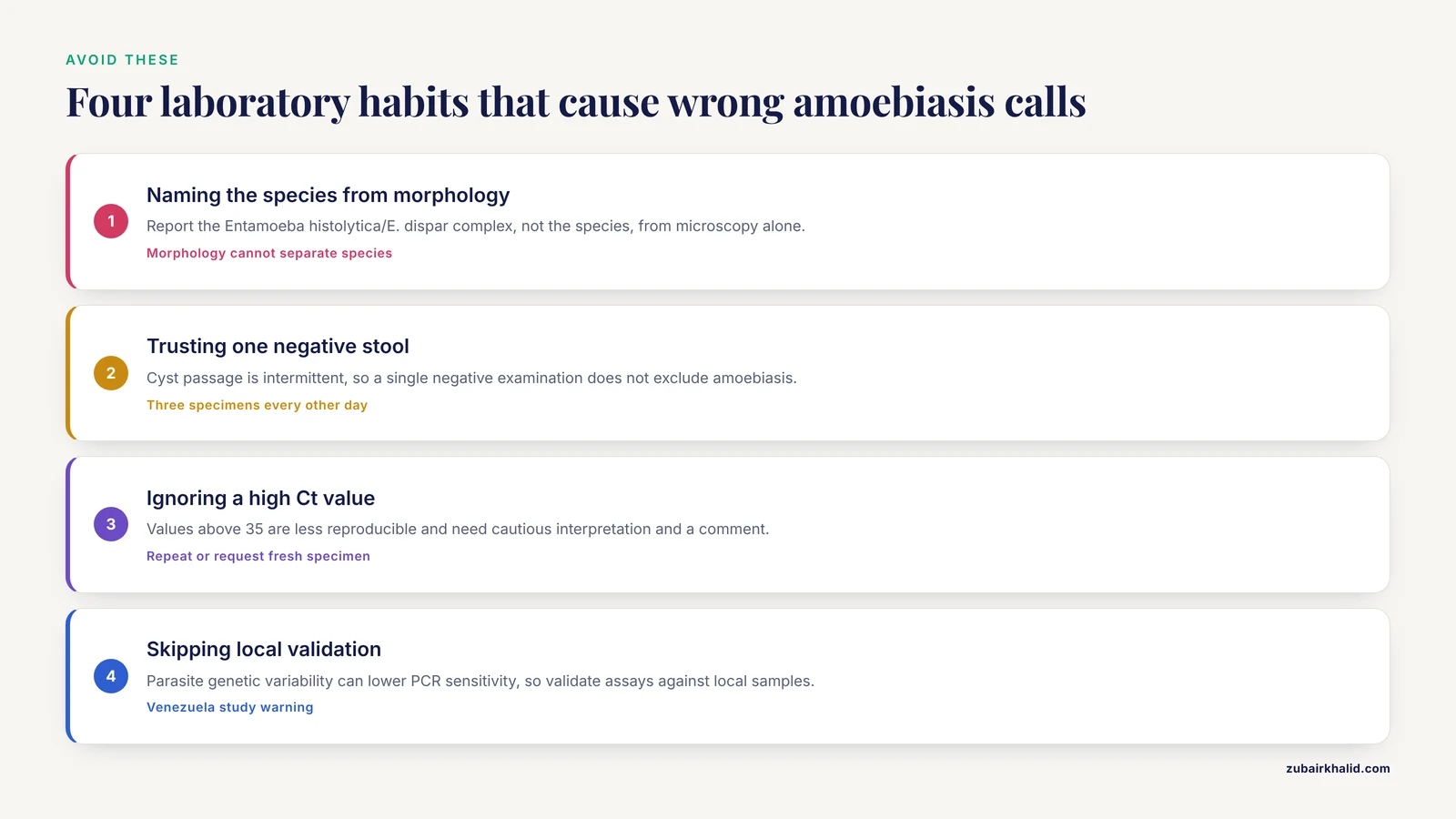
- Calling every Entamoeba histolytica cyst pathogenic. Morphology cannot separate Entamoeba histolytica from Entamoeba dispar or Entamoeba moshkovskii [2][3].
- Diagnosing from a single stool microscopy result. Cyst passage can be intermittent, and sensitivity is low when parasite loads are low [2].
- Treating a positive antibody test as proof of active intestinal disease. Serology can remain positive after past exposure.
- Ignoring the Ct value on qPCR. Values above 35 are less reproducible and need cautious interpretation [13].
- Using a rectal swab as a perfect substitute for stool. The agreement is moderate, and discordant results occur [12].
- Forgetting the luminal agent. Tissue amoebicides alone may not clear cysts, and relapse can follow [1].
- Missing extraintestinal disease. Cutaneous amoebiasis can present without diarrhea and can mimic other ulcers [10].
- Assuming diarrhea equals amoebiasis. Amoebic colitis overlaps with ulcerative colitis and other enteric infections [3].
- Overlooking drug adverse effects. Metronidazole has been linked to suspected pancreatitis in a pediatric case [14].
- Applying a PCR assay without local validation. Parasite genetic variability can affect sensitivity [2].
Individual cases need a clinician who can integrate exposure, symptoms, imaging, and laboratory results. For a pet with diarrhea, the diagnostic plan is different and belongs with a veterinarian.
Quick Review
- Entamoeba histolytica has a cyst stage for transmission and a trophozoite stage for tissue invasion.
- Transmission is fecal-oral through contaminated water, food, and sexual contact.
- Asymptomatic carriers are a major reservoir and can pass cysts for long periods.
- Microscopy cannot reliably distinguish Entamoeba histolytica from Entamoeba dispar and Entamoeba moshkovskii.
- Species-specific PCR or LAMP improves diagnosis, but assays need local validation.
- Treatment combines a tissue amoebicide with a luminal agent.
- Amoebic liver abscess and cutaneous amoebiasis are important extraintestinal syndromes.
Microscopy Bench Work: Wet Mounts, Iodine Preparations, and Permanent Stains
Microscopy remains the most accessible first-line method for amoebiasis, but its value depends entirely on technique. A poorly prepared wet mount can miss trophozoites that a careful reader would find in seconds. This section walks through the three routine preparations and the artifacts that cause false calls.
Saline Wet Mount for Trophozoites
Use a fresh, diarrheic or mucoid stool specimen. Place a small fleck of mucus or bloody mucus on a clean slide, add one drop of 0.85 percent saline, and apply a coverslip. Examine within 30 minutes of collection because trophozoites lose motility and lyse quickly at room temperature. Warming the stage or using a warm slide can preserve motility slightly longer, but the real solution is rapid transport.
Look for refractile, pleomorphic organisms with finger-like pseudopods. Entamoeba histolytica trophozoites measure roughly 12 to 60 micrometers, with most in the 15 to 25 micrometer range. The nucleus is usually not visible in an unstained wet mount. Ingested red blood cells inside the cytoplasm are a supportive clue, but they are not a species-specific finding.
Iodine Wet Mount for Cysts
Add one drop of Lugol or D'Antoni iodine to a small amount of stool, mix, and coverslip. Iodine kills trophozoites, so use it only for cyst morphology. Cysts appear yellow-brown. The mature Entamoeba histolytica cyst has four nuclei, a small central karyosome, and blunt-ended chromatoid bars. Immature cysts may show one or two nuclei and a glycogen mass.
The most common confusion is with Entamoeba coli. E. coli cysts are larger, often 15 to 25 micrometers, contain up to eight nuclei, and have chromatoid bars with pointed or splintered ends. Inexperienced readers can easily reverse the two. The nuclear chromatin pattern is the anchor: E. histolytica has fine, uniform peripheral chromatin and a small central karyosome, while E. coli has coarse, irregular chromatin and a large eccentric karyosome.
Permanent Stains: Trichrome and Iron Hematoxylin
Permanent stains are the standard for species-complex morphology. Trichrome is the workhorse. Smears are fixed in Schaudinn or SAF fixative and stained with a trichrome protocol. Cytoplasm appears blue-green or purple, and nuclear chromatin appears red or purple. Iron hematoxylin produces dark blue-black chromatin and is excellent for teaching, but it is more time-consuming.
The nuclear features that matter are the same as in iodine mounts but sharper. A central karyosome with fine, beaded peripheral chromatin favors Entamoeba histolytica. A large eccentric karyosome with coarse chromatin favors Entamoeba coli. Chromatoid bars stain dark and are easier to assess in iron hematoxylin.
Artifacts and Look-Alikes
Macrophages are the most common false positive for trophozoites in dysenteric stool. They have a single nucleus, often with a prominent nucleolus, and may contain ingested debris. The key difference is that macrophages lack the delicate peripheral chromatin rim of Entamoeba histolytica and do not show the same progressive pseudopod motility.
Polymorphonuclear leukocytes are abundant in bacillary dysentery and typically sparse in amoebic dysentery. This is a useful clinical clue but not a diagnostic rule. Yeast cells can mimic cysts but have no nuclei or chromatoid bars. Blastocystis hominis can be mistaken for a cyst by novices; it has a large central body and multiple peripheral nuclei.
Troubleshooting Common Microscopy Problems
A smear that is too thick obscures nuclear detail. A smear that is too thin makes scanning laborious. Over-decolorization in trichrome washes out chromatin. Old fixative can cause poor preservation. The remedy is a standardized protocol, a timer, and a control slide. Every batch of stain should include a known positive control to confirm that the reagents are working.
The Species Problem: Entamoeba histolytica, Entamoeba dispar, and Entamoeba moshkovskii at the Bench
Routine microscopy cannot separate Entamoeba histolytica from Entamoeba dispar or Entamoeba moshkovskii [2][3]. The three species are morphologically identical in cyst and trophozoite stages. This is not a minor academic point. Entamoeba histolytica is a pathogen that requires treatment, while Entamoeba dispar is generally considered nonpathogenic and Entamoeba moshkovskii is a free-living species that can colonize humans and occasionally cause diarrhea.
Why Morphology Fails
The cyst size ranges overlap. The nuclear number overlaps. The chromatoid bar shape overlaps. Even erythrophagocytosis, the presence of ingested red blood cells in a trophozoite, is strongly associated with Entamoeba histolytica but has been reported in other amoebae and can be mimicked by macrophages that have engulfed red cells. A careful microscopist can say that an organism is in the Entamoeba histolytica complex, but cannot say which species it is.
The Reporting Language Problem
A laboratory that reports "Entamoeba histolytica cysts seen" on the basis of an iodine mount is overstating the result. The defensible report is "Entamoeba histolytica/E. dispar complex cysts seen" or "Entamoeba species, morphology consistent with the E. histolytica complex." The clinician then needs a species-specific test to decide on treatment.
A Practical Decision Tree
When microscopy is positive for the complex, the next step is a species-specific antigen test or molecular test. In a resource-limited setting where neither is available, the clinician must weigh the local epidemiology and the patient's symptoms. In an asymptomatic patient with cysts only, the probability of Entamoeba dispar is high in many endemic settings, but it cannot be assumed.
Worked Example
A 30-year-old food handler has cysts on a routine stool examination. The iodine mount shows four-nucleated cysts with blunt chromatoid bars. The laboratory reports "Entamoeba histolytica/E. dispar complex." A species-specific PCR returns Entamoeba dispar. The patient does not need a tissue amoebicide, but the laboratory and clinician should still discuss hygiene and the possibility of mixed infections, because a person can carry both species at different times.
Preanalytical Variables: Why the Right Specimen Matters
The best PCR assay in the world cannot compensate for a stool specimen that was collected in a diaper, left on a counter for six hours, or diluted with urine. Preanalytical variables cause more false negatives than any analytical step.
Collection Devices and Preservatives
Stool should be collected in a clean, wide-mouth, leak-proof container without urine or water contamination. If the specimen cannot reach the laboratory within one hour, it should be placed in a preservative. Common options include polyvinyl alcohol (PVA), sodium acetate-acetic acid-formalin (SAF), and 10 percent formalin. SAF is versatile because it supports both permanent stains and some molecular workflows. The ratio should be roughly one part stool to three parts preservative.
Timing and Specimen Type
Trophozoites are found in diarrheic or mucoid stool and degenerate within 30 to 60 minutes at room temperature. Cysts are found in formed stool and survive longer, but they too degrade over days. A formed stool is better for cyst detection; a diarrheic stool is better for trophozoite detection. The laboratory should note the stool consistency on the report because it changes the interpretation.
The Three Stool Rule
Cyst passage is intermittent. A single negative stool examination does not exclude amoebiasis. The classic recommendation is three specimens collected every other day. This increases sensitivity but does not guarantee detection, especially when parasite loads are low [2].
Interfering Substances
Barium from a recent upper or lower gastrointestinal series coats the stool and obscures parasites. Antacids, oily laxatives, and bismuth compounds can also interfere. Antibiotics can suppress the gut microbiota and reduce trophozoite numbers. The ideal is to collect stool before such procedures or at least several days after stopping the interfering agent.
Rectal Swabs as an Alternative
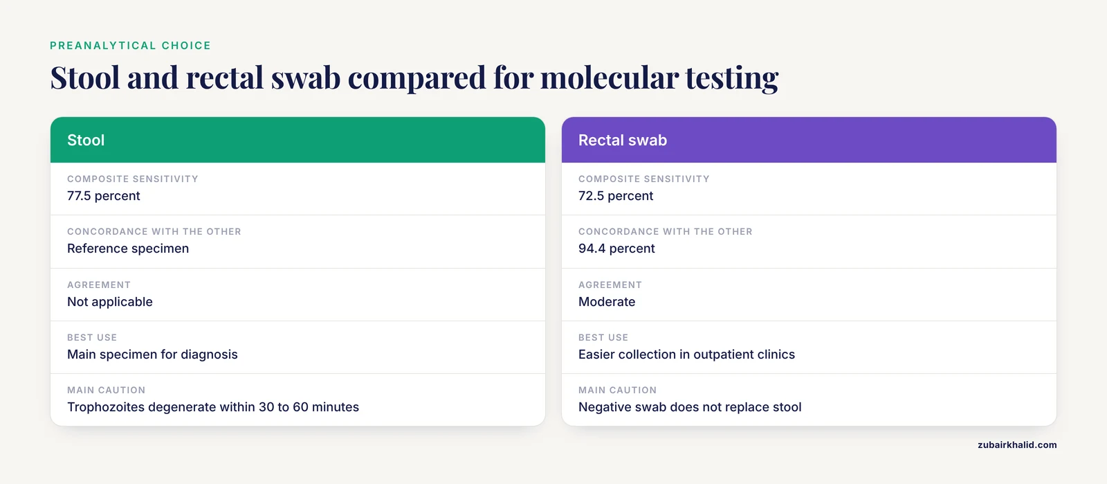
A multisite study compared 355 paired stool and rectal swab samples using Entamoeba histolytica-specific quantitative PCR. Concordance was 94.4 percent with moderate agreement. Composite sensitivity was 77.5 percent for stool and 72.5 percent for rectal swab [12]. Rectal swabs are easier to collect, especially in outpatient sexual health clinics, but a negative swab does not replace a stool test when suspicion is high [12].
Worked Example
A patient with diarrhea takes bismuth subsalicylate and mineral oil. The stool is black and greasy. Microscopy is negative. The clinician repeats the test after the patient stops the products for three days. The repeat specimen shows trophozoites. The first result was a preanalytical false negative.
Molecular Diagnostics in Practice: Extraction, Amplification, and Interpretation
Molecular testing is the only routine method that can definitively identify Entamoeba histolytica, but it has its own failure modes. Extraction, inhibition, target choice, and interpretation all matter.
Extraction Is the First Failure Point
A 2026 study standardized two PCR protocols and compared four DNA extraction methods for stool samples. The Wizard kit produced the best DNA quality [2]. This finding is a reminder that extraction kits are not interchangeable. Stool contains bile salts, bilirubin, complex polysaccharides, and plant fibers that inhibit PCR. A kit that works for blood may fail for stool.
Target Genes and Assay Design
Common targets include the 18S rRNA gene, the SREHP gene, and the LSU rRNA gene. A real-time LAMP assay targeting SREHP and LSU rRNA was evaluated in 494 stool samples from North India, including 117 patients with ulcerative colitis [3]. LAMP runs at a constant temperature, which makes it attractive for laboratories without a real-time thermocycler.
The Meaning of Ct Values
Quantitative PCR produces a cycle threshold, or Ct, value. A low Ct means a high starting amount of target DNA. A high Ct means a low starting amount. A study of 873 stool samples in Ghana compared three real-time PCR assays using latent class analysis. Sensitivity ranged from 75 to 100 percent and specificity from 94 to 100 percent. Ct values above 35 had reduced reproducibility when competitor assays were applied [13].
Worked Example: A High Ct Result
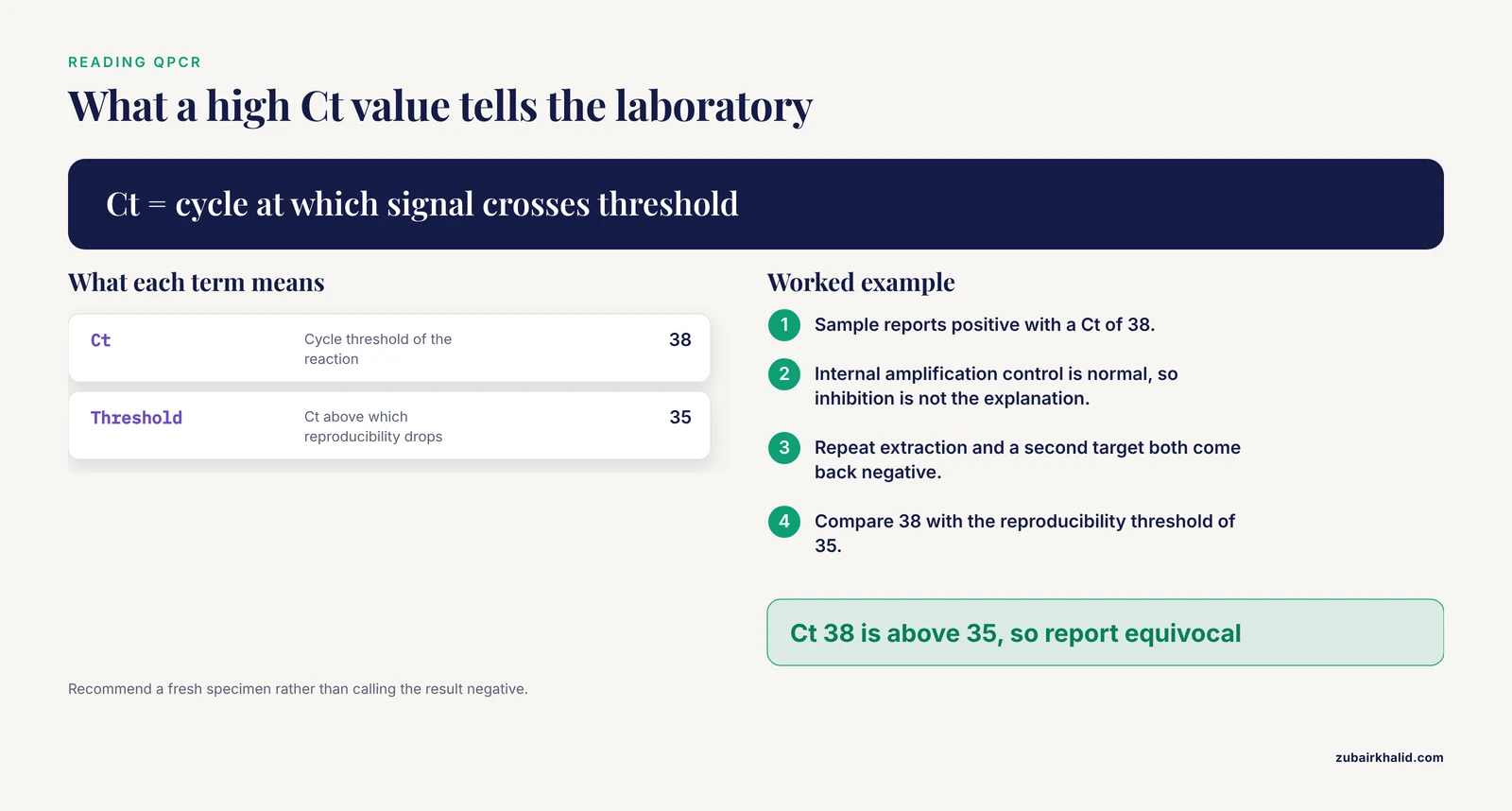
A stool sample is positive for Entamoeba histolytica with a Ct of 38. The internal amplification control is normal, so inhibition is not the explanation. The laboratory repeats the extraction from the original specimen and runs a second target. The repeat is negative. The laboratory reports the result as equivocal and recommends a fresh specimen. This is not a failure of the assay; it is an honest reflection of a low-burden sample.
Local Validation Is Essential
The Venezuela study found that PCR sensitivity was lower than coprology in 180 rural stool samples, possibly because of genetic variability in local parasites [2]. A commercial assay validated in one country may miss strains in another. Laboratories should validate their chosen assay against local samples and compare it with an independent method before relying on it for clinical decisions.
Multiplex Assays
Multiplex PCR can detect Entamoeba histolytica, Entamoeba dispar, and Entamoeba moshkovskii in a single reaction [2]. This is the ideal format because it answers the species question directly and also detects the nonpathogenic look-alikes, which explains a positive microscopy result that would otherwise be confusing.
Stool Antigen Testing: Practical Role and Limitations
Stool antigen tests detect Entamoeba histolytica proteins in fecal samples. They are faster than PCR and do not require a thermocycler. Their main value is in settings where molecular testing is unavailable but species-specific information is needed.
How Antigen Tests Fit
A positive antigen test in a patient with compatible symptoms supports the diagnosis of amoebiasis. A negative antigen test does not exclude it, especially when the parasite burden is low or the specimen is not fresh. Antigen tests also vary in performance, and some detect only Entamoeba histolytica while others cross-react with Entamoeba dispar.
Comparison With Microscopy and PCR
Microscopy cannot identify species. Antigen tests can, if the test is specific. PCR is the reference standard for species identification. The practical hierarchy is: microscopy for screening, antigen for rapid species-specific triage, and PCR for confirmation or when antigen results conflict with the clinical picture.
Worked Example
A clinic in a low-resource setting sees a patient with bloody diarrhea. The saline wet mount shows motile trophozoites with ingested red cells. A rapid antigen test is positive for Entamoeba histolytica. The clinician starts treatment with a tissue amoebicide and a luminal agent [1]. A confirmatory PCR is sent to a reference laboratory. The antigen test allowed treatment to start the same day.
Serology: Antibody Kinetics and Clinical Interpretation
Serology detects the host antibody response, not the parasite. This distinction explains both its strengths and its traps.
IgG Kinetics
IgG antibodies appear within days to weeks after tissue invasion and can persist for months to years. A single positive IgG in a patient from an endemic area may reflect past infection. In a non-endemic setting, a positive IgG in a patient with a liver abscess is strong supportive evidence of amoebic liver abscess.
Seroepidemiology in MSM
A Belgian sexual health clinic cohort of men who have sex with men used an Entamoeba histolytica-specific IgG ELISA and found a seroincidence of 1.47 per 100 person-years [5]. This is an epidemiological measure, not a diagnostic one. It tells us that transmission is ongoing in this population and that clinicians should consider amoebiasis in MSM with compatible symptoms.
Worked Example: The Traveler
A 35-year-old traveler returns from Southeast Asia with fever and right upper quadrant pain. Ultrasound shows a 5 cm liver abscess. IgG is positive. The combination strongly supports amoebic liver abscess. Stool PCR is negative, which is common because the intestinal infection may have resolved. The clinician treats with a tissue amoebicide and a luminal agent [1].
The Early Infection Trap
IgG can be negative in the first week of symptoms. If clinical suspicion is high, repeat serology in 7 to 10 days. A rising titer is more informative than a single negative result.
Amoebic Liver Abscess: Integrating Laboratory and Imaging Findings
Amoebic liver abscess is the most common extraintestinal manifestation of Entamoeba histolytica. The laboratory and radiology findings must be interpreted together.
Pathogenesis
Trophozoites invade the colonic mucosa, enter the portal circulation, and reach the liver. The host response contributes to tissue damage. In a mouse model, a pathogenic clone called B2p triggered stronger cytokine and chemokine expression, stress pathways, and tissue remodeling than a nonpathogenic A1np clone [9]. Liver pathology is therefore a product of both parasite factors and host immunity [9].
Imaging Findings
Ultrasound, CT, and MRI can identify a liver abscess, but they cannot identify the organism. A pyogenic abscess, an echinococcal cyst, and a necrotic tumor can look similar. Imaging must be combined with serology and clinical context.
Aspirate Limitations
Aspirated pus often has the classic "anchovy paste" appearance, but trophozoites are usually found at the margin of the abscess, not in the necrotic center. A negative aspirate smear does not exclude amoebic liver abscess. Drainage is indicated for large abscesses, suspected rupture, or failure to respond to medical therapy, not as a diagnostic requirement.
Worked Example
A 45-year-old man has fever, weight loss, and a tender liver. Ultrasound shows a 7 cm right lobe abscess. Serology is positive. Stool microscopy is negative. The clinician starts a tissue amoebicide. The patient improves over 72 hours. The diagnosis is amoebic liver abscess. The negative stool result is expected and does not change the diagnosis.
Treatment Follow-up, Relapse Prevention, and Test of Cure
Amoebiasis treatment has two compartments. The tissue compartment contains invasive trophozoites. The luminal compartment contains cysts and noninvasive trophozoites. A tissue amoebicide alone may clear symptoms but leave cysts behind, allowing relapse and continued transmission [1].
The Two-Drug Strategy
Standard treatment combines a tissue amoebicide such as metronidazole or tinidazole with a luminal agent such as paromomycin, diloxanide furoate, or diiodohydroxyquin [1]. Asymptomatic carriers with cysts only can be treated with a luminal agent alone.
Adverse Effects Matter
Metronidazole is generally effective but not harmless. A published case reported suspected metronidazole-induced acute pancreatitis in a three-year-old child six days after completing a 10 day oral course for amoebic gastroenteritis. Amylase was 3251 IU/L and lipase was greater than 1000 U/L [14]. New abdominal pain after treatment should not be dismissed as persistent amoebiasis without considering drug toxicity.
Why Carriage Persists
The gut environment can influence drug susceptibility. In vitro, 100 micromolar deoxycholic acid, a microbiota-modified secondary bile acid, extended Entamoeba histolytica survival under nutrient-limited conditions and reduced metronidazole susceptibility [4]. This is experimental, but it helps explain why some carriers are hard to clear.
Experimental Therapies
Aqueous Carica papaya callus extracts killed trophozoites in vitro and reduced amoebic liver abscess in hamsters [15]. Entamoeba histolytica carbonic anhydrase, EhiCA, is being studied as a future drug target [1]. Neither is standard care.
Test of Cure
Repeat stool examination or PCR after treatment is reasonable for carriers, food handlers, and patients with recurrent symptoms. Timing matters. Testing too soon after the luminal agent may detect dead organisms or residual DNA. A practical approach is to wait one to two weeks after completing the luminal agent, then collect three stool specimens over several days.
Amoebiasis in Special Populations
Children
A 2026 systematic review and meta-analysis included 119 studies and 70,259 children and found high heterogeneity in global prevalence estimates [11]. The heterogeneity reflects differences in diagnostic methods, endemicity, and study design. A prevalence estimate from one region should not be applied to another, and a child with diarrhea in a low-endemic setting does not have the same pretest probability as a child in a high-endemic setting.
Men Who Have Sex With Men
Entamoeba histolytica is recognized as a sexually transmitted infection in some populations [5]. Oral-anal contact is the route. Clinicians should consider amoebiasis in MSM with proctitis, bloody diarrhea, or unexplained gastrointestinal symptoms, especially when routine bacterial testing is negative.
Patients With Ulcerative Colitis
Amoebic colitis can mimic ulcerative colitis. A real-time LAMP study from North India specifically included 117 patients with ulcerative colitis because the two conditions overlap [3]. Starting immunosuppressive therapy in a patient with undiagnosed amoebiasis can lead to severe disease. In endemic areas, exclude amoebiasis before making a definitive diagnosis of inflammatory bowel disease.
Cutaneous Amoebiasis
Cutaneous amoebiasis is rare and usually affects the perianal or genital region. It can present as a chronic ulcer that mimics malignancy or a surgical wound infection [10]. When gastrointestinal symptoms are absent, the diagnosis is easily missed. Biopsy showing trophozoites in the ulcer base supports the diagnosis [10].
The Microbiota-Amoeba Interface: What the Laboratory Should Know
Entamoeba histolytica does not act alone. The gut microbiota shapes its growth, survival, and drug susceptibility.
Bacterial Support
The parasite feeds on bacteria and uses non-opsonic phagocytosis to engulf them. Cell-surface glycocalyx molecules help the trophozoite recognize and engulf bacteria [6]. This is not a side detail; it is central to the parasite's survival in the colon.
Microbiota Shifts in Infection
A real-time PCR case-control study of 23 confirmed Entamoeba histolytica cases in Egypt found lower Firmicutes, Bacteroidetes, Lactobacillus, and Clostridium leptum and higher Bifidobacterium than in healthy controls [7]. These shifts do not prove causation in an individual patient, but they support the view that amoebiasis is a host-parasite-microbiota interaction.
Bile Acids and Drug Susceptibility
Microbiota-modified bile acids can affect the parasite. In vitro, deoxycholic acid extended survival under nutrient-limited conditions and reduced metronidazole susceptibility [4]. This is experimental evidence, not a treatment recommendation, but it helps explain why carriage can persist despite apparently adequate therapy.
Laboratory Implication
Microbiota testing is not part of routine amoebiasis diagnosis. However, the laboratory should recognize that a patient's bacterial community can influence both the likelihood of detecting the parasite and the response to treatment. A negative stool test in a patient with a disrupted microbiome is less reassuring than a negative test in a healthy host.
Host-Parasite Interactions: Phagocytosis and Immune Evasion
The innate immune system should be able to clear a luminal parasite, but Entamoeba histolytica has evolved to avoid this.
The Kiss and Run Strategy
Human macrophages and neutrophils can phagocytose some amoebae, but live Entamoeba histolytica uses a "kiss and run" evasion strategy, making brief contact without being engulfed [8]. Formalin-killed and UV-irradiated trophozoites are phagocytosed efficiently, which shows that live parasite factors are involved in evasion [8].
Why This Matters for the Laboratory
This evasion explains why invasive disease can develop despite an intact immune system. It also explains why killed organisms in fixed specimens are easier for phagocytes to clear, a fact that is relevant to vaccine research but not to routine diagnostics.
Host Response and Severity
The host response contributes to disease severity. In a mouse model, the pathogenic B2p clone triggered stronger cytokine and chemokine expression, stress pathways, and tissue remodeling than the nonpathogenic A1np clone [9]. This means that two people infected with different parasite strains can have very different outcomes, even with the same immune status.
Quality Assurance, Biosafety, and Reporting
Biosafety
Stool specimens are potentially infectious. Trophozoites and cysts are not highly contagious by the respiratory route, but ingestion is a real risk in the laboratory. Wear gloves, wash hands, and process specimens in a biological safety cabinet when performing culture or molecular testing. Fixation in formalin or SAF inactivates the organisms for microscopy.
Reporting Language
The laboratory must not overstate morphology. If only microscopy is performed, the report should say "Entamoeba histolytica/E. dispar complex" or "Entamoeba species, morphology consistent with the E. histolytica complex." A report that says "Entamoeba histolytica" without molecular or antigen confirmation is misleading.
Proficiency Testing
Stool parasitology is a skill that decays without practice. Laboratories should participate in external quality assessment programs and maintain a library of reference images. A known positive control should be stained with every batch of trichrome or iron hematoxylin.
Communication With Clinicians
When PCR is positive for Entamoeba histolytica, the laboratory should flag the result and suggest clinical correlation. When the Ct is high, add a comment about reduced reproducibility. When microscopy is positive but PCR is negative, the laboratory should explain that the organism is likely Entamoeba dispar or Entamoeba moshkovskii [2][3].
Worked Example
A laboratory reports "Entamoeba histolytica cysts seen" on an iodine mount. The clinician treats the patient with a tissue amoebicide. A subsequent PCR identifies Entamoeba dispar. The patient received unnecessary treatment. The corrected laboratory policy is to report "Entamoeba histolytica/E. dispar complex" and recommend species-specific testing before treatment in asymptomatic carriers.
Worked Case: A Diagnostic Pathway for Bloody Diarrhea
A 28-year-old man presents with five days of bloody diarrhea, cramping, and tenesmus. He traveled to a tropical region three weeks ago. He has no fever.
Step 1. A fresh stool specimen is collected and delivered to the laboratory within 30 minutes. The saline wet mount shows motile trophozoites with finger-like pseudopods and ingested red blood cells.
Step 2. The laboratory reports "Entamoeba histolytica/E. dispar complex trophozoites seen" and recommends species-specific PCR.
Step 3. PCR returns positive for Entamoeba histolytica and negative for Entamoeba dispar and Entamoeba moshkovskii.
Step 4. The clinician prescribes a tissue amoebicide plus a luminal agent [1].
Step 5. After completing the luminal agent, the patient submits three stool specimens over several days. PCR is negative.
Troubleshooting. If the wet mount had been negative, the clinician would have submitted two more specimens and ordered antigen or PCR. A single negative wet mount does not exclude amoebiasis, especially when parasite loads are low [2].
Related Guides
- Toxoplasma gondii in Cats: Life Cycle, Zoonotic Transmission, and Veterinary Management: Another protozoan parasite with a complex life cycle and zoonotic transmission
- Avian Coccidiosis in Chickens: Prevention, Life Cycle, and Cross-Species Risks: Coccidiosis is a protozoan intestinal infection with similar life-cycle stages
- Cryptocaryon irritans in Marine Aquaculture: Life Cycle and Control Strategies: Marine protozoan parasite with direct life cycle and control strategies
- Whirling Disease in Salmonid Fish: Myxobolus cerebralis Life Cycle, Detection, and Control: Myxozoan parasite life cycle illustrates diagnostic challenges in parasitic disease
- Heartworm Disease in Dogs: Life Cycle and Treatment: Parasitic disease with life cycle and treatment parallels for clinical management
- Spirocerca lupi: Carcinogenic Esophageal Nematode of Canids, Life Cycle, Pathogenesis, Diagnostic Approaches, and Molecular Epidemiology: Nematode parasite with detailed pathogenesis, diagnostics, and molecular epidemiology
- Fish Cestodiasis (Bothriocephalus acheilognathi) in Carp Aquaculture: Life Cycle and Control: Cestode parasite with life cycle and control in aquaculture settings
- Prosthogonimus macrorchis: Oviduct Fluke in Chickens, Life Cycle, Pathogenesis, and Diagnosis: Fluke parasite with life cycle, pathogenesis, and diagnosis in poultry
- Toxoplasmosis in Cats: Life Cycle, Zoonotic Risk, and Prevention: Another protozoan zoonosis with complex life cycle
- Examples of Poultry Parasites: Morphology, Life Cycles, and Clinical Impact: Overview of poultry parasites including life cycles
- Poultry Internal Parasites: Identification, Life Cycles, and Veterinary Control Programs: Internal parasites of poultry with control programs
- Toxoplasma gondii in Cats: Life Cycle, Zoonotic Risk, and Veterinary Management: Toxoplasma gondii life cycle and zoonotic risk
Frequently Asked Questions
How is Entamoeba histolytica transmitted?
Entamoeba histolytica is transmitted by swallowing infectious cysts in contaminated food or water. Oral-anal sexual contact can also transmit the parasite.
What are the symptoms of Entamoeba histolytica infection?
Many infections cause no symptoms. Symptomatic cases can have diarrhea, bloody dysentery, abdominal pain, or liver abscess with fever and right upper quadrant pain.
How is Entamoeba histolytica diagnosed?
Diagnosis uses fresh stool microscopy, species-specific PCR or LAMP, antigen tests, serology, and tissue examination. No single test is perfect in every setting.
What is the difference between an Entamoeba histolytica cyst and a trophozoite?
The cyst is the infectious, environmentally resistant form passed in stool. The trophozoite is the motile, feeding form that invades tissue and causes disease.
What stain is used for Entamoeba histolytica?
Trichrome and iron hematoxylin are common permanent stains for Entamoeba histolytica. Iodine wet mounts help visualize cysts, and saline wet mounts help visualize motile trophozoites.
How is amoebiasis treated?
Amoebiasis is treated with a tissue amoebicide such as metronidazole or tinidazole plus a luminal agent such as paromomycin, diloxanide furoate, or diiodohydroxyquin.
Sources
- Entamoeba histolytica carbonic anhydrase.
- Evaluation of two PCR protocols for the detection of Entamoeba histolytica/E. dispar/E. moshkovskii in stool samples.
- Evaluation of real-time LAMP assay for detection of Entamoeba species in clinical stool samples from a tertiary care center in North India.
- Multi-omics profiling and bile-acid exposure assays implicate a gut microbiome-parasite axis linked to persistent Entamoeba histolytica carriage.
- Seroincidence and Clinical Manifestations of Entamoeba histolytica Among Belgian MSM in the PrEP Era: A Retrospective Cohort Study.
- Exploring glycocalyx components powering the contact between Entamoeba histolytica and bacteria during non-opsonic phagocytosis.
- Gastrointestinal microbiota dysbiosis in entamoeba histolytica infection: a real-time PCR case-control study.
- Phagocytosis of Acanthamoeba by macrophages and neutrophils and "kiss and run" evasion by Entamoeba histolytica.
- Distinct temporal patterns of liver immune responses to pathogenic and non-pathogenic Entamoeba histolytica clones.
- Perianal cutaneous amoebiasis: An unsuspected extra-intestinal manifestation of Entamoeba histolytica.
- Global Prevalence of Entamoeba histolytica in Children: A Systematic Review and Meta-Analysis.
- Diagnosis of intestinal Entamoeba histolytica infection by rectal swab: An international multisite cross-sectional study between Taiwan and Japan.
- Evaluation of Each Three Entamoeba histolytica- and Strongyloides stercoralis-Specific Real-Time PCR Assays Applying Test Comparisons Without Reference Standards.
- Suspected Metronidazole-Induced Acute Pancreatitis in a Three-Year-Old Child Following Treatment for Entamoeba histolytica Gastroenteritis: A Case Report.
- Aqueous Extracts of Carica papaya Embryogenic Callus Kill Entamoeba histolytica Trophozoites and Orally Protect against the Development of Amoebic Liver Abscesses in Hamsters.