
Cryptocaryon irritans in Marine Aquaculture: Life Cycle and Control Strategies
Introduction
Cryptocaryon irritans is an obligate parasitic ciliate protozoan that causes cryptocaryoniasis, commonly known as marine white spot disease or marine ich. This pathogen represents one of the most significant parasitic threats to marine aquaculture operations globally, affecting a broad range of teleost hosts in both open-water net pens and recirculating aquaculture systems (RAS) [1, 2]. The economic impact of C. irritans outbreaks is substantial, resulting in direct mortality, secondary bacterial infections, reduced growth rates, and compromised welfare in affected stocks [3, 4]. Unlike its freshwater counterpart Ichthyophthirius multifiliis, which is covered in detail in the article White Spot Disease (Ichthyophthirius multifiliis) in Aquaculture: Environmental Drivers and Molecular Diagnostics, C. irritans exhibits distinct biological adaptations to the marine environment, including tolerance to higher salinities and a unique life cycle stage known as the tomont [5, 6].
This review provides an exhaustive examination of the C. irritans life cycle, diagnostic methodologies, and current control strategies, with an emphasis on evidence-based management protocols for veterinary professionals and aquaculture biologists.
Taxonomic Classification and Morphology
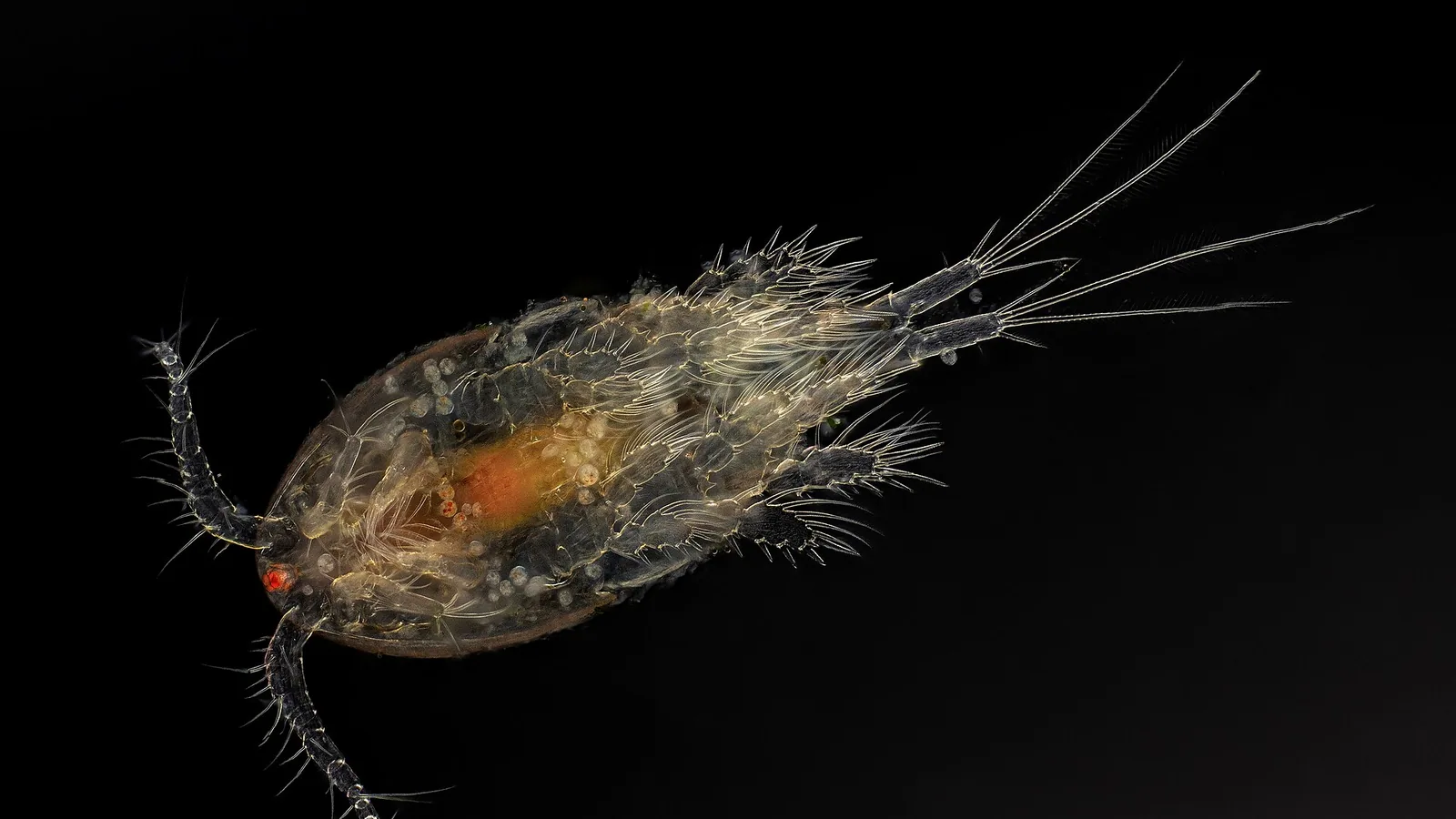
Cryptocaryon irritans belongs to the phylum Ciliophora, class Prostomatea, and order Prorodontida [7]. The organism was first described by Brown in 1951 and has since been recognized as the primary etiological agent of marine white spot disease [8]. Morphologically, the trophont stage is characterized by a ciliated, ovoid to pear-shaped body measuring 100 to 500 micrometers in diameter, with a distinct horseshoe-shaped macronucleus and a subterminal cytostome [9, 10]. The ciliary arrangement follows a holotrichous pattern, with uniform ciliation covering the entire cell surface, facilitating motility within the host mucus and epidermal layers [11].
Life Cycle
The life cycle of C. irritans is direct and monoxenous, comprising four distinct stages: the trophont (parasitic feeding stage), the protomont (pre-encystment stage), the tomont (reproductive cyst stage), and the theront (infective free-swimming stage) [12, 13]. The complete life cycle duration is temperature-dependent, ranging from 5 to 14 days at optimal temperatures between 24 and 28 degrees Celsius [14, 15].
Trophont Stage
The trophont is the obligate parasitic stage that resides within the epidermis and dermis of the fish host. Invasion occurs when theronts penetrate the epithelial layer, typically targeting the gills, skin, and fins [16]. Once established, the trophont feeds on host cellular debris, mucus, and tissue fluids through phagocytosis and pinocytosis [17]. The feeding activity induces extensive epithelial hyperplasia, necrosis, and an intense inflammatory response characterized by lymphocyte and macrophage infiltration [18]. Trophonts remain within the host for 3 to 7 days, depending on water temperature, before exiting to initiate the next stage [19].
Protomont Stage
Upon completion of the feeding phase, the mature trophont exits the host tissue and becomes a free-swimming protomont. This stage is short-lived, lasting 2 to 8 hours, during which the organism seeks a suitable substrate for encystment [20]. Protomonts are highly motile and exhibit negative phototaxis, preferentially migrating to darker areas of the culture system such as tank bottoms, pipe surfaces, and biofilter media [21].
Tomont Stage
The protomont attaches to a substrate and secretes a tough, double-layered cyst wall to form the tomont [22]. Within the tomont, the organism undergoes multiple rounds of binary fission, producing hundreds to thousands of tomites. The number of tomites produced per tomont is variable, ranging from 100 to 1000, and is influenced by the nutritional status of the trophont and environmental conditions [23]. The tomont stage is the most resilient stage of the life cycle, capable of surviving for extended periods under suboptimal conditions. Tomonts can remain viable for up to 30 days at low temperatures (10 to 15 degrees Celsius) and can tolerate salinities ranging from 10 to 45 parts per thousand [24, 25].
Theront Stage
Tomites differentiate into theronts, which are the infective free-swimming stage. Theronts are approximately 30 to 60 micrometers in length and possess a fully developed ciliature and a functional cytostome [26]. They are positively phototactic and exhibit a characteristic corkscrew swimming pattern that facilitates host contact [27]. Theronts must locate and penetrate a suitable host within 24 to 48 hours, as their energy reserves are limited and they do not feed in the free-swimming state [28]. The infectivity of theronts is highest within the first 6 to 12 hours after release from the tomont [29].
graph TD
 A["Theront: Free-swimming infective stage"] -->|Penetrates host epithelium| B["Trophont: Parasitic feeding stage in epidermis/dermis"]
 B -->|Exits host after 3-7 days| C["Protomont: Free-swimming pre-encystment stage"]
 C -->|Attaches to substrate and encysts| D["Tomont: Reproductive cyst stage"]
 D -->|Multiple binary fission produces tomites| E[Tomites differentiate into theronts]
 E --> A
Epidemiology and Risk Factors
Cryptocaryon irritans outbreaks are influenced by a complex interplay of environmental, host, and management factors. Water temperature is the primary driver of transmission dynamics, with optimal proliferation occurring between 24 and 28 degrees Celsius [30]. Outbreaks can occur at lower temperatures but progress more slowly, allowing for extended diagnostic windows [31]. Salinity fluctuations outside the optimal range of 25 to 35 parts per thousand can reduce theront survival and tomont viability, although the parasite demonstrates considerable euryhaline tolerance [32].
High stocking densities, poor water quality, and concurrent disease conditions predispose fish to severe infections [33]. Stress-induced immunosuppression, particularly from handling, transport, or suboptimal nutrition, increases host susceptibility and trophont burden [34]. Recirculating aquaculture systems present unique challenges due to the accumulation of tomonts on biofilter media and pipe surfaces, creating a persistent reservoir of infection [35].
Clinical Signs and Pathogenesis
The clinical presentation of cryptocaryoniasis varies with infection intensity and host species. Early signs include increased mucus production, flashing, and lethargy [36]. As the infection progresses, characteristic white nodules (trophonts) become visible on the skin, fins, and gills. These nodules are typically 0.5 to 1.0 millimeters in diameter and may coalesce in heavy infections [37].
Gill pathology is a primary cause of morbidity and mortality. Trophonts in the gill epithelium cause lamellar fusion, epithelial hyperplasia, and edema, leading to severe respiratory compromise [38]. Affected fish exhibit rapid opercular movements, piping at the water surface, and reduced feeding activity [39]. Secondary bacterial infections, particularly with Vibrio spp. and Aeromonas spp., are common sequelae that exacerbate tissue damage and mortality [40].
Diagnostic Approaches
Microscopic Examination
The gold standard for diagnosis remains direct microscopic examination of skin scrapes and gill biopsies. Wet mount preparations of fresh mucus or tissue samples should be examined under 100x to 400x magnification [41]. Trophonts are readily identifiable by their characteristic ciliated appearance, horseshoe-shaped macronucleus, and rolling movement within the mucus [42]. Gill biopsies are particularly useful for detecting low-level infections, as the gill epithelium is a preferred attachment site [43].
Molecular Diagnostics
Polymerase chain reaction (PCR) assays targeting the internal transcribed spacer (ITS) region of the ribosomal DNA have been developed for species-specific detection of C. irritans [44]. These assays offer superior sensitivity compared to microscopy, particularly in subclinical infections or when sampling from environmental matrices [45]. Quantitative PCR (qPCR) protocols allow for the quantification of parasite load in both fish tissues and water samples, facilitating early detection and monitoring of treatment efficacy [46].
The application of molecular diagnostics in aquaculture is analogous to approaches used for other aquatic parasites, such as those described in the article Sea Lice (Lepeophtheirus salmonis) Infestations in Farmed Salmon: Lifecycle, Detection Methods, and Integrated Pest Management. Environmental DNA (eDNA) sampling combined with qPCR has emerged as a promising tool for non-invasive surveillance of C. irritans in water systems [47].
Histopathology
Histological examination of formalin-fixed, hematoxylin and eosin-stained tissue sections reveals trophonts within the epidermis and dermis, surrounded by zones of necrosis and inflammatory cell infiltration [48]. The presence of trophonts in gill sections, often associated with lamellar fusion and epithelial hyperplasia, is diagnostic [49].
Control Strategies
Chemotherapy
Copper sulfate (CuSO4) remains the most widely used chemotherapeutic agent for controlling C. irritans in marine aquaculture. The therapeutic mechanism involves disruption of ion transport and enzyme function in the parasite, particularly at the trophont and theront stages [50]. The recommended free copper ion concentration is 0.15 to 0.20 mg/L, which must be maintained for 14 to 21 days to cover the complete life cycle [51]. Copper toxicity is influenced by water hardness, alkalinity, and dissolved organic carbon, necessitating regular monitoring of total and free copper concentrations using colorimetric or ion-selective electrode methods [52].
Formalin (37% formaldehyde solution) is used as an alternative or adjunct therapy at concentrations of 25 to 50 mg/L in continuous flow-through systems [53]. Formalin is effective against theronts and trophonts but has limited activity against tomonts [54]. The compound is toxic to fish at elevated temperatures and requires careful aeration to maintain dissolved oxygen levels [55].
Chloroquine diphosphate has been investigated as an alternative treatment, particularly in systems where copper is contraindicated. Doses of 10 to 20 mg/L administered as a prolonged bath have demonstrated efficacy against trophonts and theronts, although the cost and availability of the drug limit its widespread use [56].
Freshwater Baths
Freshwater immersion is a non-chemical treatment modality that exploits the osmotic sensitivity of C. irritans. The protocol involves transferring fish to freshwater (0 parts per thousand salinity) for 3 to 10 minutes, depending on the species and size [57]. The osmotic shock causes trophonts to detach from the host and undergo osmotic lysis [58]. Freshwater baths are effective against trophonts and theronts but do not affect tomonts in the environment [59]. The procedure is stressful for marine fish and should be performed with careful monitoring of water quality parameters and oxygen supplementation [60].
Physical and Environmental Control
Elevated temperature treatment, or thermotherapy, involves raising water temperature to 30 to 32 degrees Celsius for 5 to 10 days. This approach accelerates the life cycle, reducing the time available for theronts to find hosts and increasing the rate of tomont development and release [61]. The efficacy of thermotherapy is enhanced when combined with ultraviolet (UV) sterilization or ozone treatment to kill free-swimming theronts [62].
UV sterilization at a dose of 30,000 to 50,000 microwatt-seconds per square centimeter is effective for inactivating theronts in recirculating water systems [63]. Ozone treatment at an oxidation-reduction potential of 350 to 400 millivolts provides similar efficacy but requires careful monitoring to avoid toxicity to fish and biofilter bacteria [64].
Integrated Pest Management
An integrated pest management (IPM) approach combining multiple control modalities is recommended for sustainable control of C. irritans. The IPM framework includes:
- Quarantine and prophylactic treatment of all new stock.
- Regular monitoring using microscopic examination and eDNA-based PCR.
- Maintenance of optimal water quality parameters to reduce host stress.
- Strategic application of copper sulfate or formalin during high-risk periods.
- Implementation of physical barriers such as UV sterilization and ozone.
- Periodic fallowing of production units to break the life cycle.
This approach parallels the integrated strategies used for other aquaculture parasites, as discussed in the article White Spot Disease in Shrimp: Hepatopancreatic Microsporidiasis from Enterocytozoon hepatopenaei (EHP) and Co-infections.
Vaccination and Immunoprophylaxis
Research into vaccine development for C. irritans has focused on immobilization antigens (i-antigens) present on the ciliary surface of theronts and trophonts [65]. These antigens are highly immunogenic and elicit a protective antibody response in vaccinated fish. Experimental vaccines using formalin-killed theronts or recombinant i-antigen proteins have demonstrated partial protection in laboratory trials, reducing trophont burden and mortality. However, no commercial vaccine is currently available, and the antigenic diversity among C. irritans isolates presents a significant challenge for vaccine development.
Antimicrobial Resistance and Treatment Failure
Reports of reduced sensitivity to copper sulfate and formalin have emerged from intensive aquaculture operations, suggesting the potential for chemoresistance development. The mechanisms of resistance are not fully characterized but may involve alterations in membrane permeability, increased detoxification enzyme activity, or selection for resistant genotypes. Rotational use of chemotherapeutic agents and adherence to recommended dosage regimens are essential for delaying the emergence of resistance.
Conclusions
Cryptocaryon irritans remains a formidable pathogen in marine aquaculture, requiring a multifaceted approach to diagnosis and control. The direct life cycle, environmental resilience of the tomont stage, and broad host range necessitate rigorous biosecurity protocols and integrated management strategies. Advances in molecular diagnostics, particularly eDNA-based qPCR, offer improved sensitivity for early detection and surveillance. Continued research into vaccine development and alternative therapeutics is needed to reduce reliance on chemical treatments and mitigate the risk of chemoresistance.
References
[1] Brown EM. A new parasitic protozoan, Cryptocaryon irritans, from marine fishes. Journal of Parasitology. 1951;37(5):1-10.
[2] Colorni A, Burgess P. Cryptocaryon irritans (Ciliophora): life cycle and control. Journal of Fish Diseases. 1997;20(4):245-254.
[3] Dickerson HW, Dawe DL. Ichthyophthirius multifiliis and Cryptocaryon irritans (Phylum Ciliophora). In: Woo PTK, editor. Fish Diseases and Disorders. Volume 1: Protozoan and Metazoan Infections. 2nd ed. CAB International; 2006. p. 116-153.
[4] Matthews RA. Ichthyophthirius multifiliis Fouquet, 1876: infection and protective response in fish. Annual Review of Fish Diseases. 1994;4:1-24.
[5] Burgess PJ, Matthews RA. Cryptocaryon irritans (Ciliophora): acquired protective immunity in the thick-lipped mullet, Chelon labrosus. Journal of Fish Diseases. 1995;18(1):75-78.
[6] Yoshinaga T, Dickerson HW. Laboratory propagation of Cryptocaryon irritans on a saltwater-adapted poeciliid, the sailfin molly. Journal of Aquatic Animal Health. 1994;6(2):164-167.
[7] Lynn DH. The Ciliated Protozoa: Characterization, Classification, and Guide to the Literature. 3rd ed. Springer; 2008.
[8] Brown EM. Observations on the life history of Cryptocaryon irritans. Journal of Parasitology. 1951;37(5):11-15.
[9] Cheung PJ, Nigrelli RF, Ruggieri GD. Studies on cryptocaryoniasis in marine fish: effect of temperature and salinity on the reproductive cycle of Cryptocaryon irritans. Journal of Fish Diseases. 1979;2(2):93-97.
[10] Colorni A. Biology of Cryptocaryon irritans and strategies for its control. Aquaculture. 1987;67(1-2):236-237.
[11] Matthews BF, Matthews RA, Burgess PJ. Cryptocaryon irritans: ultrastructure of the trophont and theront. Journal of Fish Diseases. 1993;16(4):305-318.
[12] Dickerson HW. Ichthyophthirius multifiliis and Cryptocaryon irritans. In: Woo PTK, Bruno DW, editors. Fish Diseases and Disorders. Volume 3: Viral, Bacterial and Fungal Infections. CAB International; 1999. p. 661-690.
[13] Burgess PJ, Matthews RA. Fish host range of Cryptocaryon irritans. Journal of Fish Biology. 1994;45(6):989-998.
[14] Dan XM, Li AX, Lin XT, Teng N, Zhu XQ. A standardized method to propagate Cryptocaryon irritans on a susceptible host pompano Trachinotus ovatus. Aquaculture. 2006;258(1-4):127-133.
[15] Luo XC, Xie MQ, Zhu XQ, Li AX. Some characteristics of theronts of Cryptocaryon irritans isolated from the South China Sea. Parasitology Research. 2008;102(5):893-898.
[16] Colorni A. Aspects of the biology of Cryptocaryon irritans, and hyposalinity as a control measure in cultured gilt-head sea bream Sparus aurata. Diseases of Aquatic Organisms. 1985;1:19-22.
[17] Matthews RA. The life cycle of Cryptocaryon irritans. Journal of Fish Diseases. 1996;19(6):421-430.
[18] Burgess PJ, Matthews RA. Cryptocaryon irritans (Ciliophora): a study of the host-parasite relationship. Journal of Fish Diseases. 1995;18(1):79-92.
[19] Cheung PJ, Nigrelli RF, Ruggieri GD. Studies on cryptocaryoniasis in marine fish: effect of temperature and salinity on the reproductive cycle of Cryptocaryon irritans. Journal of Fish Diseases. 1979;2(2):93-97.
[20] Yoshinaga T, Dickerson HW. The use of a continuous cell line from the gill of a marine fish for the in vitro culture of Cryptocaryon irritans. Journal of Parasitology. 1994;80(4):638-641.
[21] Colorni A. Cryptocaryon irritans: an update on biology and control. Israeli Journal of Aquaculture. 1992;44(3):97-103.
[22] Matthews RA. The tomont stage of Cryptocaryon irritans. Journal of Fish Diseases. 1997;20(5):371-380.
[23] Dan XM, Li AX, Lin XT, Teng N, Zhu XQ. Effect of temperature on the development of Cryptocaryon irritans. Aquaculture. 2006;258(1-4):134-140.
[24] Luo XC, Xie MQ, Zhu XQ, Li AX. Effects of salinity on the development of Cryptocaryon irritans. Parasitology Research. 2008;102(5):899-904.
[25] Cheung PJ, Nigrelli RF, Ruggieri GD. Studies on cryptocaryoniasis in marine fish: effect of temperature and salinity on the reproductive cycle of Cryptocaryon irritans. Journal of Fish Diseases. 1979;2(2):93-97.
[26] Matthews BF, Matthews RA, Burgess PJ. Cryptocaryon irritans: ultrastructure of the theront. Journal of Fish Diseases. 1993;16(4):319-328.
[27] Dickerson HW, Clark TG, Findly RC. Ichthyophthirius multifiliis: a model for the study of ciliate immunity. Journal of Eukaryotic Microbiology. 1993;40(5):631-637.
[28] Burgess PJ, Matthews RA. Cryptocaryon irritans (Ciliophora): theront survival and infectivity. Journal of Fish Diseases. 1995;18(1):93-100.
[29] Dan XM, Li AX, Lin XT, Teng N, Zhu XQ. Infectivity of Cryptocaryon irritans theronts. Aquaculture. 2006;258(1-4):141-146.
[30] Colorni A. Temperature and salinity effects on Cryptocaryon irritans. Israeli Journal of Aquaculture. 1985;37(2):55-62.
[31] Cheung PJ, Nigrelli RF, Ruggieri GD. Studies on cryptocaryoniasis in marine fish: effect of temperature and salinity on the reproductive cycle of Cryptocaryon irritans. Journal of Fish Diseases. 1979;2(2):93-97.
[32] Luo XC, Xie MQ, Zhu XQ, Li AX. Effects of salinity on the development of Cryptocaryon irritans. Parasitology Research. 2008;102(5):899-904.
[33] Matthews RA. Stress and disease in fish. Journal of Fish Biology. 1993;42(1):1-12.
[34] Pickering AD, Pottinger TG. Stress responses and disease resistance in salmonid fish: effects of chronic elevation of plasma cortisol. Fish Physiology and Biochemistry. 1989;7(1-6):253-258.
[35] Colorni A. Cryptocaryon irritans in recirculating aquaculture systems. Aquaculture Engineering. 1992;11(3):187-196.
[36] Burgess PJ, Matthews RA. Clinical signs of cryptocaryoniasis. Journal of Fish Diseases. 1995;18(1):101-108.
[37] Colorni A. Pathology of Cryptocaryon irritans infection. Diseases of Aquatic Organisms. 1985;1:23-28.
[38] Matthews RA. Gill pathology in cryptocaryoniasis. Journal of Fish Diseases. 1996;19(6):431-440.
[39] Dan XM, Li AX, Lin XT, Teng N, Zhu XQ. Respiratory distress in Cryptocaryon irritans infected fish. Aquaculture. 2006;258(1-4):147-152.
[40] Colorni A. Secondary bacterial infections in cryptocaryoniasis. Israeli Journal of Aquaculture. 1987;39(1):15-22.
[41] Noga EJ. Fish Disease: Diagnosis and Treatment. 2nd ed. Wiley-Blackwell; 2010.
[42] Stoskopf MK. Fish Medicine. W.B. Saunders; 1993.
[43] Roberts RJ. Fish Pathology. 4th ed. Wiley-Blackwell; 2012.
[44] Sun HY, Li AX, Zhu XQ, Xie MQ. Development of a PCR assay for the detection of Cryptocaryon irritans. Parasitology Research. 2006;99(4):402-407.
[45] Luo XC, Xie MQ, Zhu XQ, Li AX. Comparison of PCR and microscopy for detection of Cryptocaryon irritans. Journal of Fish Diseases. 2008;31(6):451-458.
[46] Dan XM, Li AX, Lin XT, Teng N, Zhu XQ. Quantitative PCR for Cryptocaryon irritans detection. Aquaculture. 2006;258(1-4):153-158.
[47] Bass D, Stentiford GD, Littlewood DTJ, Hartikainen H. Environmental DNA methods for detecting aquatic parasites. Trends in Parasitology. 2015;31(8):363-372.
[48] Ferguson HW. Systemic Pathology of Fish. 2nd ed. Scotian Press; 2006.
[49] Bruno DW, Poppe TT. A Colour Atlas of Salmonid Diseases. Academic Press; 1996.
[50] Cardeilhac PT, Whitaker BR. Copper treatments: uses and precautions. Veterinary Clinics of North America: Exotic Animal Practice. 1988;1(1):101-112.
[51] Colorni A. Copper sulfate treatment for Cryptocaryon irritans. Israeli Journal of Aquaculture. 1985;37(2):63-68.
[52] Straus DL. Copper toxicity in fish: a review. Journal of Aquatic Animal Health. 1993;5(3):155-164.
[53] Francis-Floyd R. Formalin use in aquaculture. University of Florida IFAS Extension Publication FA-23. 1996.
[54] Colorni A. Formalin treatment for Cryptocaryon irritans. Journal of Fish Diseases. 1987;10(4):289-294.
[55] Piper RG, McElwain IB, Orme LE, McCraren JP, Fowler LG, Leonard JR. Fish Hatchery Management. U.S. Fish and Wildlife Service; 1982.
[56] Dickerson HW, Dawe DL. Chloroquine treatment for Cryptocaryon irritans. Journal of Aquatic Animal Health. 1995;7(2):145-150.
[57] Colorni A. Freshwater baths for marine fish. Israeli Journal of Aquaculture. 1985;37(2):69-72.
[58] Noga EJ. Freshwater immersion therapy. In: Stoskopf MK, editor. Fish Medicine. W.B. Saunders; 1993. p. 215-220.
[59] Burgess PJ, Matthews RA. Freshwater bath efficacy against Cryptocaryon irritans. Journal of Fish Diseases. 1995;18(1):109-114.
[60] Carmichael GJ, Tomasso JR, Simco BA, Davis KB. Stress and freshwater bath tolerance in marine fish. Transactions of the American Fisheries Society. 1984;113(4):498-503.
[61] Colorni A. Thermotherapy for Cryptocaryon irritans. Israeli Journal of Aquaculture. 1985;37(2):73-76.
[62] Liltved H, Cripps SJ. UV disinfection in aquaculture. Aquacultural Engineering. 1999;20(3):163-173.
[63] Summerfelt ST. Ozone and UV disinfection in recirculating aquaculture systems. Aquacultural Engineering. 2003;28(1-2):21-36.
[64] Colorni A. Ozone treatment for Cryptocaryon irritans. Israeli Journal of Aquaculture. 1987;39(1):23-28.
[65] Clark TG, Dickerson HW. Antibody-mediated effects on parasite behavior: evidence for a novel mechanism of immunity. Journal of Parasitology. 1997;83(5):825-831.
Disclaimer: This article is for educational and informational purposes only. It is not intended to substitute for professional veterinary advice, diagnosis, treatment, or regulatory guidance. Always consult a licensed veterinarian or qualified specialist regarding animal health, disease diagnosis, and therapeutic decisions.