Dog Pink Eye: Causes, Symptoms, and Treatment
By Dr. Zubair Khalid, DVM, MS, PhD ·
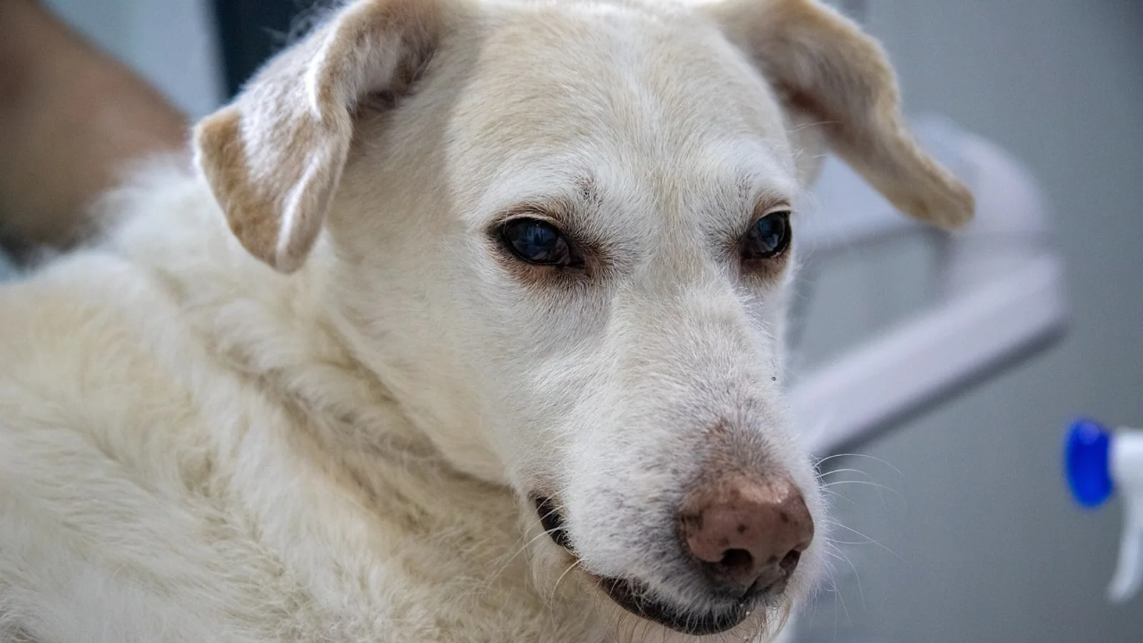
Dog pink eye is a red, inflamed eye with discharge, and it is a sign rather than a single disease. In dogs the redness usually comes from conjunctivitis, keratoconjunctivitis sicca (dry eye), a corneal ulcer, a foreign body, entropion, or an allergic reaction, and each of these needs a different treatment. The single most important fact for owners is this: never put a human antibiotic or steroid eye drop into a dog's eye before a veterinarian has stained the cornea with fluorescein. A steroid drop on a corneal ulcer can make the ulcer dramatically worse, sometimes turning a surface scratch into a melting eye that threatens vision.
This guide explains how veterinarians sort out the causes of red, discharging eyes in dogs, which tests matter, and when a red eye is an emergency. It is written for US pet owners who want to understand what their veterinarian is doing and why.
This article is educational and is not a substitute for veterinary diagnosis or treatment.
At a Glance
| What you see | Most likely cause | First-line test | Urgency |
|---|---|---|---|
| Red eye, clear or watery discharge, itchy, both eyes, seasonal | Allergic conjunctivitis | History and full eye exam, rule out other causes | Routine visit within a few days |
| Red eye, thick yellow-green discharge, one or both eyes | Bacterial conjunctivitis, often secondary to another problem | Ocular exam, Schirmer tear test, fluorescein stain, sometimes culture | Visit within 24 to 48 hours |
| Red eye, thick sticky mucoid discharge, dull cornea, no tears | Keratoconjunctivitis sicca (dry eye) | Schirmer tear test 1 (under 15 mm/min wetting is abnormal) | Visit within 24 to 48 hours |
| Squinting, pawing at the eye, tearing, blue or cloudy spot on the cornea | Corneal ulcer | Fluorescein stain | Same-day emergency |
| Sudden redness, swelling, blinking, often after a walk in tall grass or a car ride | Foreign body (seed, hair, grit) | Fluorescein stain plus close exam under magnification | Same-day emergency |
| Redness along the eyelid margin, eye discharge, squinting, young dog with a fever or neurologic signs | Canine distemper virus | Physical exam, history, supportive testing | Same-day emergency |
| Redness with eyelid rolled inward, lashes rubbing the eye | Entropion | Fluorescein stain plus eyelid exam | Visit within 24 hours |
| Red eye with a dark plaque on the cornea in a dog with long-standing dry eye | Corneal sequestrum | Ocular exam, Schirmer tear test, fluorescein stain | Visit within a few days |
Why Dog Pink Eye Is Not Human Pink Eye
Owners often assume that a red eye in a dog is the same problem as pink eye in a child. It is not. Human infectious conjunctivitis is frequently caused by adenovirus, a virus that spreads easily between people and usually clears on its own. Dogs have their own set of eye diseases, and the most common causes of a red, discharging canine eye are dry eye, corneal ulcers, allergies, eyelid problems, and bacterial infections that are usually secondary to one of those underlying issues.
That difference matters because the treatments are different. A human over-the-counter pink eye drop is not formulated for a dog's tear film, and more importantly, some human drops contain steroids that are dangerous for a dog with a corneal ulcer. Bacterial conjunctivitis in dogs is also often a complication rather than the primary disease, so treating only the bacteria while ignoring the dry eye or the ulcer underneath will not fix the eye.
The Main Causes of Red, Discharging Eyes in Dogs

_eye_of_dog.png)
Infectious Causes
Several infectious agents can cause conjunctivitis in dogs, and they behave very differently from one another.
Canine distemper virus. Distemper is a systemic viral disease that affects the respiratory, gastrointestinal, and nervous systems, and it also attacks the eye. A study comparing conjunctival tissue from dogs with distemper-induced keratoconjunctivitis sicca to dogs with non-infectious KCS found that the two conditions produce very similar histopathological changes, including low tear production, conjunctival hyperemia, and mucopurulent discharge [1]. In other words, distemper can cause dry eye by damaging the tear-producing tissue, and the resulting eye looks much like immune-mediated dry eye. Viral inclusion bodies were found sporadically in the conjunctival samples of the distemper group [1]. A dog with distemper-related eye disease usually has other signs of illness, such as fever, nasal discharge, coughing, vomiting, diarrhea, or neurologic changes, and the eye problem is part of a much larger picture.
Canine herpesvirus. Herpesvirus is best known as a cause of fading puppy syndrome, but it can also cause ocular disease. In one case report, a dog with chronic dry eye developed a corneal sequestrum, a dark plaque of dead corneal tissue. Testing for canine herpesvirus by virus isolation and PCR was negative in that dog, and electron microscopy found no viral particles [2]. That case shows that not every corneal problem in a dog with dry eye is viral, and it also shows how thoroughly veterinarians investigate these eyes.
Chlamydia and Mycoplasma. These organisms are more commonly discussed in cats, but they can be involved in canine conjunctivitis. A microbiological study of conjunctival swabs from dogs and cats with conjunctivitis found that no Mycoplasma organisms grew in culture and no fungal agents were identified, while bacterial culture was positive in about a third of all samples [3]. That study also found that the most frequently isolated bacteria were Staphylococcus epidermidis, Corynebacterium mucifaciens, Psychrobacter species, and Streptococcus lutetiensis [3]. The practical takeaway is that most canine conjunctivitis that has a bacterial component involves common environmental or skin bacteria rather than exotic organisms, and culture is usually reserved for eyes that are not responding to treatment.
Staphylococcus pseudintermedius. This bacterium is a common opportunistic pathogen in dogs and is frequently found in eyes with keratoconjunctivitis sicca. A 2025 study isolated multidrug-resistant Staphylococcus pseudintermedius from canine KCS cases and tested natural antibacterial compounds against it [4]. The relevance to owners is that bacterial infection in a dry eye can be stubborn and may involve resistant organisms, which is one reason veterinarians sometimes culture the eye rather than guessing at an antibiotic.
Toxoplasma gondii. Ocular toxoplasmosis is rare in dogs, but it has been reported. One case described a 12-year-old Pug with dry eye and pigmentary keratitis that developed a conjunctival mass. Histopathology showed necrotizing conjunctivitis with protozoal parasites, and Toxoplasma gondii was confirmed with immunohistochemical staining. The conjunctivitis resolved after a six-week course of oral clindamycin, and a later recurrence responded to oral ponazuril [5]. This is an unusual cause, but it belongs on the list for eyes that do not respond to standard treatment.
Allergic and Irritant Conjunctivitis
Allergic conjunctivitis is common in dogs and is frequently underdiagnosed. A 2023 study of 122 dogs with canine allergic conjunctivitis found that 64 percent had moderate disease, 24 percent had mild disease, and 12 percent had severe disease. All of the dogs had bilateral involvement, meaning both eyes were affected. The most common signs were chemosis (swelling of the conjunctiva) in 84 percent, hyperemia in 83 percent, and ocular pruritus (itchiness) in 79 percent. Seromucoid to mucopurulent discharge occurred in 62 percent and epiphora (excess tearing) in 69 percent [6].
Allergic conjunctivitis in dogs is often associated with canine atopic dermatitis, the itchy skin disease that affects many dogs. The severity of the eye disease did not correlate with the dog's sex or age at diagnosis [6]. Because the disease is bilateral and itchy, a dog that rubs both eyes after being outdoors in pollen season is a classic presentation. Irritants such as smoke, dust, shampoo, and swimming pool water can produce a similar picture.
Keratoconjunctivitis Sicca (Dry Eye)
Keratoconjunctivitis sicca, usually called KCS or dry eye, is one of the most important causes of a red, discharging canine eye, and it is the cause most often mistaken for simple conjunctivitis. KCS results from a deficiency in one or more components of the precorneal tear film [7]. The most prevalent form in dogs is immune-mediated, but congenital, metabolic, infectious, drug-induced, neurogenic, radiation-induced, iatrogenic, and idiopathic forms also occur [7].
The diagnosis rests on the Schirmer tear test. A Schirmer tear test 1 value under 15 mm/min of wetting is abnormal, and values of 10 mm/min or lower are clearly consistent with KCS [8][9]. In a study of Pugs and Shih-Tzus, the KCS group had a mean Schirmer tear test 1 of 4.46 mm/min compared with 18.80 mm/min in the control group [9]. Tear film osmolarity was also significantly higher in the KCS group at 353.02 mOsm/L versus 315.27 mOsm/L in controls, and the relationship between the two values was strong enough that osmolarity could be predicted from the Schirmer value with a coefficient of determination of 0.71 [9].
Clinical signs of KCS include blepharospasm from ocular pain, mucoid to mucopurulent discharge, and conjunctival hyperemia. With chronicity, the cornea develops epithelial hyperplasia, pigmentation, neovascularization, and ulceration [7]. Secondary bacterial infection is common [7]. A dog with KCS that goes untreated can lose vision as pigment spreads across the cornea.
Several specific situations cause KCS in dogs. Removal of the nictitating membrane gland, which sits in the third eyelid, is a known cause. One case report described an English Cocker Spaniel with a Schirmer tear test 1 of 3 mm/min, severe follicular conjunctivitis, and a third eyelid gland mass that was removed because malignancy was suspected. The final histopathology showed only chronic inflammation, and surprisingly the tear production after surgery remained higher than before surgery [10]. That case shows how closely tear production is tied to the third eyelid gland and how carefully veterinarians weigh surgery near it.
Radiation therapy for intranasal tumors can also cause KCS. A 2025 study of 15 dogs treated with hypofractionated intensity-modulated radiation therapy found that 6 dogs (33 percent) developed KCS within three months of treatment. The study identified dose thresholds predictive of KCS at 13.8 Gy for the eyeball, 14.9 Gy for the cornea, and 17.0 Gy for the retina, with the retina showing the highest odds ratio at 28.33 [11].
Neurogenic KCS is a distinct form in which the nerve supply to the tear glands is interrupted. A case series of 34 dogs with neurogenic KCS found that all had dry eye plus ipsilateral xeromycteria, meaning the same side of the nose was also dry. Concurrent neurologic deficits included facial neuropathy in 38 percent, peripheral vestibular syndrome in 29 percent, and Horner's syndrome in 15 percent. Etiologies were idiopathic in 53 percent, endocrinopathy in 18 percent, otitis interna in 12 percent, head trauma in 9 percent, and a few other causes [8]. A separate report described 29 dogs that developed neurogenic KCS within one day of receiving an otic medication containing terbinafine and florfenicol for ear infections. Tear production returned to normal in 24 of 29 dogs, with a median time of 86 days, and 68 percent developed a corneal ulcer in the meantime [12]. This is a striking example of how a medication applied to the ear can affect the eye through the nerves that run near the ear canal.
Foreign Bodies
A seed, a grass awn, a piece of grit, or even a stray eyelash can lodge under the third eyelid or against the cornea and cause sudden redness, squinting, and tearing. Foreign bodies are one of the reasons a red eye that appears overnight should be examined the same day. The cornea can be scratched by the object itself or by the dog rubbing the eye, and a scratch that is not treated can become infected.
Entropion and Eyelid Problems
Entropion is a condition in which the eyelid rolls inward so that the hair and skin rub against the cornea. It is common in certain breeds with loose facial skin or heavy brow folds. The constant rubbing causes redness, discharge, squinting, and eventually corneal ulceration. The diagnosis is made by examining the eyelid margin and staining the cornea to check for damage. Entropion often requires surgical correction, but the eye must be protected and the ulcer treated first.
Corneal Ulcers
A corneal ulcer is a defect in the clear surface of the eye, and it is the most important diagnosis to rule out in any dog with a red, painful eye. Ulcers are diagnosed with fluorescein stain, a dye that turns bright green where the corneal surface is missing. A dog with an ulcer typically squints, holds the eye closed, pawing at the face, and has excessive tearing. The eye may look cloudy or blue. Because steroid-containing eye drops can worsen an ulcer, fluorescein staining must be done before any steroid is applied.
Corneal Sequestrum
A corneal sequestrum is a dark plaque of dead corneal tissue that develops most often in dogs with chronic dry eye. One case report described a 14-year-old Cairn Terrier with chronic KCS in the left eye that developed severe blepharospasm and a black plaque over three weeks. The plaque was removed by superficial keratectomy with a conjunctival graft, and histology showed ulceration, stromal necrosis, and chronic suppurative keratitis with fibrosis and neovascularization. No bacteria, mineralization, or viral particles were found [2]. Sequestra are uncommon but they illustrate how chronic dry eye can damage the cornea over time.
Immune-Mediated Nodular Disease
Not every red eye lump is an infection. A case report described a dog with sterile granuloma and pyogranuloma syndrome, an immune-mediated disease that usually causes skin nodules, that developed a firm nodule on the lower palpebral conjunctiva along with hyperemia of the bulbar, palpebral, and third-eyelid conjunctivae and mucopurulent discharge. Topical and systemic corticosteroids improved the skin lesions but not the conjunctival mass, which required surgical excision. Histopathology showed a well-demarcated conjunctival stromal nodule with an infiltrate of T cells, B cells, and plasma cells [13]. This is a reminder that a non-healing conjunctival mass should be sampled rather than treated blindly.
The Decision Table: Matching Signs to Causes and Tests
The table below organizes the most common presentations by sign, likely cause, first-line test, and urgency. It is a starting point for a conversation with a veterinarian, not a substitute for an examination.
| Sign pattern | Likely cause | First-line test | Urgency |
|---|---|---|---|
| Both eyes red and itchy, watery discharge, seasonal, dog has skin allergies | Allergic conjunctivitis | Full ocular exam, history, Schirmer tear test, fluorescein stain to rule out ulcer | Routine, within a few days |
| One or both eyes red with thick yellow-green discharge | Bacterial conjunctivitis, often secondary | Ocular exam, Schirmer tear test, fluorescein stain, culture if not responding | Within 24 to 48 hours |
| Both eyes red with thick sticky discharge, dull cornea, dog seems uncomfortable in bright light | Keratoconjunctivitis sicca | Schirmer tear test 1, fluorescein stain | Within 24 to 48 hours |
| Sudden squinting, pawing, tearing, cloudy spot on the cornea | Corneal ulcer | Fluorescein stain | Same day |
| Sudden redness and swelling after a walk or car ride, dog rubs the eye | Foreign body | Fluorescein stain plus magnified exam of the conjunctival sac | Same day |
| Redness with eyelid rolled inward, hair contacting the eye | Entropion | Fluorescein stain plus eyelid exam | Within 24 hours |
| Red eye with a dark plaque on the cornea in a dog with long-standing dry eye | Corneal sequestrum | Ocular exam, Schirmer tear test, fluorescein stain | Within a few days |
| Red eye plus fever, nasal discharge, cough, vomiting, or neurologic signs | Canine distemper virus | Physical exam, history, supportive testing | Same day |
| Red eye that does not improve after several days of treatment | Resistant infection, underlying dry eye, or missed ulcer | Repeat Schirmer tear test, fluorescein stain, culture | Within 24 to 48 hours |
How Veterinarians Diagnose the Red Eye
The diagnostic process for a red, discharging eye follows a logical sequence. Each step answers a specific question.
Step 1: History and Visual Inspection
The veterinarian asks when the redness started, whether one or both eyes are affected, what the discharge looks like, whether the dog is squinting or pawing at the face, whether there has been any recent trauma, travel, boarding, or new medication, and whether the dog has other signs of illness. A bilateral, itchy eye in a dog with a history of skin allergies points toward allergic conjunctivitis. A unilateral, painful eye that appeared suddenly points toward a foreign body or ulcer.
Step 2: Schirmer Tear Test
The Schirmer tear test measures tear production by placing a small strip of filter paper in the lower eyelid for one minute. The distance the moisture travels up the strip is measured in millimeters per minute. A value under 15 mm/min is abnormal, and a value of 10 mm/min or lower is strongly consistent with KCS [8][9]. This test is quick, painless, and essential. Skipping it is one of the most common reasons a dry eye is misdiagnosed as simple conjunctivitis.
Step 3: Fluorescein Stain
Fluorescein dye is applied to the eye and the surface is examined under a blue light. Where the corneal epithelium is missing, the dye turns bright green. This test identifies corneal ulcers, which change the treatment plan completely. A steroid-containing drop must never be used before this test is done.
Step 4: Ocular Examination Under Magnification
The veterinarian examines the conjunctiva, the third eyelid, the eyelid margins, and the cornea under magnification. This is when foreign bodies, entropion, conjunctival masses, and corneal sequestra are found.
Step 5: Additional Testing When Needed
If the eye is not responding to treatment, the veterinarian may collect a conjunctival swab for bacterial culture and sensitivity. A study of conjunctival swabs from dogs and cats with conjunctivitis found that culture was positive in about a third of samples and that multiple bacterial isolates were found in some animals [3]. Culture helps identify resistant organisms and guide antibiotic selection. In dogs with suspected neurogenic KCS, advanced imaging may be used to look for underlying causes such as otitis interna, head trauma, or a brainstem mass [8].
The flowchart below shows the main decision path a veterinarian follows when a dog presents with a red, discharging eye.
flowchart TD
    A[Dog has red eye and discharge] --> B[Full ocular exam]
    B --> C[Schirmer tear test]
    C --> D{Tear value under 15}
    D -->|Yes| E[Diagnose keratoconjunctivitis sicca]
    D -->|No| F[Fluorescein stain]
    F --> G{Stain positive}
    G -->|Yes| H[Diagnose corneal ulcer]
    G -->|No| I[Examine for foreign body and entropion]
    I --> J{Foreign body or entropion found}
    J -->|Yes| K[Treat cause]
    J -->|No| L[Consider allergy or infection]
    E --> M[Start tear stimulation and lubrication]
    H --> M
    K --> M
    L --> M
Treatment Approaches by Cause
Treating Keratoconjunctivitis Sicca
Treatment of KCS aims to stimulate natural tear production and to replace the missing tear film. Topical immunomodulators are the mainstay. A study comparing topical tacrolimus 0.03 percent with homologous platelet-rich plasma found that the tacrolimus group had significant increases in Schirmer tear test 1 values, strip meniscometry values, and goblet cell counts in the palpebral conjunctiva by the end of the study. The platelet-rich plasma group showed significant decreases in lymphocyte and neutrophil counts in the palpebral conjunctiva, and both groups improved equivalently in tear film break-up time, osmolarity, and lissamine green staining [14].
A separate double-blind randomized study compared a subconjunctival injection of liposome-encapsulated sirolimus given every two weeks with daily cyclosporine or tacrolimus. After 14 weeks, there was no statistically significant difference between the groups in Schirmer tear test 1 scores, conjunctival hyperemia scores, or corneal opacity scores, and no clinically significant side effects occurred in either group [15].
Mesenchymal stem cell therapy is an emerging option. A systematic review and meta-analysis of six studies involving 151 eyes from 98 dogs found that stem cell therapy significantly increased tear production at one and six months after treatment, though the quality of evidence was graded as level C [16]. This is a promising area but not yet a standard first-line treatment.
Supportive care for KCS includes ocular lubricants, mucolytics to break up thick discharge, and antibiotics for secondary bacterial infection [7]. The goal of therapy is to stimulate natural tear production, because that provides the highest recovery in clinical signs and the best prevention of vision loss [7].
Treating Corneal Ulcers
A corneal ulcer requires treatment of the underlying cause plus protection of the cornea while it heals. Bacterial infection is common in ulcers, and a recent study characterized the tear film pharmacokinetics of parenterally administered cefovecin in dogs, finding that tear concentrations remained detectable for 336 hours after a single injection and exceeded the minimum inhibitory concentrations for Staphylococcus pseudintermedius, Streptococcus canis, and Pseudomonas aeruginosa [17]. This kind of pharmacokinetic data helps veterinarians choose systemic antibiotics when topical treatment is difficult.
The critical rule for ulcers is that steroids must be avoided. A steroid drop on an ulcer can inhibit corneal healing and allow the ulcer to deepen. This is why fluorescein staining must always precede any steroid use.
Treating Bacterial Conjunctivitis
When bacterial infection is the primary problem, topical antibiotics are the usual treatment. Culture and sensitivity testing is reserved for eyes that do not respond, because most infections involve common bacteria that respond to standard antibiotics [3]. In dry eye, bacterial infection is usually secondary, and treating the dry eye is the priority.
Treating Allergic Conjunctivitis
Allergic conjunctivitis is managed by removing the allergen when possible, using cool compresses, and applying medications that reduce inflammation. Because the disease is often linked to atopic dermatitis, treating the skin disease can help the eyes. The severity grading system developed for canine allergic conjunctivitis, which scores conjunctival hyperemia, chemosis, ocular pruritus, epiphora, discharge, and keratitis from 0 to 3, gives veterinarians a way to track response to treatment [6].
Treating Foreign Bodies and Entropion
A foreign body must be removed, sometimes under topical anesthesia with magnification. After removal, the cornea is stained to check for damage and the eye is treated supportively. Entropion usually requires surgical correction, but the cornea must be protected and any ulcer treated before surgery.
Treating Neurogenic KCS
Neurogenic KCS is treated with oral pilocarpine and lacrimostimulants in most cases. In a case series of 34 dogs, treatment was initiated in 88 percent of cases, with 19 dogs receiving both oral pilocarpine 2 percent and a lacrimostimulant, 3 receiving oral pilocarpine only, and 8 receiving a lacrimostimulant only [8]. When the cause is a medication such as a long-acting otic product, tear production often returns after the drug is discontinued, but this can take months. In the report of 29 dogs with medication-associated neurogenic KCS, tear production returned to normal in 24 of 29 dogs with a median time of 86 days, and 68 percent developed a corneal ulcer while waiting [12]. That is why these dogs need aggressive lubrication and monitoring during recovery.
Supplies and Selection Criteria for Home Eye Care
Owners can support treatment at home with a few basic supplies. The selection criteria below help you choose wisely.
- Artificial tear lubricants. Look for preservative-free formulations if the dog needs drops more than four to six times daily, because preservatives can irritate an already inflamed eye. Lubricating ointments last longer but blur vision temporarily.
- Saline eye wash. A sterile saline flush can help remove discharge and small irritants. Do not use contact lens solutions or homemade salt water.
- Soft cloths or cotton pads. Use these to gently clean discharge from the eyelid margins. Wipe from the inner corner outward, and use a fresh pad for each eye to avoid spreading infection.
- Elizabethan collar. An e-collar prevents the dog from rubbing or scratching the eye, which can turn a minor problem into a serious one. Soft collars work for some dogs, but a rigid collar is more reliable for a determined scratcher.
- Medication log. Record the time of each dose, which eye was treated, and any change in discharge, redness, or squinting. This information helps the veterinarian adjust treatment at the recheck.
Do not use human antibiotic or steroid eye drops on a dog without veterinary guidance. This is the single most common and most dangerous mistake owners make with red eyes.
Common Mistakes Owners Make With Dog Pink Eye
Using leftover human eye drops. Human drops may contain steroids, preservatives, or antibiotics that are not appropriate for dogs. A steroid drop on an undiagnosed corneal ulcer can cause the ulcer to worsen rapidly.
Assuming both eyes means allergies. While allergic conjunctivitis is bilateral, so is KCS, and so is distemper-related eye disease. Bilateral redness does not rule out serious causes.
Waiting to see if it clears up. A red eye that is painful, that has thick discharge, or that has been present for more than a day should be examined. Corneal ulcers can worsen quickly.
Cleaning the eye too aggressively. Rubbing the eye can worsen inflammation and dislodge a healing corneal epithelium. Gentle wiping is enough.
Stopping treatment early. Dry eye and allergic conjunctivitis are chronic conditions. Stopping medication when the eye looks better often leads to a relapse.
Missing the underlying cause. Treating discharge with an antibiotic while ignoring a low Schirmer tear test means the eye will keep producing discharge because the tear film is still inadequate.
Welfare and Safety Points
Eye pain in dogs is real and underrecognized. A dog that squints, paws at the face, or avoids bright light is uncomfortable. Relieving that pain is part of treatment, not an optional extra.
Dogs with vision loss from chronic dry eye or corneal disease may become anxious in unfamiliar environments. Keep the home layout consistent, use night lights, and speak to the dog before touching it so it is not startled.
If a dog has a corneal ulcer or has had eye surgery, an Elizabethan collar should stay on until the veterinarian says it can come off. A few seconds of rubbing can undo weeks of healing.
Any dog with a red eye that is also systemically ill, with fever, vomiting, diarrhea, or neurologic signs, needs prompt evaluation because the eye problem may be part of a serious systemic disease such as distemper.
When to See a Veterinarian
See a veterinarian the same day if the dog is squinting, holding the eye closed, pawing at the face, or has a cloudy or blue spot on the cornea. These signs suggest a corneal ulcer or foreign body.
See a veterinarian within 24 to 48 hours if the eye has thick yellow-green or mucoid discharge, if the eye looks dull rather than shiny, or if the redness has not improved after a day of gentle home cleaning.
See a veterinarian within a few days if both eyes are red and itchy and the dog has a history of skin allergies, or if the redness is mild and the dog is comfortable.
Limitations and When to Contact a Veterinarian
This guide covers the common causes of red, discharging eyes in dogs, but individual cases vary. A dog with a red eye may have more than one problem at once, such as dry eye plus a corneal ulcer plus a bacterial infection. The only way to know which problems are present is an examination with a Schirmer tear test and a fluorescein stain.
Contact a veterinarian promptly if the dog's eye does not improve within 24 to 48 hours of starting treatment, if the eye becomes more painful, if the cornea becomes cloudy, if the dog stops eating or becomes lethargic, or if new neurologic signs such as head tilt, facial droop, or unsteady walking appear. These signs can indicate a deeper problem such as neurogenic KCS, otitis interna, or systemic infection.
Do not adjust or stop prescribed eye medication without talking to the veterinarian first. Many eye conditions in dogs are chronic and require ongoing treatment even when the eye looks normal.
Frequently Asked Questions
Is dog pink eye contagious to other dogs?
Some causes of red eye in dogs are contagious, including viral diseases such as distemper and bacterial infections, while others such as allergies and dry eye are not. A veterinarian can tell you which category your dog's eye problem falls into after an examination.
Can I use human pink eye drops on my dog?
No. Human eye drops may contain steroids or other ingredients that can harm a dog's eye, especially if there is a corneal ulcer. Wait for a veterinary diagnosis before putting anything in the eye.
How do I know if my dog has dry eye or just conjunctivitis?
The only reliable way to tell is a Schirmer tear test. A value under 15 mm/min of wetting indicates inadequate tear production, which points to dry eye rather than simple conjunctivitis.
Why does my dog's eye keep producing thick discharge?
Thick mucoid or mucopurulent discharge is common in dry eye because the tear film is not clearing debris and bacteria normally. It can also indicate a bacterial infection. Both possibilities need veterinary evaluation.
Is a red eye in a dog always an emergency?
No. Mild bilateral redness in an itchy dog with a history of allergies is usually not an emergency. But squinting, pawing at the eye, corneal cloudiness, or sudden severe pain are emergency signs that need same-day care.
Can allergies cause pink eye in dogs?
Yes. Allergic conjunctivitis is a common cause of red, itchy eyes in dogs, and it often occurs in both eyes at once. It is frequently associated with atopic dermatitis.
What should I do if my dog's eye is red after a walk?
Flush the eye gently with sterile saline and check for a foreign body such as a seed or grass awn. If the dog is squinting or the eye does not look normal within a few hours, see a veterinarian the same day.
How long does dog pink eye take to heal?
Healing time depends on the cause. Bacterial conjunctivitis may improve within days of starting antibiotics, while dry eye and allergic conjunctivitis are often chronic conditions that require long-term management.
Related Articles
- Reptile Respiratory Infections: Causes, Symptoms, and Treatment
- Betta Fish Popeye: Causes, Symptoms, and Treatment
- Canine Hyperthyroidism: Causes, Diagnosis, and Treatment
- Aquarium Fish Fin Rot: Causes, Symptoms, and Treatment
- Koi Fish Gill Disease: Causes, Symptoms, and Treatment
- Snake Respiratory Infection: Symptoms, Causes, Diagnosis and Veterinary Treatment
- Dog Eye Ulcer: Symptoms, Causes and Treatment
- Mange in Cats: Symptoms, Types, and Treatment
- Dog Digestive Issues: Causes, Symptoms, and When to Worry
Sources
- Conjunctival effects of canine distemper virus-induced keratoconjunctivitis sicca.
- Corneal sequestrum in a dog with chronic unilateral keratoconjunctivitis sicca.
- Microbiological investigation of conjunctivitis in cats and dogs from Kars, Türkiye (2024-2025): culture- and MALDI-TOF MS-based identification and antimicrobial susceptibility testing of bacterial isolates.
- Synergistic Antibacterial Effect of Eugenol and Biogenic Silver Nanoparticles on Staphylococcus pseudintermedius Isolated from Canine Keratoconjunctivitis Sicca.
- Keratoconjunctivitis associated with Toxoplasma gondii in a dog.
- Diagnostic approach and grading scheme for canine allergic conjunctivitis.
- Immune-mediated keratoconjunctivitis sicca in dogs: current perspectives on management.
- Neurogenic keratoconjunctivitis sicca in 34 dogs: A case series.
- Assessment of tear film osmolarity using the IPen(®) Vet osmometer in Pug and Shih-Tzu dogs with and without keratoconjunctivitis sicca.
- Changes in tear production in a dog with keratoconjunctivitis sicca for a year after inevitable nictitating membrane gland resection due to suspicion of a malignant tumour.
- Risk Factors for Radiation-Induced Keratoconjunctivitis Sicca in Dogs Treated with Hypofractionated Intensity-Modulated Radiation Therapy for Intranasal Tumors.
- Long-lasting otic medications may be a rare cause of neurogenic keratoconjunctivitis sicca in dogs.
- Nodular conjunctivitis as a novel ocular manifestation of canine sterile granuloma/pyogranuloma syndrome.
- Comparison of topical 0.03% tacrolimus and homologous injectable platelet-rich plasma in the treatment of keratoconjunctivitis sicca in dogs.
- Subconjunctival liposomal sirolimus vs. cyclosporine or tacrolimus as treatment of keratoconjunctivitis sicca in dogs: A double-blind, randomized study.
- Clinical Outcomes of Stem Cell Therapy in Dogs With Keratoconjunctivitis Sicca; A Systematic Review and Meta-Analysis.
- Sustained Tear Film Exposure After Parenteral Cefovecin Administration in Dogs: Pharmacokinetics and Relevance to Canine Bacterial Keratitis.