Dog Eye Ulcer: Symptoms, Causes and Treatment
By Dr. Zubair Khalid, DVM, MS, PhD ·
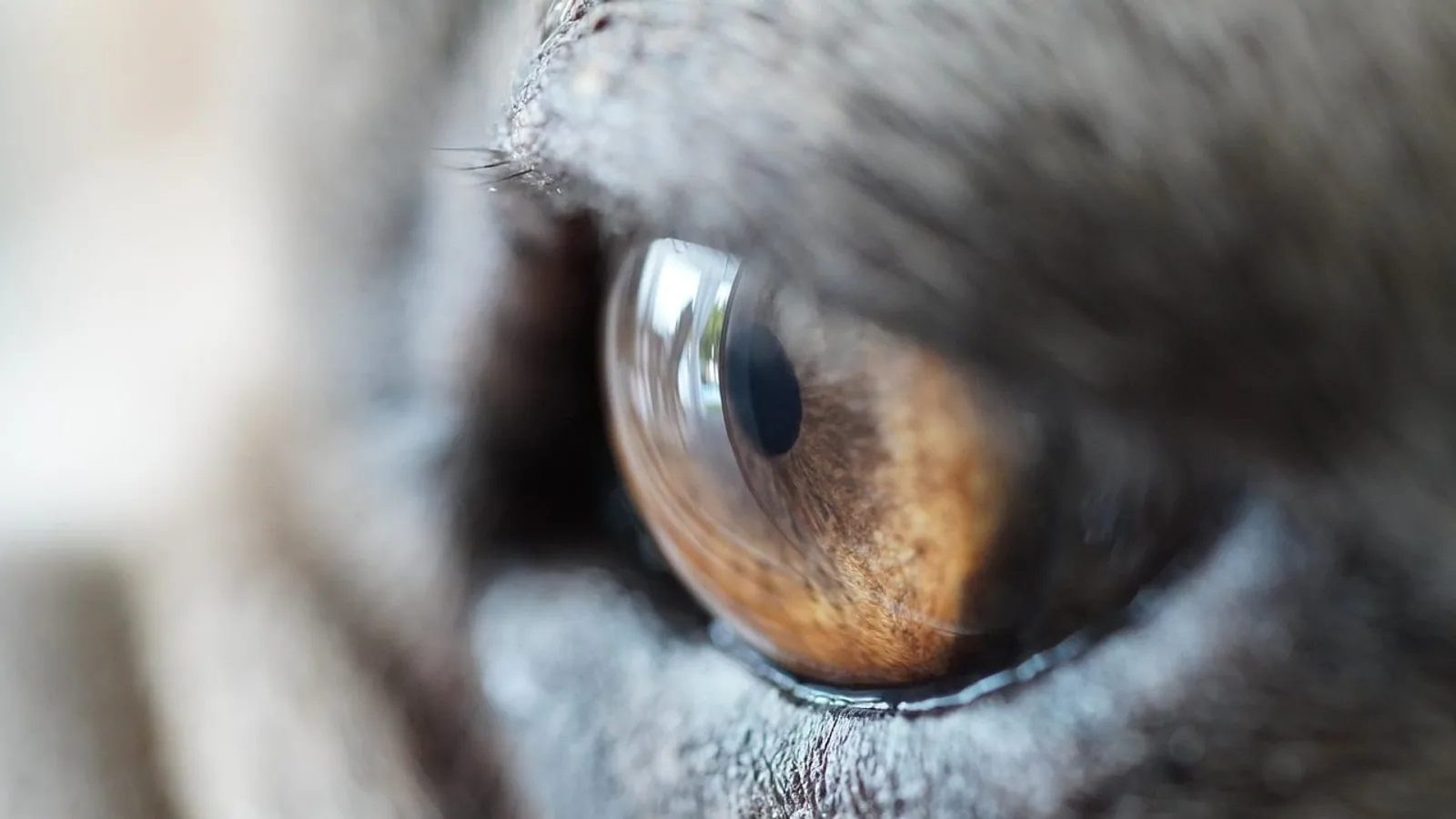
A dog corneal ulcer is a break in the clear outer surface of the eye. The medical term is ulcerative keratitis. Owners usually say their dog has an eye ulcer, an ulcer on the cornea, or a sore on the eyeball. The cornea is one of the most sensitive tissues in the body, so even a small defect causes a big amount of pain, squinting and tearing.
Treatment depends almost entirely on depth. A superficial ulcer that only removes the top layer can heal in days with medication. An ulcer that eats into the middle layer of the cornea can melt, thin and perforate the eyeball within hours to days. That difference is why a veterinarian needs to stain and examine the eye rather than wait and see.
This guide explains corneal anatomy, the four practical severity grades, how fluorescein staining works, the common causes, evidence-based treatment and the red flags that mean an emergency referral. Where research supports specific predispositions, healing times or outcomes, those findings are included so you can understand what your veterinarian is likely to recommend.
This article is educational and is not a substitute for veterinary diagnosis or treatment.
At a Glance: Owner Triage Summary
- A dog with a painful, squinting, tearing eye needs a same-day veterinary visit.
- A cloudy or bluish eye, a visible dent, or a dark spot inside the cornea needs emergency referral.
- Do not use leftover eye drops from a previous episode. Some medications, such as topical steroids, can worsen certain ulcers, including herpesvirus-related dendritic ulcers [1].
- Fluorescein dye is the standard test that shows whether the surface is broken and how large the defect is.
- Most superficial ulcers are managed medically. Deep ulcers, descemetoceles and perforations usually need surgery or an advanced stabilizing procedure [2].
| Finding at Home | What It Suggests | Urgency |
|---|---|---|
| Squinting, tearing, redness, no cloudiness | Possible superficial ulcer or erosion | See a vet within 24 hours |
| Cloudy or hazy cornea, marked squinting | Possible infection or stromal involvement | Same-day veterinary visit |
| Bluish or gray deep defect, visible dent | Possible descemetocele | Emergency referral now |
| Sudden bulge, dark tissue, blood in eye | Possible perforation | Emergency referral now |
| Ulcer that has not healed after weeks of treatment | Possible indolent or complicated ulcer | Urgent recheck and referral |
Corneal Anatomy: Why the Layers Matter
The cornea is a transparent dome made of layers stacked from outside to inside. Each layer behaves differently when injured, and the name of the layer involved determines how dangerous the ulcer is.
Epithelium
The epithelium is the outermost layer. It is a thin, multi-layered sheet of cells that is water-repellent and acts as a barrier. The epithelium regenerates quickly when the underlying surface is healthy. A defect limited to the epithelium is called an erosion or a superficial ulcer. The most familiar example is the spontaneous chronic corneal epithelial defect, often shortened to SCCED. In these eyes the epithelium fails to stick to the layer beneath it, so the defect persists despite appearing shallow [3].
Stroma
The stroma is the thick middle layer and the structural core of the cornea. It is made mostly of collagen arranged in precise parallel fibers that keep the cornea clear. Because the stroma provides thickness and tensile strength, any loss of stromal tissue is the key event that separates a simple ulcer from a complex one. A study of 347 dogs with corneal ulceration found that complex ulcers, meaning deep ulcers, ulcers with keratomalacia, descemetoceles and corneal lacerations or foreign bodies, were the largest single category, making up about 38.5 percent of cases [4]. That statistic matters because stromal loss is exactly what threatens the eye's integrity.
Stromal tissue is also vulnerable to collagen enzymes. When inflammation drives those enzymes, the stroma can liquefy in a process called keratomalacia or melting [2]. Melting ulcers lose tissue quickly and are one of the most sight-threatening forms.
Descemet's Membrane
Descemet's membrane is a thin, tough, elastic sheet that sits between the stroma and the endothelium. It resists collagen enzymes better than the stroma does. When infection or melting consumes all the stroma above it, the remaining clear bubble of Descemet's membrane is called a descemetocele. A descemetocele is the last barrier before the eye ruptures. In the same 347-dog study, descemetoceles accounted for 17.0 percent of cases, which makes them common enough that every painful eye needs careful depth assessment [4].
Endothelium
The endothelium is the innermost single layer of cells. It is a pump that keeps the cornea dehydrated and clear by moving fluid out of the stroma. Endothelial cells do not regenerate effectively in dogs. If the endothelium is damaged, fluid enters the stroma and the cornea becomes cloudy and waterlogged. That is why a perforation is so serious. It breaches every layer, allows fluid and sometimes intraocular tissue to escape, and puts the inside of the eye at risk of infection.
Symptoms of a Dog Eye Ulcer
The signs of a corneal ulcer in a dog follow a recognizable pattern because the cornea is so richly supplied with pain nerves.
Common Symptoms Owners Notice
- Squinting or holding the eye closed, often called blepharospasm.
- Excessive tearing or a wet streak below the eye.
- Pawing at or rubbing the face.
- Redness of the white of the eye.
- Reluctance to go outside in bright light or into sunlight.
- A cloudy, hazy or slightly blue-gray patch on the eye.
- Discharge that may be watery at first and thicker later.
- Reduced willingness to be handled around the head.
Symptoms That Point to a Deeper Problem
- A visible dent or crater in the cornea.
- A dark or bluish bubble at the base of the defect.
- Sudden swelling or bulging of the eyeball.
- Blood or dark material inside the front of the eye.
- A tear or flap of tissue on the cornea suggesting a laceration.
- Marked cloudiness that develops over hours.
A dull, hazy cornea and a very painful eye should push you toward a same-day or emergency visit rather than routine scheduling. Older dogs are more likely to develop SCCEDs and keratomalacia than younger dogs, so age is a relevant detail to mention when you call [4].
What Causes a Dog Eye Ulcer?
Most ulcers have one or more contributing causes. Identifying the cause matters because an ulcer caused by an eyelid that rubs the cornea will keep coming back until the eyelid problem is addressed.
Trauma and Foreign Bodies
Blunt trauma, a scratch from a cat claw, a stick, plant material, or a foreign body trapped under the eyelids can all injure the cornea. Corneal lacerations and retained foreign bodies are classified as complex ulcers, and they occurred in 4.0 percent of dogs in one referral population [4]. A case report described diffuse corneal ulceration after a dog was exposed to the defensive spray of a walkingstick insect, which shows that chemical injury can also cause an ulcer [5].
Entropion and Eyelid Abnormalities
Entropion is a rolling inward of the eyelid so that hair rubs directly on the cornea with every blink. This continuous abrasion both creates ulcers and prevents them from healing. Eyelid conformation matters more widely too. Dogs with a nasal fold, meaning a skin wrinkle pressing near the inner corner of the eye, were nearly five times more likely to be affected by corneal ulcers in a United Kingdom study of 700 dogs [6].
Dry Eye (Keratoconjunctivitis Sicca)
Keratoconjunctivitis sicca, usually called KCS or dry eye, means the tear film is inadequate. Tears lubricate, rinse away debris and carry protective factors. A poor tear film leaves the cornea vulnerable to repeated ulceration and slows healing. Reduced tear production is measurable with a Schirmer tear test, and even a short period of reduced tear production under general anesthesia was associated with faint corneal erosions in dogs in one prospective study [7].
Infection
Bacteria commonly colonize an ulcer once the surface barrier is broken. In one study of dogs with corneal ulcers, 67 percent showed bacterial growth, and most isolates were gram-positive organisms, with Staphylococcus, Streptococcus and Pseudomonas the most frequent [8]. A separate retrospective review from Berlin found positive bacterial growth in 79.5 percent of samples, with Staphylococcus pseudintermedius and beta-hemolytic Streptococcus as the main agents [9]. Infection does not just sit on the surface. Bacterial enzymes and the immune response they trigger can drive melting and deepen the ulcer [2].
Herpesvirus is another cause. A case report described dendritic ulcerative keratitis, meaning branching ulcers, in an adult dog with suspected acute canine herpesvirus-1 infection after the dog had been on long-term topical immune-suppressing medication [1]. Dendritic patterns are a distinctive appearance your veterinarian will look for.
Breed and Conformation Risk
Conformation is one of the strongest risk factors. Brachycephalic breeds, the short-muzzled dogs such as Pugs and Shih Tzus, had about 2.76 times greater odds of presenting for corneal ulceration and about 2.7 times greater odds of having a complex ulcer compared with other dogs [4]. Shih Tzus were the most common breed for nearly every ulcer type in that study, except for Boxers, which were most common for SCCEDs [4]. Small body weight and increasing age were also associated with higher odds of diagnosis [4].
Severity Grading: Superficial, Deep, Descemetocele and Perforation
Grading an ulcer by depth and by the presence of melting is the single most useful step in deciding treatment. The four practical grades below are the basis for the urgency table that follows.
Superficial Ulcer
A superficial ulcer or erosion involves the epithelium and possibly the very outer stroma. The eye is painful but the globe keeps its shape. Many of these heal with medical treatment. An SCCED is a special superficial ulcer in which the epithelium does not adhere, and these may need a procedure such as debridement or keratotomy to heal [3].
Deep Stromal Ulcer
A deep ulcer extends well into the stroma. The stroma is thinner and weaker at that point, and the eye is at risk of progressing to a descemetocele or perforation. Melting may be present, which makes the situation more urgent because tissue is actively being lost [2].
Descemetocele
A descemetocele is a deep ulcer in which all the stroma over a region has been lost and only Descemet's membrane remains. The eye is on the verge of rupturing. Descemetoceles made up 17.0 percent of ulcers in one referral series, so they are not rare [4].
Perforation
A perforation means the cornea has ruptured through all layers. Intraocular contents may leak, the eye may lose its shape and internal infection becomes a serious risk. A case series described using a hyaluronic acid-based photocurable glue in dogs with a descemetocele, a corneal perforation and a deep laceration, with corneal integrity maintained in the descemetocele case over nine months of repeated applications [10]. That illustrates the kind of advanced stabilization that referral centers can offer.
Severity Grade Table
| Grade | Clinical Signs | What It Means | Urgency |
|---|---|---|---|
| Superficial | Squinting, tearing, positive stain, clear globe | Epithelium involved, stroma largely intact | Veterinary visit within 24 hours |
| Deep stromal | Cloudiness, deeper stain, possible dent, marked pain | Significant stromal loss, risk of progression | Same-day or emergency visit |
| Descemetocele | Bluish or gray bubble in a deep defect | Only Descemet's membrane remains | Emergency referral now |
| Perforation | Collapsed or bulging globe, dark tissue, blood | Full-thickness rupture | Emergency referral now |
The Fluorescein Stain: How It Works and What It Shows


Fluorescein is a dye that reveals where the corneal surface is broken. Healthy epithelium repels the dye, so it rinses away from intact cornea. Where epithelium is missing, the underlying stroma absorbs the dye and glows green under cobalt blue light.
The Steps Your Veterinarian Follows
- Apply the dye. The veterinary team uses either a fluorescein-impregnated strip moistened with sterile saline or a liquid fluorescein solution applied to the eye.
- Allow the dye to contact the surface. A few seconds of contact is enough for the dye to enter any defect.
- Rinse the eye. A gentle flush of sterile saline removes excess dye so no free dye confuses the picture.
- Examine under cobalt blue light. With magnification, the blue light makes retained dye fluoresce bright green.
- Note the uptake pattern. A discrete spot means a focal ulcer. A diffuse wash of green suggests widespread erosion, as was seen in the walkingstick spray case [5]. Branching or dendritic uptake suggests a herpesvirus pattern [1].
Why the Pattern Matters
Uptake under the third eyelid or at the inner corner can be missed unless the eyelids are lifted and the whole surface is checked. An ulcer hidden under the third eyelid still needs treatment and can still deepen, so an eye that is painful but shows no stain on a quick look warrants a careful repeat examination. Fluorescein staining is used repeatedly over the course of treatment. In clinical studies, the cornea is judged healed when the stain is negative, and this is the standard endpoint for deciding when treatment can stop [3].
How Veterinarians Evaluate a Dog With an Ulcer
The examination is systematic because the depth and the cause both drive the plan.
- History: when signs started, any known trauma, prior eye disease, medications used, and general health problems.
- Vision and menace response: to gauge how the eye is functioning.
- Schirmer tear test: to detect KCS, since reduced tear production both predisposes to and delays healing of ulcers [7].
- Fluorescein stain: to confirm and map the defect.
- Magnified examination: to grade depth, look for melting, and search for foreign bodies or eyelid abnormalities.
- Intraocular pressure: to check for conditions that can accompany corneal disease.
- Cytology and culture: when infection is suspected, since the bacterial spectrum includes Staphylococcus, Streptococcus and Pseudomonas and resistance is common enough that culture-based therapy is emphasized [8][9].
- Examination of the other eye: because the cause may be bilateral, as with KCS or entropion.
Evidence-Based Treatment of Canine Corneal Ulcers
Treatment has several goals at once. The veterinary team aims to control infection, stop corneal destruction, support the corneal structure, control the uveal reaction and pain, and limit scarring [11]. The specific plan is chosen by your veterinarian based on depth, cause and examination findings.
Medical Management of Superficial Ulcers
Superficial ulcers are usually treated with topical antibiotics to prevent or treat infection, lubricants to support the tear film, and pain control. A common approach in SCCED management pairs topical antibiotics with procedures to encourage epithelial adhesion. In one study of dogs after diamond burr debridement for SCCEDs, median time to healing was about 9.4 days, and use of a bandage contact lens significantly improved healing times [12].
Topical non-steroidal anti-inflammatory drugs deserve caution. In a retrospective study of SCCED eyes, healing times were significantly longer in eyes that received topical NSAIDs, though those eyes also tended to receive more medications overall, so the effect could not be separated fully from the number of treatments used [13]. This is one reason your veterinarian may choose systemic pain control rather than a topical NSAID.
Managing Melting and Deep Ulcers
Melting ulcers need aggressive anticollagenase therapy and, often, surgical stabilization. A mechanism-based framework groups adjunctive therapy into structural preservation, inflammatory modulation, regenerative augmentation and functional optimization, with precision stratification guiding which approach fits which eye [2]. In practical terms, this means the treatment is chosen based on the dominant biological process, not just on how the ulcer looks.
Corneal collagen cross-linking has been studied as a way to stabilize melting corneas. In a controlled trial of dogs and cats with melting keratitis, cross-linking did not eliminate the need for rescue stabilization, but ulcer deepening during follow-up was less pronounced in the treated dogs than in controls, and no treatment-related complications were observed [14].
Procedures for Non-Healing Ulcers
SCCEDs often need more than drops. Options studied in dogs include diamond burr debridement, grid keratotomy and superficial keratectomy. One retrospective study of 107 dogs with 121 SCCEDs found that superficial keratectomy with bandage lens placement healed 99 percent of defects within 21 days with no post-operative complications [3]. Another study found that bandage contact lens retention was common and that lens wear improved healing times after diamond burr debridement [12].
Adjunctive Therapies Under Study
Several adjuncts have been investigated. Topical hyaluronic acid was tested in a small pilot study of dogs with experimentally created corneal wounds, and no statistical difference was found in clinical scores or healing rate compared with control [15]. Topical heterologous serum was evaluated in a randomized, double-masked trial of dogs with SCCEDs, and median time to reepithelialization was two weeks in both the serum and saline groups, meaning no additional benefit was demonstrated in that trial [16]. A separate study compared autologous, homologous and heterologous blood serums in chemically induced ulcers and found treatment-related improvements over time, with heterologous serum reducing thickness and ulcer diameter earlier than the others from day seven [17]. Calcareous corneal degeneration, a mineral deposit condition that can be associated with ulceration, has been treated by dissolving the mineral with a chelating solution, with resolution in some eyes and improvement in most [18].
Treatment of the Underlying Cause
No ulcer treatment succeeds long term if the cause persists. Entropion may need surgical correction, KCS needs ongoing tear support and monitoring, and foreign bodies must be removed. Brachycephalic conformation and nasal folds increase risk, so your veterinarian may discuss eyelid or facial fold management in recurrent cases [4][6].
flowchart TD
    A[Painful red eye in a dog] --> B[Veterinary examination]
    B --> C[Fluorescein stain applied]
    C --> D{Stain uptake present}
    D -->|No| E[Look for other causes]
    D -->|Yes| F[Grade the ulcer depth]
    F --> G[Superficial ulcer]
    F --> H[Deep stromal ulcer]
    F --> I[Descemetocele]
    F --> J[Perforation]
    G --> K[Medical treatment and recheck]
    H --> L[Intensive treatment or surgery]
    I --> M[Emergency referral]
    J --> M
What Not to Do: Unsafe Home Remedies
The most common mistake owners make is reaching for leftover medication. Two specific dangers stand out.
- Topical steroids: corticosteroids suppress the immune response. A dog that developed dendritic herpesvirus ulcers had been receiving topical prednisolone and tacrolimus for months, and the ulcers resolved once these were discontinued and antiviral treatment started [1]. Steroids can also worsen melting.
- Human eye drops: products made for people may contain preservatives, anesthetics or antibiotics that are not appropriate for a dog's eye and may delay healing.
Other things to avoid include trying to remove a foreign body yourself, using a makeshift collar substitute to stop rubbing, or waiting to see if the eye improves overnight. A dog with an ulcer on the eye is in genuine pain, and delay allows a superficial ulcer to become deep.
An Elizabethan collar is the one home measure that is consistently useful because it prevents self-trauma while you arrange veterinary care.
When to Seek Emergency Care
Some findings mean the eye may be lost without rapid intervention.
- A deep ulcer or a melting ulcer.
- A descemetocele, seen as a bluish or gray bubble in the base of a defect.
- A perforation, with a changing shape of the globe, blood or dark tissue.
- A positive fluorescein stain that extends under the third eyelid, because a hidden deep component may be present.
- An ulcer that has suddenly become more cloudy, more painful or larger.
- Sudden onset of severe squinting with a cloudy eye in a brachycephalic dog.
Brachycephalic breeds have both higher odds of corneal ulceration and higher odds of complex ulcers, so a Pug, Shih Tzu or similar dog with an acutely painful eye deserves a low threshold for emergency assessment [4]. Multiple complex ulcer types, including deep ulcers, melting, descemetoceles and lacerations, made up a large share of referral cases, which reinforces that depth must be assessed early rather than assumed [4].
Prognosis and Follow-Up
Prognosis depends on depth, cause, the presence of infection and how quickly treatment starts. Superficial ulcers that are treated promptly generally heal well. SCCEDs treated with a procedure such as superficial keratectomy healed in nearly all eyes within three weeks in one large series [3]. Deep ulcers, melting ulcers and descemetoceles carry a guarded prognosis and often need surgery or advanced stabilization, and even then the eye may develop scarring that affects vision.
Follow-up is not optional. Rechecks with repeat fluorescein staining confirm healing and catch complications. In one study of Staphylococcus pseudintermedius keratitis, eyes with multidrug-resistant infection took longer to re-epithelialize and required significantly longer follow-up, which is a reminder that infection control shapes the timeline [19]. Median healing after diamond burr debridement in another study was around nine days, but individual eyes vary widely [12].
Expect your veterinarian to recheck the eye until the stain is negative and the eye is comfortable. Do not stop medication early because the eye looks better, and do not restart old drops without guidance.
Prevention
Prevention focuses on the causes that can be modified.
- Control tear production problems. Regular monitoring of dogs with KCS helps prevent ulcer formation and recurrence [7].
- Address eyelid conformation. Nasal folds and eyelid abnormalities that rub the cornea increase risk and can be managed [6].
- Protect brachycephalic eyes. These dogs have higher odds of both ulcers and complex ulcers, so prompt attention to any eye sign is worthwhile [4].
- Use eye protection during anesthesia. Even with lubricating gel, some dogs develop faint corneal erosions after general anesthesia, so discuss ocular protection if your dog is having a procedure [7].
- Manage systemic disease. Comorbidities were associated with repeat ulcer diagnoses, and diabetes mellitus specifically appeared as a relevant comorbidity, so good control of underlying conditions supports eye health [4].
- Avoid unsupervised exposure to irritants. Chemical injuries such as the walkingstick insect spray case show that environmental exposures can cause corneal damage [5].
Limitations and When to Contact a Veterinarian
This article covers general principles. Every eye is different, and the depth, cause and infection status of an ulcer can only be determined by a veterinarian who examines the eye with magnification and a fluorescein stain.
Contact a veterinarian the same day if your dog is squinting, tearing or pawing at an eye, even if the eye looks normal to you. Seek emergency care immediately if the cornea looks cloudy, if you see a dent, a bluish bubble or a dark spot in the eye, if the eyeball looks like it has changed shape, or if a previously treated ulcer suddenly worsens. If an ulcer has not healed after the expected course of treatment, ask for a recheck rather than continuing the same drops. If your dog has a brachycephalic skull shape, a known dry eye diagnosis or an eyelid that rolls inward, use a lower threshold for seeking care.
Frequently Asked Questions
Is a dog eye ulcer an emergency?
It can be. A superficial ulcer is urgent but usually manageable, while a deep ulcer, a descemetocele or a perforation is a true emergency that threatens the eye.
How do I know if my dog has an ulcer on the cornea?
You often cannot tell from the outside. Squinting, tearing, redness and pawing at the face are the main clues, and a fluorescein stain at the veterinary clinic confirms the diagnosis.
Can a dog eye ulcer heal on its own?
Some superficial erosions can heal with supportive lubrication, but you cannot predict which will progress. Untreated ulcers can deepen, melt and perforate, so veterinary assessment is the safer path.
Why does my dog keep getting ulcers in the same eye?
Recurring ulcers usually mean an ongoing cause such as entropion, dry eye, a nasal fold or an indolent epithelial defect. Comorbidities have also been linked to repeat ulcer diagnoses, so a full evaluation matters [4].
What does a melting ulcer mean?
A melting ulcer is one where collagen enzymes are destroying stromal tissue. It can progress quickly and is one of the most serious forms of ulceration [2].
Is a cloudy eye always an ulcer?
No. Cloudiness can come from many causes, including fluid build-up in the stroma after endothelial damage. A fluorescein stain and an examination are needed to tell the difference.
How long does a dog eye ulcer take to heal?
It varies by type and treatment. In one study, median healing after a debridement procedure was about nine days, and SCCEDs treated with superficial keratectomy healed in nearly all eyes within 21 days [12][3].
Can I use human eye drops on my dog?
No. Human products may contain medications or preservatives that are inappropriate or harmful, and topical steroids in particular can worsen certain ulcers [1].
Related Articles
- Cattle Corneal Ulcer and Infectious Keratoconjunctivitis: Examination, Treatment, and Outbreak Control
- Koi Fish Ulcer Disease: Causes, Treatment, and Prevention
- Dog Skin Ulcers: Causes, Treatment, and When to See a Vet
- Reptile Respiratory Infections: Causes, Symptoms, and Treatment
- Betta Fish Popeye: Causes, Symptoms, and Treatment
- Canine Hyperthyroidism: Causes, Diagnosis, and Treatment
- Dog Pink Eye: Causes, Symptoms, and Treatment
- Mange in Cats: Symptoms, Types, and Treatment
- Dog Digestive Issues: Causes, Symptoms, and When to Worry
Sources
- Acute primary canine herpesvirus-1 dendritic ulcerative keratitis in an adult dog.
- Mechanistic domains in canine corneal ulcer progression: a conceptual framework for adjunctive management.
- Superficial keratectomy for the treatment of spontaneous chronic corneal epithelial defects in dogs.
- Evaluation of corneal ulcer type, skull conformation, and other risk factors in dogs: A retrospective study of 347 cases.
- Corneal ulceration in a dog following exposure to the defensive spray of a walkingstick insect (Anisomorpha spp.).
- Impact of facial conformation on canine health: corneal ulceration.
- A prospective study of the prevalence of corneal surface disease in dogs receiving prophylactic topical lubrication under general anesthesia.
- Identification and antimicrobial susceptibility of bacteria isolated from corneal ulcers and healthy eyes of canines in Ibagué-Tolima, Colombia.
- Patterns of Bacterial Infection and Antibiotic Resistance in Canine Corneal Ulcers: A Retrospective Analysis (Berlin, Germany, 2021-2024).
- Hyaluronic Acid-Based Photocurable Glue Application as an Alternative Surgical Approach for Corneal Injuries in Canine Patients: A Case Series.
- Bacterial corneal diseases in dogs and cats.
- Effect of bandage contact lens wear and postoperative medical therapies on corneal healing rate after diamond burr debridement in dogs.
- Effect of topical non-steroidal anti-inflammatory drugs on healing times and complications in dogs with spontaneous chronic corneal epithelial defects.
- Corneal collagen cross-linking as treatment for infectious and noninfectious corneal melting in cats and dogs: results of a prospective, nonrandomized, controlled trial.
- Effects of topical hyaluronic acid on corneal wound healing in dogs: a pilot study.
- Effects of topically applied heterologous serum on reepithelialization rate of superficial chronic corneal epithelial defects in dogs.
- Efficacy of autologous, homologous, and heterologous blood serums in healing of chemically induced corneal ulcers in dogs: a clinical and ultrasonographic study.
- Dissolution of presumed calcareous corneal degeneration with 13.8% ethylenediaminetetraacetic acid solution in 17 dogs.
- Impact of multi-drug resistance on clinical outcomes of dogs with corneal ulcers infected with Staphylococcus pseudintermedius.