Skull Sutures: Types, Names, and Functions
By Dr. Zubair Khalid, DVM, MS, PhD ·
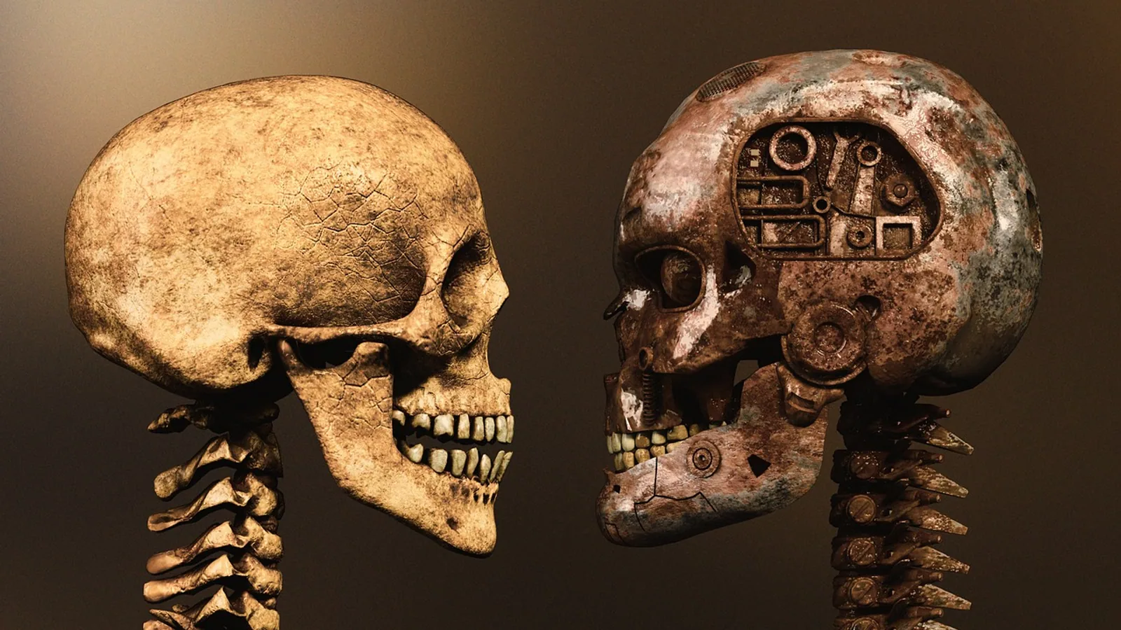
Skull sutures are fibrous joints (synarthroses) that knit the flat bones of the cranium together, and they stay patent, meaning open and unossified, through the period of rapid brain growth so the skull can expand and mold. The main sutures of the skull are the coronal, sagittal, lambdoid, and squamous sutures, plus the metopic or frontal suture that closes early in most species and persists into adulthood in a minority of individuals [1].
This article is educational and is not a substitute for veterinary diagnosis or treatment.
What Is a Suture of the Skull?
A suture of the skull is a joint in which two cranial bones meet through a thin layer of fibrous connective tissue rather than through cartilage or a fluid-filled cavity. Anatomists classify it as a synarthrosis, a joint that permits essentially no movement. The collagen fibers of the suture ligament interdigitate with the mineralized bone edges, and the whole structure acts as a shock absorber, a growth site, and a seam that keeps the skull as a single functional unit.
The skull sutures differ from the other two major joint classes in the body. A synovial joint, such as the temporomandibular joint or the stifle, has a joint capsule, synovial fluid, and free movement. A cartilaginous joint, such as the growth plate of a long bone, has a cartilage interface. A suture has neither. It is bone, fibrous tissue, and bone, with no cavity and no cartilage in the normal adult state. That design is exactly what a protective brain case needs: rigidity without brittleness, and a small amount of compliance during the years when the brain is still growing.
In the neonate, the same suture is a wide, soft, fibrous gap. The parietal and frontal bones are separated by a membrane, and this membrane allows the cranial vault to deform slightly during birth and to expand as the brain enlarges. Over months to years, the osteogenic front at each bone edge deposits new bone, the fibrous gap narrows, and the suture gradually becomes a tightly interlocked adult joint. The transition is not a single event. It is a gradient that runs from a wide-open, soft suture in a newborn puppy to a fully ossified, immovable joint in a mature dog or cat.
Types of Skull Sutures by Morphology
Cranial sutures are not all shaped alike. Comparative anatomists sort them into morphological types based on the way the bony edges meet. A detailed anatomical and radiographic study of sixteen sheep skulls identified 28 cranial sutures and classified them into four morphological types: serrated, plane, squamosal, and foliate [2]. The same categories apply broadly across domestic mammals.
Serrated sutures. The bone edges carry interlocking finger-like projections, like the teeth of a jigsaw. Serrated sutures resist tensile and shear forces well and are common where the cranial vault must withstand pulling from muscle attachments. The coronal and sagittal sutures are classically serrated.
Plane sutures. The bone edges are essentially straight and meet end to end with a thin fibrous layer between them. Plane sutures are less resistant to shear but are simple and quick to form. They are common in smaller, flatter regions of the skull.
Squamosal sutures. The two bones overlap like roof shingles, with a beveled edge sliding over a matching bevel on the other bone. The squamous suture between the parietal and temporal bones is the classic example. An overlapping joint is well suited to resisting compression.
Foliate sutures. The bone edges are shaped like a leaf with a central ridge and beveled sides, a leaf-like interlock that provides a broad contact surface.
This morphological variety is not cosmetic. A 3D geometric morphometric study spanning 22 mammal species and sampling from fetal to adult stages found that suture complexity decreases through ontogeny for the anteroposterior sutures (the interfrontal and sagittal sutures) but actually increases after birth for the transversely oriented coronal suture [3]. The authors interpret the difference as a response to mechanical loading. Anteroposterior sutures experience tensile stress and tend to simplify and obliterate with age, consolidating the skull once brain growth is done. The coronal suture, which is oriented across the skull and absorbs compressive forces from cranial mechanics, stays complex [3].
The Main Sutures of the Skull: Names and Articulating Bones


Coronal Suture
The coronal suture runs transversely across the top of the skull and joins the frontal bone to the parietal bones. It is one of the most clinically important sutures because the coronal suture is the one most frequently affected in syndromic craniosynostosis, the premature fusion of a cranial suture [4]. In combined sagittal and unilateral coronal synostosis, a case series of ten patients found fusion of the left coronal suture in 80% of cases, and 70% of the patients were male [5]. In healthy children, incidentally discovered coronal suture closure on CT is uncommon, appearing in only 2.8% of a pediatric imaging cohort [6].
The coronal suture is also a focus of developmental biology. A transient cartilage adjacent to the coronal suture, the tectum transversum, has long been considered absent in humans but is now shown to be a conserved structure present in human embryonic datasets [7]. In mice, this cartilage acts as a physical and molecular barrier that restricts BMP signaling within the coronal suture, helping keep it patent [7]. A second mechanism involves dural cells migrating into the coronal suture mesenchyme under the control of TGFβ signaling. When this migration fails, the Gli1-positive suture progenitor pool shrinks and the coronal suture fuses prematurely [4].
Sagittal Suture
The sagittal suture lies in the midline along the top of the skull and joins the two parietal bones to each other. It is the longest of the cranial vault sutures and the one most often measured in forensic work. A study of 80 adult autopsy cases measured coronal, sagittal, and lambdoid suture lengths ectocranially using a thread-and-caliper method and evaluated their relationship to stature [8].
The sagittal suture does not exist in every species. The sheep skull study specifically reported the absence of the sagittal suture in sheep [2]. That is a useful comparative point. Do not assume every domestic mammal has the same midline suture arrangement.
Sagittal suture biology has been studied in genetically modified mice. In sclerostin knockout mice, loss of the sclerostin gene (Sost) caused increased bone formation and thickening of the calvaria, with suture stenosis and partial bone obliteration accompanied by higher β-catenin expression, though the sagittal suture retained patency into the late developmental stage [9]. In healthy children, complete or partial sagittal suture closure was observed in 11.1% of a pediatric imaging cohort, and the closure was associated with a decreased cephalic index [6].
Lambdoid Suture
The lambdoid suture forms the back of the cranial vault and joins the occipital bone to the two parietal bones. Its name comes from the lambda, the Greek letter that the suture's inverted-V shape resembles. Like the coronal suture, it can fuse prematurely, and lambdoid synostosis produces a characteristic flattening on one side of the back of the head. In pediatric CT imaging, incidental lambdoid suture closure appeared in 1.6% of patients, the least common of the major vault sutures [6]. Postoperative helmet therapy after open cranial vault remodeling has been shown to improve the cephalic index in patients with unicoronal and lambdoid synostosis [10].
Squamous Suture
The squamous suture is the overlapping joint between the parietal bone and the temporal bone on the lateral wall of the skull. Its beveled, shingle-like construction is what gives the squamosal suture type its name. It is a compression-resistant joint located where the skull wall takes the forces of jaw muscle action and head impact.
Metopic (Frontal) Suture
The metopic suture, also called the frontal suture or the interfrontal suture, runs down the midline of the frontal bone between its two halves. In most species it closes during early childhood. In humans, the metopic suture closes following a predictable pattern, with more than 75% closure rates by 21 months of age [6]. Persistence of the metopic suture into adulthood is called metopism. An observational study of 50 dry adult human skulls found the metopic suture present in 6 skulls, or 12%, of which 5 were complete and 1 was incomplete, and linear morphology was the most common pattern [1]. Metopism has clinical, forensic, and anthropological relevance because a persistent metopic suture can be mistaken for a fracture on radiographs [1].
Other Named Sutures
Beyond the five main sutures, the skull contains many smaller named joints. The temporozygomatic suture lies on the zygomatic arch between the temporal and zygomatic bones. A morphometric study of 55 temporozygomatic sutures in dry adult human skulls identified four joint shape types (angular, curvy, oblique, horizontal) and five suture margin patterns (linear, denticulate, serrated, mixed, fused), with denticulate being the most common margin pattern at 34.55% [11]. The study also found a statistically significant side difference, with the suture located more anteriorly on the left side than the right [11].
The midpalatal suture joins the two halves of the maxilla in the roof of the mouth. A CBCT study of 120 adults found that facial growth pattern was significantly associated with the maturation stage of the midpalatal suture, with dolichofacial individuals more frequently showing earlier maturation [12].
Summary Table: Skull Sutures, Articulating Bones, and Function
| Suture | Bones joined | Suture type | Function and timing |
|---|---|---|---|
| Coronal | Frontal to parietal | Serrated | Transverse vault growth. Most frequently fused in syndromic craniosynostosis [4]. Incidental closure in 2.8% of pediatric CT scans [6] |
| Sagittal | Parietal to parietal | Serrated, midline | Longitudinal vault growth. Absent in sheep [2]. Incidental closure in 11.1% of pediatric CT scans [6]. Sclerostin loss causes stenosis in mice [9] |
| Lambdoid | Occipital to parietal | Serrated | Posterior vault growth. Incidental closure in 1.6% of pediatric CT scans [6] |
| Squamous | Parietal to temporal | Squamosal (overlapping) | Lateral wall stability. Resists compressive load [3] |
| Metopic (frontal) | Frontal to frontal, midline | Plane or serrated | Anterior frontal growth. Closes in early childhood (>75% by 21 months); persists as metopism in 12% of adult skulls [6][1] |
| Temporozygomatic | Temporal to zygomatic | Variable (angular, curvy, oblique, horizontal) | Zygomatic arch articulation. Shows side asymmetry in humans [11] |
| Midpalatal | Maxilla to maxilla | Plane | Palatal growth. Maturation stage linked to facial growth pattern [12] |
Functions of Skull Sutures
Growth Centers for the Cranial Vault
Cranial sutures are dynamic growth sites that balance bone growth with mesenchymal patency to accommodate cranial expansion during development [13]. Each suture contains a population of skeletal stem cells that can differentiate into bone-forming osteoblasts. As long as the suture stays open, the skull can grow. Once the suture fuses, growth at that site stops.
This is why suture timing matters so much in clinical practice. A suture that fuses too early, a condition called craniosynostosis, traps the growing brain and forces the skull to expand in abnormal directions. Craniosynostosis is defined by premature cranial suture fusion and is biologically heterogeneous [14]. It affects roughly 5.2 cases per 10,000 live births in human populations and can result in impaired neurodevelopment if not treated early [15].
Load Distribution and Shock Absorption
Sutures distribute mechanical loads across the skull rather than concentrating them at a single point. Serrated and squamosal sutures are mechanically optimized for this. The 22-species comparative study showed that anteroposterior sutures facing tensile stress simplify with age while the transversely oriented coronal suture, which faces compressive stress, increases in complexity after birth [3]. Suture morphology is therefore tuned to the forces the skull actually experiences.
Molding at Birth and in the Neonate
The patent, soft sutures of a neonate allow the cranial vault to deform during passage through the birth canal and then to re-expand. In domestic species with particularly large neonatal heads, or in brachycephalic neonates with short, broad skulls, this flexibility is developmentally important. The same softness means a neonatal skull can be molded by external pressure, which is the biological principle behind cranial remolding orthoses used after craniosynostosis repair [16].
Barriers Between Bone Compartments
A suture is not just a gap. It is an active boundary that keeps adjacent bones from fusing prematurely. The tectum transversum cartilage near the coronal suture restricts BMP signaling within the suture, acting as a molecular barrier [7]. Dural cells migrating into the coronal suture mesenchyme maintain the Gli1-positive progenitor pool that keeps the suture open [4]. When these barriers fail, the result is premature fusion.
Comparative Species Differences
Sheep
The sheep skull is a useful reference because it has been described in detail. A study of sixteen Najdi breed sheep skulls identified 28 cranial sutures across four morphological types and four cranial regions (dorsal, ventral, lateral-vertical, internal) [2]. The authors note that this intraspecies diversity reflects species-specific cranial architecture and developmental factors, and that suture patterns function as regulatory units in cranial development [2]. The absence of the sagittal suture in sheep is a reminder that suture nomenclature developed largely from human anatomy does not map perfectly onto every species.
Dogs
Dog breeds show dramatic variation in skull shape, from the long dolichocephalic skulls of sight hounds to the short brachycephalic skulls of pugs and bulldogs. Suture timing and patency vary accordingly. The general principle that holds across breeds is that sutures remain patent while the brain and skull are growing and ossify progressively afterward. Brachycephalic skulls achieve their short, broad shape through altered growth rates at the sutures and synchondroses, and clinical problems in brachycephalic dogs often involve the interaction of skull base, airway, and vault geometry. The specific suture fusion timings for individual dog breeds are not standardized in the veterinary literature, so breed-level predictions about exact fusion ages should be treated with caution and evaluated case by case.
Cats
The domestic cat has a more conservative skull shape than most dog breeds, and its sutures follow the standard carnivore pattern. The coronal, sagittal, lambdoid, and squamous sutures are present, and the metopic suture closes in early life as in other mammals. Cats less commonly present with the extreme suture-related skull deformities seen in brachycephalic dog breeds, though congenital skull anomalies do occur.
Cetaceans and Artiodactyls
Cetaceans provide a striking counterexample to the general mammalian pattern. A study of 47 cetacean species and 15 terrestrial artiodactyls found that cetaceans have a lower rate of cranial suture ossification than most terrestrial artiodactyls, with the exception of deer and mouse deer, which show somewhat similar patterns [17]. Incomplete ossification of cranial sutures throughout life in cetaceans correlates with the evolution of rarely reported accessory bones in the skull [17]. The authors note this represents a reversal of the general mammalian trend toward reduction in bone number and the development of new accessory cranial bones [17].
Clinical Relevance, Limitations and Common Mistakes
Cranial sutures matter in veterinary practice for several reasons.
Fracture interpretation. A persistent suture can be mistaken for a skull fracture on radiographs, and a fracture can be mistaken for a suture. The metopic suture is a classic trap. Persistent metopism occurs in about 12% of adult human skulls and can mimic a frontal bone fracture [1]. The same principle applies in veterinary radiography: know which sutures are normally present in the species and at what age they usually close before calling a lucent line a fracture.
Surgical landmarks. Cranial sutures are reference points in veterinary surgery and radiography [2]. Surgeons use them to orient approaches to the calvaria and to plan osteotomies.
Age estimation. Suture closure patterns contribute to age estimation in forensic and archaeological contexts [2]. In veterinary medicine, suture appearance on imaging can support but not replace a full age assessment.
Craniosynostosis. Premature fusion of a cranial suture is a congenital anomaly that disrupts craniofacial development and produces characteristic skull deformities [18]. In humans, isolated synostosis of the sagittal, metopic, coronal, or lambdoid sutures is treated surgically, and neurodevelopmental outcomes vary by suture type, with isolated coronal and metopic synostosis showing higher odds of developmental delay in one series [19]. The underlying biology involves abnormal fate specification of skeletal stem cells in the suture niche [18].
Mechanobiology and potential therapy. Mechanical loading can affect suture biology. In Crouzon mice carrying an Fgfr2cC342Y/+ mutation, in vivo cyclic loading increased the number of proliferative cells in both wild-type and mutant coronal sutures, though it did not change skull shape or suture patency in that experiment [20]. This is an experimental finding, not a clinical protocol, and the authors call for further work on the molecular mechanotransduction pathways involved [20].
Common Mistakes
Assuming all sutures are the same. Serrated, plane, squamosal, and foliate sutures have different mechanical properties and different fusion timelines [2].
Assuming every species has the same sutures. The sagittal suture is absent in sheep [2]. Cetaceans retain incompletely ossified sutures for life [17].
Assuming a closed suture is always abnormal. Incidental suture closure occurs in healthy pediatric patients, with sagittal closure seen in 11.1%, coronal in 2.8%, and lambdoid in 1.6% of one imaging cohort [6]. Most of these patients remained normocephalic [6].
Assuming a suture that looks open is functioning normally. A suture can be patent but have abnormal cellular or signaling activity that will lead to dysfunction later. Suture biology is not visible on plain imaging.
Limitations
Suture fusion timing varies by species, breed, individual, and imaging modality. Human data on suture closure percentages and morphological types are far more detailed than veterinary data, and extrapolating from human studies to dogs and cats requires caution. Individual animals with suspected skull abnormalities need a veterinarian for diagnosis and treatment planning.
Frequently Asked Questions
What are skull sutures made of?
Skull sutures are made of dense fibrous connective tissue that bridges the gap between two cranial bones. The tissue is continuous with the periosteum and the dura mater, and it contains skeletal stem cells and osteogenic progenitor cells that drive bone growth at the suture edges.
Which bones does the sagittal suture connect?
The sagittal suture connects the two parietal bones along the midline of the skull roof. It does not exist in every species. The sheep skull study reported the absence of the sagittal suture in sheep.
Is the metopic suture the same as the frontal suture?
Yes. The metopic suture is also called the frontal suture or the interfrontal suture. It runs down the midline of the frontal bone and normally closes in early childhood, though it persists into adulthood in a minority of individuals, a condition called metopism.
Why are skull sutures immovable in adults?
Skull sutures are immovable in adults because the fibrous gap between the bones ossifies and the joint becomes a solid synarthrosis. Immobility protects the brain by making the cranial vault a rigid case once growth is complete.
Do dogs have a coronal suture?
Yes. Dogs have a coronal suture between the frontal and parietal bones, just as humans do. The coronal suture is oriented transversely across the skull roof and is one of the major cranial vault sutures.
What happens if a skull suture fuses too early?
Premature fusion of a cranial suture causes craniosynostosis, which traps the growing brain and forces the skull to expand in abnormal directions. This produces characteristic skull deformities and, in humans, can impair neurodevelopment if not treated early.
Why do newborn animals have soft spots on the skull?
Newborn animals have soft spots where sutures and fontanelles are wide and fibrous rather than ossified. These open areas let the skull mold during birth and expand as the brain grows during the first months of life.
Are skull sutures the same as fontanelles?
No. Fontanelles are the wider membranous gaps at suture intersections, most noticeably where the coronal and sagittal sutures meet. Sutures are the narrower fibrous joints along the length of the bone edges. Both are fibrous and both close over time.
Related Articles
- Suture Patterns for Specific Tissue Types in Veterinary Surgery
- Plasmid in Bacteria: Structure, Types, and Functions
- Histone Acetyltransferase: Function, Types, and Mechanism
- Plasmid Definition: Structure, Types, and Functions in Genetics
- tRNA Modification: Types, Functions, and Mechanisms
- Non-Coding RNA: Types, Functions, and Mechanisms
- Skull of Horse: Anatomy and Landmarks
- Lacrimal Bone: Anatomy and Location in the Skull
- Cat Skull Anatomy: Bones, Teeth, and Features
Sources
- Unveiling the Enigma: A Comprehensive Morphological Study of Metopic Suture in Adult Human Skulls (An Observational Study).
- Cranial Suture Joints in Sheep Ovis aries (Najdi breed): Anatomical and Radiographic Findings.
- A suture in time: The ontogeny of cranial suture morphology in mammals.
- TGFβ-mediated dural progenitor cell migration into the coronal suture is crucial for preventing craniosynostosis.
- Morphology and outcomes in combined sagittal and unilateral coronal synostosis.
- Incidental cranial suture closure in pediatric patients: a rigorous evaluation of suture closure timing in the era of high-resolution CT.
- The tectum transversum maintains patency of the developing coronal suture.
- Exploratory Evaluation of Cranial Suture Lengths As Supplementary Indicators of Stature: An Autopsy-Based Morphometric Study in a North Indian Population.
- Remodeling of the sagittal suture development in sclerostin knockout male mice associated with cranial flat bone growth.
- The Efficacy of Postoperative Helmet Therapy After Open Cranial Vault Remodeling for Nonsyndromic Craniosynostosis.
- Morphometric and morphological evaluation of temporozygomatic suture anatomy in dry adult human skulls.
- Facial Growth Patterns and Midpalatal Suture Maturation in Adults: A Retrospective CBCT-Based Observational Study.
- Divergent Mechanisms of Cranial Suture Ossification in Normal Development and Pathologic Fusion.
- Mitochondria-related gene and protein changes in craniosynostosis: integrated transcriptomics and Fgfr2C361Y/+ cranial suture proteomics.
- Machine Learning-Based Identification of Craniosynostosis From Nonsynostotic Cranial Deformities.
- Evaluating compliance with postoperative cranial remolding orthotic treatment in infants with single suture craniosynostosis: A comparative study of objective and subjective approaches.
- Evolution of accessory bones in cetacean skull coupled with decreasing rate of ossification of cranial sutures.
- A tissue engineering approach to regenerate the cranial suture skeletal stem cell niche with a multicompartment biomaterial scaffold.
- Neurodevelopmental Outcomes Following Open and Endoscopic Repair of Isolated Sagittal, Coronal, Metopic, and Lambdoid Craniosynostosis.
- Mechanical bone loading effects on morphology and mechanobiology in the coronal suture of Crouzon mice.