Lacrimal Bone: Anatomy and Location in the Skull
By Dr. Zubair Khalid, DVM, MS, PhD ·
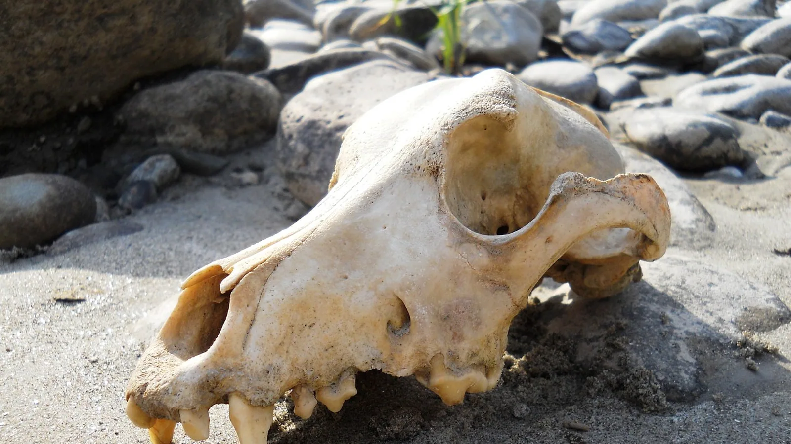
The lacrimal bone is a small, paired facial bone that sits at the front (rostral) part of the medial wall of the orbit, the bony eye socket. It holds the lacrimal fossa, a shallow depression that cradles the lacrimal sac, and it gives rise to the lacrimal foramen where the nasolacrimal duct begins its run toward the nose.
Despite its size, the lacrimal bone is a keystone of the tear drainage system. It bridges the orbit and the nasal cavity, and its shape changes noticeably between species. In dogs and cats it is a thin, modest plate with a shallow fossa. In horses and cattle it is a larger bone with a prominent extension onto the face. This guide covers its anatomy, its four main articulations, its landmarks, and the species differences that matter for anyone studying or working with skulls.
Where the Lacrimal Bone Sits in the Skull
The skull divides into two broad regions. The neurocranium is the braincase. The viscerocranium, also called the splanchnocranium, is the facial skeleton. The lacrimal bone belongs to the viscerocranium, along with the maxilla, nasal, zygomatic, incisive, palatine, pterygoid, vomer, and mandible. Comparative skull studies in deer list the lacrimal bone among the paired facial bones of the viscerocranium, alongside the maxilla, zygomatic, and palatine bones [1][2].
Each lacrimal bone is positioned at the rostral medial corner of the orbit. Picture the eye socket as a cone. Its medial wall faces the nasal cavity, and the lacrimal bone occupies the front portion of that wall. The bone is thin, flat, and roughly rectangular or oval depending on the species.
Two surfaces matter most:
- The orbital surface faces into the eye socket and carries the lacrimal fossa.
- The facial surface, present in some species, appears on the outer skull near the rim of the orbit.
The bone also contributes to the nasolacrimal canal, the bony tunnel that carries tears from the eye toward the nasal cavity. In this way the lacrimal bone links two separate spaces, the orbit and the nasal cavity, through a single small structure.
The Four Articulations of the Lacrimal Bone
The lacrimal bone meets four neighboring bones in most domestic mammals. Each joint is a suture, a fibrous seam that locks the bones together.
Maxilla
The maxilla is the largest facial bone and forms most of the upper jaw. The lacrimal bone articulates with the frontal process of the maxilla along its rostral and ventral borders. This is the most substantial of the lacrimal articulations, and in many species the maxilla wraps around the front edge of the lacrimal bone.
The maxillary contribution to the lacrimal fossa is clinically relevant. In humans, the frontal process of the maxilla forms part of the wall of the lacrimal sac fossa, and its thickness varies with ancestry and with position along the fossa. A CT study of 72 patients found that the frontal process of the maxilla was thickest inferiorly, and that maximum maxillary thickness was significantly greater in black Africans than in Caucasians at all measured planes [3]. At the midfossa level, the proportion of maxillary bone forming the lacrimal fossa wall was also greater in black Africans [3]. These findings describe human anatomy, but they illustrate a general principle: the maxilla and lacrimal bone share the fossa wall, and the split between them is not fixed.
Frontal Bone
The frontal bone forms the forehead and the roof of the orbit. The lacrimal bone articulates with the frontal bone along its dorsal border. In the horse, the suture between the lacrimal and frontal bones is a useful landmark. A morphometric study of 30 horses used a line drawn level with the palpable notches caudal to the caudal lacrimal process of each orbit to estimate the suture line of the nasal and frontal bones, which helped separate the neurocranium from the splanchnocranium [4].
Ethmoid Bone
The ethmoid bone sits deep in the skull between the orbits and forms part of the medial wall of the orbit and the roof of the nasal cavity. The lacrimal bone articulates with the ethmoid along its caudal border. In the grey heron, researchers described an ectethmoid-lacrimal complex, an osseous frame linking the ectethmoid plate to the lacrimal bone, which shows how tightly these two bones can be associated in some species [5].
Zygomatic or Palatine Bone
The fourth articulation varies. In many species the lacrimal bone meets the zygomatic bone (also called the malar bone) along its ventral or lateral border. In others, the palatine bone takes that position. Comparative deer studies list both the zygomatic and palatine bones as facial bones that sit near the lacrimal in the orbital region [1][2]. The exact partner depends on the species and sometimes on the individual.
Table: Lacrimal Bone Articulations, Landmarks, and Species Variation
| Feature | Description | Notes |
|---|---|---|
| Maxilla | Rostral and ventral border | Forms part of the lacrimal fossa wall. Maxillary thickness varies with ancestry and position in humans [3] |
| Frontal bone | Dorsal border | Suture used as a landmark in horses [4] |
| Ethmoid bone | Caudal border | Forms an ectethmoid-lacrimal complex in some birds [5] |
| Zygomatic or palatine | Ventral or lateral border | Partner varies by species |
| Lacrimal fossa | Depression on the orbital surface | Cradles the lacrimal sac. Shallow in dogs and cats |
| Lacrimal foramen | Opening at the fossa floor | Origin of the nasolacrimal duct |
| Nasolacrimal canal | Bony tunnel to the nasal cavity | Formed partly by the lacrimal bone |
| Dogs and cats | Small bone, shallow fossa | Modest facial contribution |
| Horses and cattle | Larger bone, prominent facial extension | Lacrimal bone reaches the face |
| Sheep and dromedary | Lacrimal sinus present | Air-filled cavity within the bone [6][7] |
The Lacrimal Fossa and Lacrimal Foramen
The lacrimal fossa is a shallow depression on the orbital surface of the lacrimal bone. It holds the lacrimal sac, the upper dilated end of the nasolacrimal duct system. Tears flow across the eye, enter two small openings at the inner corner of the eyelid, and drain into the sac, which sits in this fossa.
The floor of the fossa is thin. A study of 69 lacrimal bones from 48 patients measured the thickness of the lacrimal bone at the lacrimal sac fossa and found a mean thickness of 106 microns [8]. In 67 percent of patients the mean thickness of an individual lacrimal bone was less than 100 microns, and in 4 percent it exceeded 300 microns [8]. The thinnest cross section measured 11 microns and the thickest 722 microns [8]. The bone was composed of a thin plate of lamellar bone [8]. These numbers come from human anatomy, but they show how delicate this part of the skull can be.
At the bottom of the fossa is the lacrimal foramen, the opening that marks the start of the nasolacrimal duct. The duct continues through the nasolacrimal canal, a bony tunnel that runs through the maxilla and other bones to reach the nasal cavity. The lacrimal bone forms the upper part of this canal.
The lacrimal foramen is not unique to mammals. A study of a juvenile green turtle skull identified a lacrimal foramen among the main cranial structures [9]. The presence of this opening across such different species shows how fundamental the tear drainage pathway is.
The Nasolacrimal Duct and Its Origin
The nasolacrimal duct begins at the lacrimal foramen and ends in the nasal cavity. Along the way it passes through the nasolacrimal canal, a passage built from the lacrimal bone, the maxilla, and sometimes the palatine bone.
The duct is more than a simple tube. It is lined with mucosa and surrounded by mucosa-associated lymphoid tissue, or MALT. MALT is a collection of immune cells that guards mucosal surfaces exposed to the outside world. In a mouse model of autoimmune disease, researchers identified lacrimal duct-associated lymphoid tissue (LDALT) and nasopharynx-associated lymphoid tissue (NALT) as the major MALT structures in the head [10]. Mice with an autoimmune-prone genetic background had more lymphoid tissue diffusely localized beneath the mucosa of their lacrimal tracts and nasal cavity than healthy controls [10]. In some of these mice, LDALT developed by connecting directly to the bone marrow surrounding the nasolacrimal duct [10]. This work shows that the bony canal around the duct is not just a passive conduit. It sits next to active immune tissue.
The same study found that autoimmune-prone mice had larger NALT, more non-ciliated epithelial cells, and more goblet cells in the nasal mucosa than controls [10]. Goblet cells produce mucus. More of them suggests a change in the mucosal barrier. The findings point to a crosstalk between autoimmune disorders, mucosal epithelium, and MALT in the head [10].
Species Differences in the Lacrimal Bone
The lacrimal bone varies more across species than most facial bones. Size, shape, and the depth of the fossa all change.
Dogs and Cats
In dogs and cats the lacrimal bone is small and the lacrimal fossa is shallow. The bone makes only a modest contribution to the face. Most of the medial orbital wall in these species is formed by the maxilla and frontal bone, with the lacrimal bone filling a narrow strip at the front.
Even so, the lacrimal bone can be affected by disease. A study of four dogs with dacryops, a cyst of the lacrimal gland or duct, found extensive defects in the lacrimal and maxillary bones on CT scans in all cases [11]. The cysts appeared ventromedial to the globe and were treated by aspiration with a sclerosing agent or by cystorhinostomy [11]. The bone defects were extensive enough to be visible on imaging, which shows that even a small lacrimal bone can be the site of significant pathology.
Horses and Cattle
In horses and cattle the lacrimal bone is larger and has a prominent facial extension. This means the bone appears on the outer surface of the skull, not just inside the orbit. The facial extension forms part of the nasolacrimal groove and contributes to the bony rim of the orbit.
The horse lacrimal bone is also a surgical landmark. The suture between the lacrimal and frontal bones, estimated by a line level with the notches caudal to the caudal lacrimal process, helps divide the neurocranium from the splanchnocranium in morphometric studies [4]. This suture is palpable in live horses, which makes it useful for both research and clinical work.
Sheep and Goats
In sheep the lacrimal bone contains a lacrimal sinus, an air-filled cavity. A CT study of five Ile de France sheep heads identified the paranasal sinuses as maxillary, frontal, palatine, sphenoid, lacrimal, and ethmoidal [6]. The lacrimal sinus was one of the named sinuses visible on CT images and corresponding anatomical cross sections [6]. In Saanen goats, the lacrimal bone was perceived as a distinct bone on radiographs [12].
Dromedary Camels
The dromedary has a lacrimal sinus that occupies a small cavity in the rostro-medial lacrimal bone of the orbit [7]. A study of 12 dromedary heads used dissection, radiography, and CT to map the paranasal sinuses [7]. The lacrimal sinus was small compared with the frontal sinus, which is a large compartment divided by thick partitions [7]. The study found no palatal sinus and no ventral conchal sinus in any of the dissected heads [7].
Alpacas
Alpacas have a lacrimal sinus in most individuals. A study of seven alpacas found bilateral lacrimal sinuses in five of the seven specimens [13]. The ethmoidal bone contained air-filled cells divided into lateral and medial groups, and the lateral cells connected to the frontal, lacrimal, and maxillary sinuses [13]. This shows how the lacrimal bone can be part of a connected sinus network.
Other Species
The lacrimal bone appears across a wide range of mammals and other vertebrates. In barking deer and sambar deer, it is one of the paired facial bones of the viscerocranium [1][2]. In the African elephant, the ethmoturbinate, nasal, and lacrimal bones are exceptionally small relative to the massive skull [14]. In the Western European hedgehog, the lacrimal bone is part of the facial skeleton, though the study focused on other features such as the primitive tympanic bullae [15]. In the grey heron, the lacrimal bone forms part of the orbital region and links to the ectethmoid plate [5]. In turkeys, the nasal gland runs underneath the lacrimal, frontal, and nasal bones, which places the lacrimal bone near an important salt-secreting organ [16].
Why the Lacrimal Bone Matters
The lacrimal bone does three jobs.
First, it forms part of the medial wall of the orbit. This wall separates the eye from the nasal cavity and the brain. A thin plate of bone is enough to do this job because the forces on the medial wall are modest.
Second, it holds the lacrimal sac in the lacrimal fossa. The sac collects tears and passes them into the nasolacrimal duct. Without a stable fossa, the sac would not have a fixed position, and tear drainage would be less efficient.
Third, it forms the upper part of the nasolacrimal canal. This canal protects the duct as it travels from the eye to the nose. The canal also gives the duct a fixed route, so tears always drain to the same place.
The bone is also a site of immune activity. The mucosa-associated lymphoid tissue around the nasolacrimal duct guards against pathogens that enter through the eye and nose [10]. This immune tissue sits right next to the bony canal, which means the lacrimal bone is part of a larger mucosal defense system.
Common Mistakes and Limitations
Several misunderstandings about the lacrimal bone come up often.
The first mistake is assuming the lacrimal bone is the same size in all species. It is not. In dogs and cats it is small and the fossa is shallow. In horses and cattle it is larger and reaches the face. In the African elephant it is exceptionally small [14]. Anyone comparing skulls across species needs to account for this variation.
The second mistake is confusing the lacrimal bone with the lacrimal gland. The gland produces tears. The bone holds the lacrimal sac and forms part of the tear drainage canal. They are different structures with different jobs.
The third mistake is assuming the lacrimal bone always articulates with the same four bones. The maxilla, frontal, and ethmoid are consistent partners. The fourth partner varies. It can be the zygomatic bone or the palatine bone, depending on the species.
The fourth mistake is treating the lacrimal fossa as a deep cup. In dogs and cats it is shallow. In humans it is also relatively shallow, though the bone around it can be thin. The mean thickness of the lacrimal bone at the fossa in one human study was 106 microns, which is thinner than a sheet of paper [8].
The fifth mistake is overlooking the lacrimal sinus. In sheep, goats, dromedaries, and alpacas, the lacrimal bone contains an air-filled cavity [13][6][7]. This sinus can become involved in disease, and it shows up on CT scans.
A final limitation is that most detailed measurements of the lacrimal bone come from humans or from a small number of animal species. The thickness values, the split between the maxilla and lacrimal bone in the fossa, and the position of the lacrimal sac relative to nearby landmarks all vary with ancestry, sex, and disease status in humans [3][17]. Similar data for dogs, cats, horses, and cattle are less complete. Individual cases need a veterinarian for diagnosis and treatment.
Practical Implications for Owners and Keepers
For pet owners and animal keepers, the lacrimal bone rarely comes up by name. It matters indirectly.
If an animal has excessive tearing, a blocked tear duct, or a swelling near the inner corner of the eye, the problem may involve the nasolacrimal duct or the lacrimal sac. The lacrimal bone forms the canal that carries the duct, so any condition that affects the bone can affect tear drainage.
In dogs, dacryops can cause a cystic swelling ventromedial to the eye and can be associated with defects in the lacrimal and maxillary bones [11]. This is uncommon, but it shows that the lacrimal bone can be involved in orbital disease.
In horses and cattle, the larger lacrimal bone and its facial extension make it easier to palpate and to see on radiographs. This can help veterinarians locate the source of a tear drainage problem.
In sheep and camelids, the lacrimal sinus can be a site of infection or inflammation. CT imaging can show the sinus and its connections to the frontal and maxillary sinuses [13][6][7].
For anyone preparing a skull for study, the lacrimal bone is easiest to find by looking at the front of the medial orbital wall. The lacrimal fossa is the shallow depression on its orbital surface. The lacrimal foramen is at the bottom of the fossa.
What Is Still Uncertain
Several questions about the lacrimal bone remain open.
The exact split between the maxilla and the lacrimal bone in the lacrimal fossa varies between individuals and between populations. A CT study of 72 patients found significant differences in maxillary thickness and in the proportion of the fossa wall formed by the maxilla between black Africans and Caucasians [3]. Similar comparative data for domestic animals are limited.
The position of the lacrimal sac relative to nearby landmarks also varies. A study of 227 Chinese patients with nasolacrimal duct obstruction measured the distance from the lacrimal sac to the middle turbinate axilla and the anterior skull base [17]. Male patients showed more superior-anterior displacement of the lacrimal sac than female patients, and patients with chronic dacryocystitis had an inferior and posterior lacrimal sac position compared with those with simple obstruction [17]. These findings show that the lacrimal sac is not fixed in place, even within one population.
The role of the lacrimal bone in immune responses is another active area. Mouse studies show that autoimmune disease can alter the mucosal immunity of the lacrimal tract and nasal cavity, and that lymphoid tissue can connect directly to the bone marrow around the nasolacrimal duct [10]. How these findings translate to other species is not yet clear.
Finally, the lacrimal bone is understudied in many species. Detailed morphometric data exist for horses, deer, sheep, goats, and dromedaries [12][6][7][2][4]. For many other species, the lacrimal bone is described only as present, without measurements.
Frequently Asked Questions
What is the lacrimal bone?
The lacrimal bone is a small, paired facial bone that forms the front part of the medial wall of the orbit. It holds the lacrimal fossa, which cradles the lacrimal sac, and it gives rise to the lacrimal foramen where the nasolacrimal duct begins.
Where is the lacrimal bone located?
It sits at the rostral medial corner of the orbit, between the maxilla, frontal bone, and ethmoid bone. In horses and cattle it also extends onto the face.
How many bones does the lacrimal bone articulate with?
It typically articulates with four bones: the maxilla, the frontal bone, the ethmoid bone, and either the zygomatic or the palatine bone, depending on the species.
What is the lacrimal fossa?
The lacrimal fossa is a shallow depression on the orbital surface of the lacrimal bone. It holds the lacrimal sac, the upper part of the tear drainage system.
Where does the nasolacrimal duct start?
The nasolacrimal duct starts at the lacrimal foramen, an opening at the bottom of the lacrimal fossa. From there it runs through the nasolacrimal canal to the nasal cavity.
Is the lacrimal bone the same in dogs and horses?
No. In dogs and cats the lacrimal bone is small and the lacrimal fossa is shallow. In horses and cattle the bone is larger and has a prominent facial extension.
Do all animals have a lacrimal sinus?
No. Sheep, goats, dromedaries, and alpacas have a lacrimal sinus, an air-filled cavity within the lacrimal bone. Dogs, cats, and horses do not have this sinus.
How thick is the lacrimal bone?
In humans, the mean thickness of the lacrimal bone at the lacrimal sac fossa is about 106 microns, though it ranges from 11 to 722 microns in individual samples. Thickness in other species is less well documented.
Related Articles
- Equine Hindlimb Anatomy: Bones, Joints, and Ligaments
- Do Frogs Have Bones? Skeletal Anatomy and Pet Care
- Bioinformatics Salary by Location
- Swine Barn Location and Site Selection
- RNA Location: Types, Functions, and Cellular Distribution
- Z-DNA Found in Biology: Location, Function, and Detection
- Cat Skull Anatomy: Bones, Teeth, and Features
- Dog Skull Anatomy: Bones, Foramina, and Landmarks
Sources
- A comparative study on the morphology, radiography and computed tomography of the skull bones of barking deer (Muntiacus muntjak) and sambar deer (Rusa unicolor).
- Comparative morphometrical studies on the skull bones of barking deer (Muntiacus muntjak) and sambar deer (Rusa unicolor).
- Radiological Comparison of the Lacrimal Sac Fossa Anatomy Between Black Africans and Caucasians.
- Conformation of the equine skull: a morphometric study.
- Skull of the grey heron (Ardea cinerea): Detailed investigation of the orbital region.
- CT anatomy of the head in the Ile de France sheep.
- Macroscopic anatomy, radiography and computed tomography of normal paranasal sinuses of the adult one-humped dromedary (Camelus dromedarius).
- Lacrimal bone thickness at the lacrimal sac fossa.
- Morphological and Radiologic Features of the Skull of a Juvenile Green Turtle (Chelonia mydas, Linnaeus, 1758) from Saint Kitts and Nevis, West Indies.
- Altered morphology of mucosa-associated lymphoid tissues and epithelium in the nasal cavity and lacrimal apparatus in autoimmune disease-prone MRL/MpJ-Fas(lpr/lpr) mice.
- Dacryops with extensive lacrimal and maxillary bone defects in four dogs.
- Evaluation of radiological and anatomical features of skull bones in adult Saanen goat.
- Computed Tomography, Magnetic Resonance, Cross-Sectional Anatomy, and 3D Volume Rendering of the Nasal Cavity and Paranasal Sinuses of the Alpaca (Vicugna pacos).
- The skull and mandible of the African elephant (Loxodonta africana).
- Anatomy, digital radiography and cone-beam computed tomography of Western European hedgehog (Erinaceus europaeus) skull.
- The Nasal Gland in Turkeys (Meleagris gallopavo): Anatomy, Histology, and Ultrastructure.
- Anatomic Variations of the Lacrimal Sac Relative to Middle Turbinate Axilla and Skull Base in Chinese Patients with Nasolacrimal Duct Obstruction: A CT-Dacryocystography.