Skull of Horse: Anatomy and Landmarks
By Dr. Zubair Khalid, DVM, MS, PhD ·
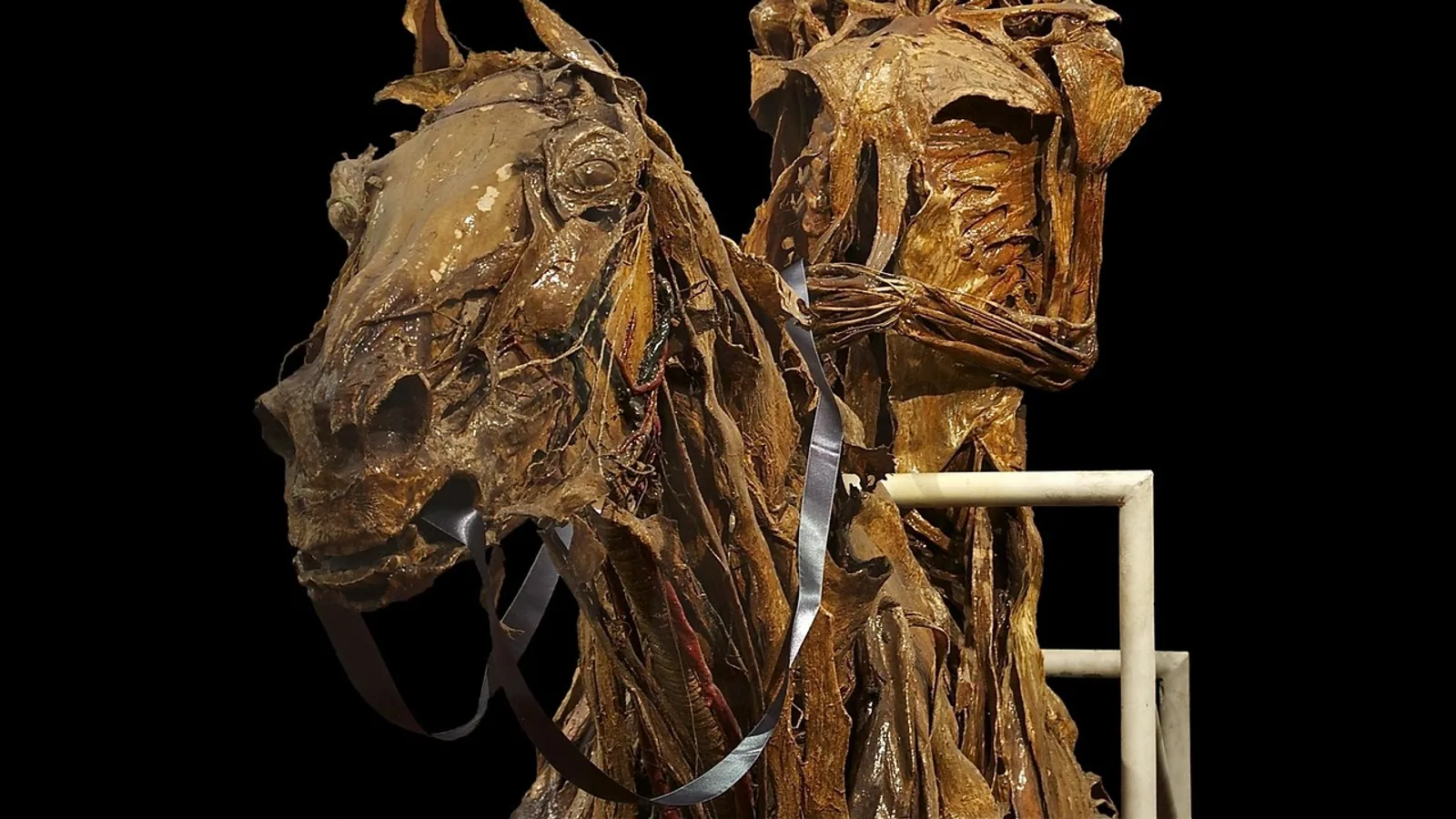
The skull of the horse is the elongated bony framework of the head that houses the brain, special sense organs, and the roots of the teeth, and that transmits the cranial nerves through named foramina. Its most recognizable features are a long facial (splanchnic) cranium, a prominent facial crest, a deep facial fossa, and a dental arcade built for continuous grazing.
The equine skull matters because it is the anatomical map for regional anesthesia, dental surgery, sinus surgery, and imaging interpretation. A clinician who can place the infraorbital and mental foramina by palpation can block the maxillary and mandibular divisions of the trigeminal nerve without entering the orbit or the mandibular canal. A student who understands the dental arcade can predict which tooth is diseased from the surface anatomy of the face. This article is educational and is not a substitute for veterinary diagnosis or treatment.
Orientation and Nomenclature
Before naming any landmark, fix the standard anatomical directions. Rostral means toward the nose, caudal toward the tail, dorsal toward the back, ventral toward the belly, medial toward the midline, and lateral away from the midline. In the skull, "rostral" replaces "anterior" and "caudal" replaces "posterior" because the head sits at a right angle to the rest of the body.
The skull is divided into two functional regions. The neurocranium is the box that surrounds the brain. The splanchnocranium (viscerocranium) is the facial skeleton, which in the horse is dramatically elongated to support the grazing dentition and the large paranasal sinuses. A morphometric study of 23 dry skulls from the Pyrenean Horse Breed found clear modularity between the splanchnocranium and the basicranium, meaning these two regions covary as semi-independent units rather than as a single rigid block [1]. That finding explains why facial length can vary between breeds without proportionally reshaping the base of the skull.
The horse skull also changes shape with age. A three-dimensional craniometric study of warmblood horses compared young animals (6 weeks) with old animals (14 and 17 years) and found that some landmarks, such as those at the zygomatic arch, grew in size but kept stable relative proportions [2]. Other landmarks shifted position as the skull matured. This age-related drift is why a foramen that is easy to find in a yearling may sit slightly differently in a 20-year-old.
The Facial Crest and Facial Fossa
The facial crest is a thick, curved ridge of bone on the lateral surface of the maxilla. It runs roughly horizontally from the caudal edge of the zygomatic arch toward the rostral face and serves as the attachment for the masseter muscle. In the living horse, the facial crest is the single most useful palpable landmark on the side of the face.
Just ventral and caudal to the facial crest lies the facial fossa (also called the malar fossa or the maxillary fossa). This is a deep, smooth depression in the maxilla. The fossa is not a foramen and transmits nothing. Its clinical value is positional: it marks the territory of the maxillary sinus and the roots of the caudal cheek teeth, and it is the surface landmark that tells you where the infraorbital foramen is relative to the orbit.
The facial crest and facial fossa are the two features that make the equine skull instantly recognizable as equine. In the dog and cat, the facial crest is absent or rudimentary and the face is short. In the horse, the crest is a load-bearing ridge and the fossa is a deep excavation, both consequences of a long face and a large masseter.
Foramina of the Equine Skull
A foramen is an opening in bone that transmits a nerve, a vessel, or both. The equine skull has many, but four are essential for the regional anesthesia and imaging work a veterinary student will actually perform.
Infraorbital Foramen and Infraorbital Canal
The infraorbital foramen is the rostral opening of the infraorbital canal on the lateral surface of the maxilla. It sits dorsal to the roots of the third and fourth premolars and ventral to the facial crest, roughly halfway between the orbit and the nostril. The infraorbital canal runs caudally from this foramen through the maxilla and opens caudally as the maxillary foramen, which is the exit of the maxillary nerve from the skull.
The canal is not a straight tube. A CT and dissection study of 13 cadaveric skulls found that each infraorbital canal follows a serpentine-curved pathway, and that anatomical dissection revealed gaps between the infraorbital nerve, the accompanying vessels, and the bony wall of the canal [3]. Those gaps matter for any clinician attempting a retrograde injection: the needle tip can sit in the canal without directly contacting the nerve, and the injectate can spread along the nerve rather than around it.
The maxillary nerve is the second division of the trigeminal nerve (cranial nerve V). It supplies sensation to the skin of the mid-face, the upper lip, the nostril, and the upper cheek teeth. Magnetic resonance imaging and CT of equine cadaver heads consistently allowed visualization of cranial nerve V and its main divisions in all five horses studied, which confirms that the maxillary nerve and its branches are identifiable structures, not abstract textbook lines [4].
Mental Foramen and Rostral Mandibular Canal
The mental foramen is the rostral opening of the mandibular canal on the lateral surface of the mandible. It sits roughly one-third of the horizontal distance from the second premolar along the incisor-premolar space (the diastema), and about one-third of the vertical distance from the dorsal surface of the interproximal space at that level [5]. In plain terms, it is on the side of the jaw, below and behind the corner incisor, in the gap between the incisors and the cheek teeth.
The mental nerve is the terminal branch of the inferior alveolar nerve, which is itself a branch of the mandibular division of the trigeminal nerve (cranial nerve V3). The inferior alveolar nerve enters the mandible at the mandibular foramen on the medial surface of the ramus, runs rostrally through the mandibular canal, and exits at the mental foramen as the mental nerve.
The mental foramen is not a fixed point. A retrospective CT study of 41 horses weighing 136 to 820 kg found that foramen width averaged 6.4 mm (range 2.3 to 17.1 mm) and height averaged 5.6 mm (range 2.1 to 10.3 mm) [5]. Age was negatively correlated with horizontal positioning, meaning the foramen drifts caudally relative to the incisor-premolar space as the horse ages, and age was positively correlated with both foramen height and width. Thoroughbred and Warmblood or Draft horses had taller foramina than Quarter Horses [5]. A clinician who uses a single fixed measurement for every horse will miss the foramen in a substantial number of patients.
Supraorbital Foramen
The supraorbital foramen is an opening in the frontal bone at the dorsal rim of the orbit. It transmits the supraorbital nerve, a branch of the ophthalmic division of the trigeminal nerve (cranial nerve V1). The supraorbital nerve supplies sensation to the skin of the upper eyelid and the forehead. The equine orbit is a complete bony ring, unlike the open orbit of the dog, and the supraorbital foramen sits within that ring [6][7]. The horse also has a well-developed set of eyelid muscles and a complete bony orbit, features that distinguish equine ocular anatomy from that of small animals [7].
Other Foramina Worth Knowing
The optic canal transmits cranial nerve II. The orbital fissure and round foramen transmit branches of cranial nerve V. The stylomastoid foramen transmits cranial nerve VII. The jugular foramen transmits cranial nerves IX, X, and XI. The hypoglossal canal transmits cranial nerve XII. A cadaver study using MRI and CT identified cranial nerves II, V, VII, VIII, and XII in all five horses examined, and identified cranial nerves III, IV, and VI as a group, while the group of IX, X, and XI was identified in four of five horses even though the region of exit was seen in all cases [4]. These are imaging landmarks rather than palpation landmarks, and they belong to the neurocranium rather than the facial skeleton.
Summary Table: Key Foramina and Their Clinical Use
| Foramen | Bone | Nerve transmitted | Clinical block or use |
|---|---|---|---|
| Infraorbital foramen | Maxilla | Maxillary nerve (V2), terminal branches | Maxillary nerve block, retrograde approach to the maxillary foramen |
| Mental foramen | Mandible | Mental nerve (terminal branch of inferior alveolar nerve, V3) | Mental foramen block, rostral inferior alveolar nerve block |
| Supraorbital foramen | Frontal | Supraorbital nerve (V1) | Desensitization of upper eyelid and forehead |
| Mandibular foramen | Mandible (medial ramus) | Inferior alveolar nerve (V3) | Intraoral inferior alveolar nerve block |
| Maxillary foramen | Maxilla (caudal) | Maxillary nerve (V2) | Target of retrograde infraorbital injection |
| Stylomastoid foramen | Temporal | Facial nerve (VII) | Imaging landmark, not a routine block site |
The Equine Dental Arcade
The horse is a grazing herbivore with hypsodont teeth, meaning the crowns are tall and continue to erupt throughout life as the occlusal surface is worn down by abrasive forage. The full permanent arcade on each side of each jaw contains six incisors, six premolars, and six molars, for a total of 36 to 40 teeth depending on whether wolf teeth are present.
Incisors
The six upper and six lower incisors sit at the rostral end of the mouth. They are used for cropping grass. The incisors are numbered from the midline outward: I1 (central), I2 (middle), and I3 (corner). The occlusal surface of the incisors changes shape with age, which is the basis of the traditional "aging by teeth" method. The upper incisors meet the lower incisors at an angle that varies with skull conformation, and the incisor arcade is the rostral boundary of the oral cavity.
The Diastema
The diastema is the toothless gap between the incisors and the first cheek tooth. It is not a defect. It is the space where the bit sits in a bridled horse, and it is the space a clinician uses to pass a stomach tube or an endoscope. The diastema is also the surface landmark for the mental foramen, which sits roughly one-third of the way along it from the second premolar [5].
Premolars and Molars
The premolars and molars together form the cheek teeth, often called the dental arcade in the strict sense. The horse has six premolars and six molars per arcade. The first premolar is the wolf tooth, abbreviated PM1. Wolf teeth are small, vestigial, and often absent, especially in the lower jaw. When present, they sit immediately rostral to the second premolar (PM2) and can interfere with the bit. The remaining cheek teeth, PM2 through M3, are the grinding teeth. They have complex infundibula (enamel folds) that trap food and wear unevenly, which is why equine dentistry focuses on floating (rasping) sharp enamel points.
Dental Formula
The permanent dental formula for the horse is:
- Incisors: 3/3 per side
- Canines: 0 or 1/0 or 1 per side (usually present in males, often absent in females)
- Premolars: 3 or 4/3 or 4 per side (the fourth is the wolf tooth, PM1)
- Molars: 3/3 per side
The variation in the canine and premolar counts is why the total ranges from 36 to 40. The dental arcade is not a flat line. The upper cheek teeth are wider than the lower, and the occlusal surfaces are angled, which produces the characteristic lateral excursion of the jaw during chewing.
Clinical Landmarks for Nerve Blocks
Regional anesthesia of the equine head is performed under standing sedation for dental extraction, sinus surgery, wound repair, and ocular procedures. The two blocks a student must know are the maxillary block via the infraorbital foramen and the mandibular block via the mental foramen or the mandibular foramen.
Maxillary Nerve Block via the Infraorbital Foramen
The traditional approach to the maxillary nerve targets the maxillary foramen through the zygomatic arch. That approach carries a risk of injury to delicate structures in the target area [3]. A retrograde approach from the infraorbital foramen was investigated in 13 cadaveric skulls using CT and dissection [3]. The technique involves inserting a needle into the infraorbital foramen and advancing it caudally along the serpentine canal toward the maxillary foramen, then injecting local anesthetic. The study measured canal length and volume, foramen diameters, and the spread of contrast medium. Two Tuohy needles were randomly inserted at 12 infraorbital foramina, and 10 mL of contrast medium was injected. CT verified the spread of the solution and any complications [3]. The retrograde approach is feasible, but the serpentine path and the gaps between nerve and bone mean that needle selection and insertion depth matter.
Mental Foramen Block and Rostral Inferior Alveolar Nerve Block
The mental foramen block desensitizes the mental nerve and the rostral portion of the inferior alveolar nerve. A cadaveric and in vivo study compared two techniques. In technique T1, the needle was directed rostrocaudally into the mental foramen for 3 cm. In technique T2, the needle was directed dorsolaterally to ventromedially into the foramen for 1 cm. Two injectate volumes, 3 mL and 5 mL, were tested. Neither technique nor volume had a significant impact on circumferential nerve staining, which had a median of 15 mm (range 0 to 33 mm) for T1 and 10 mm (range 0 to 42 mm) for T2. Injectate was more likely to thread alongside the inferior alveolar nerve with T1 (9 of 12) and to form a bolus around the rostral inferior alveolar nerve with T2 (9 of 12) [8]. The practical takeaway is that the injection technique changes the diffusion pattern, not the total length of nerve stained.
The clinical efficacy of the rostral inferior alveolar nerve block via the mental foramen was tested in a blinded trial of 10 horses assigned to left unilateral, right unilateral, or bilateral blocks, with 5 bilateral controls. Mechanical nociceptive stimulus was applied before sedation, after sedation, and at 10, 30, 60, and 90 minutes after injection. All groups were significantly less likely to respond at post-sedation, 10-minute, and 30-minute time points, but only blocked sites were less likely to respond at 60 and 90 minutes compared with pre-sedation. There was no significant difference in response between the three blocked regions (labial mucosa, alveolar mucosa, and teeth) [9]. The block works, and it lasts at least 90 minutes in the blocked sites, but sedation alone accounts for some of the early analgesia.
Intraoral Inferior Alveolar Nerve Block
A separate technique targets the inferior alveolar nerve at the mandibular foramen from inside the mouth. Measurements in 26 adult equine skulls of various ages and breeds established the distance of the mandibular foramen from the distal edge of the mandibular third molar, the rostral edge of the mandibular ramus, and the ventral margin of the mandible. CT verified placement of local anesthetic with a custom-made device on 4 cadaver heads. The technique was then applied in 43 clinical cases, and patients tolerated invasive dental procedures after a 5 mL dose of local anesthetic without evidence of self-inflicted lingual trauma [10]. The intraoral approach desensitizes the inferior alveolar nerve with a smaller volume than a blind approach and avoids the risk of lingual trauma.
Supraorbital and Ocular Blocks
The supraorbital foramen transmits the supraorbital nerve, which supplies the upper eyelid. Blocking it is part of the regional anesthesia protocol for ocular examination and surgery. The horse has a complete bony orbit and well-developed eyelid muscles, and a systematic ocular examination should be performed before sedation or nerve blocks so that baseline findings are not lost [6][7]. The appropriate order of diagnostic tests matters, and referral to an ophthalmologist should be considered if a diagnosis is elusive [7].
How the Skull Is Examined in Practice
Palpation is the first examination. Run a finger along the facial crest from the zygomatic arch rostrally. The crest ends near the infraorbital foramen. Press gently ventral to the crest and you can often feel the foramen as a small depression. On the mandible, follow the ventral border rostrally until you reach the diastema, then palpate the lateral surface about one-third of the way along the diastema and one-third of the way down from the dorsal edge. The mental foramen is there, but it is small and may be covered by the depressor labii inferioris muscle.
Imaging is the second examination. Radiography of the skull uses lateral, dorsoventral, and oblique views. CT is superior for foramina and dental roots because it removes superimposition. MRI is used for cranial nerves and soft tissue, and a cadaver study confirmed that MRI can identify cranial nerves II, V, VII, VIII, and XII in the horse [4]. The same study found that the main divisions of cranial nerve V could be distinguished in all cases, which is why MRI is the modality of choice when a maxillary nerve lesion is suspected [4].
The third examination is the oral examination. A full mouth speculum and a dental mirror or endoscope allow inspection of the arcade, the diastema, and the wolf teeth. The occlusal surfaces are checked for sharp points, wave mouth, step mouth, and missing or fractured teeth. The incisors are checked for alignment and wear.
Comparative Notes Across Domestic Species
The equine skull differs from the dog and cat skull in several ways that matter for clinical practice. The horse has a complete bony orbit, while the dog and cat have an incomplete orbit with a ligamentous orbital rim. The horse has a long facial crest, which the dog and cat lack. The horse has hypsodont teeth that erupt continuously, while the dog and cat have brachydont teeth that stop erupting after eruption is complete. The horse has a diastema, which the dog and cat do not. The horse has a large paranasal sinus system that communicates with the nasal cavity and the maxillary cheek teeth roots, while the dog and cat have much smaller sinuses.
The modularity study found clear modularity between the splanchnocranium and the basicranium in the horse, and noted that the lack of disproportions in domestic horse breeds compared with dog and cat breeds may reflect a lack of single modules to evolve independently [1]. In other words, the horse skull is relatively constrained in shape compared with the dog skull, which has been under strong artificial selection for breed-specific head shapes.
Common Misconceptions
Misconception 1: The infraorbital foramen is a fixed landmark. It is not. The canal is serpentine, and the foramen position varies with age and breed [3][5].
Misconception 2: The mental foramen is always in the same place. It drifts caudally with age, and its height and width increase with age [5].
Misconception 3: A mental foramen block anesthetizes the whole lower jaw. It anesthetizes the mental nerve and the rostral inferior alveolar nerve. The caudal inferior alveolar nerve and the molar teeth require a block at the mandibular foramen [8][10].
Misconception 4: Wolf teeth are always present. They are often absent, especially in the lower jaw. When present, they are PM1 and sit rostral to PM2.
Misconception 5: The facial fossa is a foramen. It is a depression, not an opening. Nothing passes through it.
Misconception 6: Sedation alone is enough for dental surgery. Sedation reduces response at early time points, but only a properly placed nerve block eliminates response at 60 and 90 minutes [9].
Quick Review
- The equine skull has a long splanchnocranium, a prominent facial crest, and a deep facial fossa.
- The infraorbital foramen transmits the maxillary nerve (V2) and is the entry point for a retrograde maxillary block.
- The mental foramen transmits the mental nerve (terminal branch of V3) and is the entry point for a rostral inferior alveolar block.
- The supraorbital foramen transmits the supraorbital nerve (V1) and sits at the dorsal orbital rim.
- The permanent dental arcade has 6 incisors, 6 premolars, and 6 molars per side, with a diastema between incisors and cheek teeth and a wolf tooth (PM1) when present.
- The infraorbital canal is serpentine, and the mental foramen position changes with age.
- The intraoral inferior alveolar nerve block at the mandibular foramen desensitizes the whole mandibular quadrant with a 5 mL dose [10].
Clinical Relevance, Limitations and Common Mistakes
The clinical relevance of equine skull anatomy is concentrated in three procedures: dental extraction, sinus surgery, and ocular surgery. Each depends on accurate landmark identification. A maxillary block placed too far rostral will miss the maxillary nerve. A mental foramen block placed too shallow will not reach the inferior alveolar nerve. An intraoral block placed too far caudal will miss the mandibular foramen and may injure the lingual tissues.
The limitations are anatomical. Foramen position varies with age, breed, and body weight [5]. The infraorbital canal is curved, not straight [3]. The mental foramen can be covered by muscle. Imaging is often required to confirm needle placement, and CT is the standard for verification in cadaver studies [3][8][10]. No single measurement works for every horse.
Common mistakes include using a fixed depth for every patient, failing to palpate before injecting, confusing the facial fossa with a foramen, and assuming that sedation provides surgical anesthesia. A clinician who palpates the facial crest, finds the infraorbital foramen, and confirms the mental foramen position with imaging when needed will place blocks accurately. Individual cases require veterinary assessment, and this article is educational only.
Frequently Asked Questions
What is the infraorbital foramen and what does it transmit?
The infraorbital foramen is the rostral opening of the infraorbital canal on the lateral maxilla. It transmits the maxillary nerve (cranial nerve V2) and its terminal branches.
Where is the mental foramen located on the horse?
The mental foramen sits on the lateral mandible, about one-third of the horizontal distance along the incisor-premolar space from the second premolar and about one-third of the vertical distance from the dorsal surface of the interproximal space at that level [5].
How many teeth does a horse have?
A horse has up to 40 permanent teeth: 6 incisors, 6 premolars, and 6 molars per arcade, plus up to 4 canine teeth and up to 4 wolf teeth (PM1). The total ranges from 36 to 40 depending on whether canines and wolf teeth are present.
What is the diastema in a horse?
The diastema is the toothless gap between the incisors and the first cheek tooth. It is where the bit sits and where a stomach tube or endoscope is passed.
What nerve does the mental foramen block target?
The mental foramen block targets the mental nerve, which is the terminal branch of the inferior alveolar nerve (a branch of cranial nerve V3). It desensitizes the rostral lower jaw.
Can the infraorbital canal be used for a maxillary nerve block?
Yes. A retrograde approach from the infraorbital foramen toward the maxillary foramen has been evaluated in cadaveric skulls using CT and dissection, and the canal follows a serpentine path that must be followed carefully [3].
Related Articles
- Bovine Heart Anatomy and Auscultation Landmarks
- Horse Hoof Care: Anatomy, Trimming, and Common Problems
- Equine Digestive System: Anatomy and Physiology of the Hindgut
- Equine Hoof Care: Anatomy, Trimming, and Shoeing
- Comparative Anatomy of the Mammalian Kidney
- Comparative Anatomy of the Mammalian Heart
- Dog Skull Anatomy: Bones, Foramina, and Landmarks
Sources
- No modularity at ventral level in the horse skull.
- The challenge of extra-intra craniometry: a computer-assisted three-dimensional approach on the equine skull.
- Retrograde maxillary nerve perineural injection: A tomographic and anatomical evaluation of the infraorbital canal and evaluation of needle type and size in equine cadavers.
- ANATOMICAL STUDY OF CRANIAL NERVE EMERGENCE AND SKULL FORAMINA IN THE HORSE USING MAGNETIC RESONANCE IMAGING AND COMPUTED TOMOGRAPHY.
- Anatomic analysis of the equine mental foramen and rostral mandibular canal using computed tomography.
- Equine ocular anatomy and ophthalmic examination.
- Normal equine ocular anatomy and eye examination.
- Evaluation of the equine mental foramen block: cadaveric and in vivo injectate diffusion.
- Evaluation of the Rostral Inferior Alveolar Nerve Block via the Mental Foramen in Equids: In Vivo Efficacy Testing.
- Evaluation and clinical use of an intraoral inferior alveolar nerve block in the horse.