Corneal Ulcer in Dogs: Healing Stages and Treatment
By Dr. Zubair Khalid, DVM, MS, PhD ·
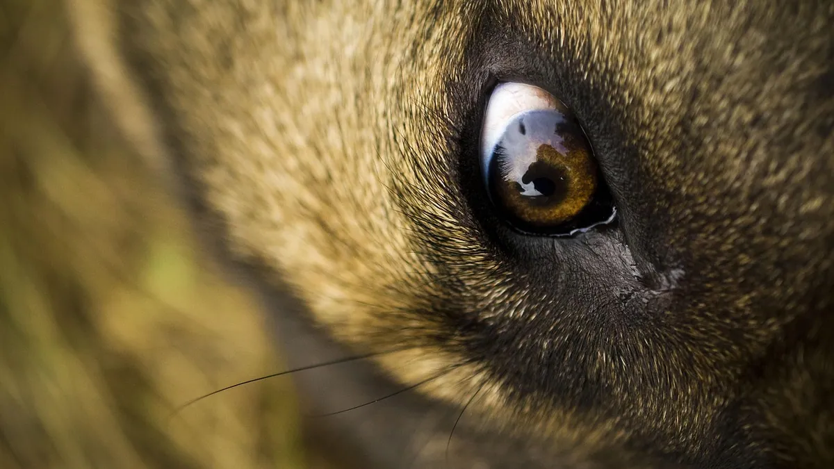
The corneal ulcer in dogs healing process usually takes about one to three weeks for a simple, superficial ulcer, but deep or infected ulcers can take much longer. Healing happens in stages, and your veterinarian needs to check the eye along the way because some ulcers get worse instead of better. Treatment almost always involves prescription eye medicine, not home remedies.
This article is educational and is not a substitute for veterinary diagnosis or treatment.
Quick Answer
- A corneal ulcer is a scratch or sore on the clear front surface of the eye, and it is painful [1].
- Most superficial ulcers heal in roughly 1 to 3 weeks with topical antibiotics and pain control, but deep ulcers or ones with infection take longer [2].
- Never put anything in your dog's eye that a veterinarian did not prescribe. Human eye drops, breast milk, honey, and herbal rinses can delay healing or cause infection.
- A vet recheck within a few days is standard because an ulcer can look the same or better to you while quietly getting deeper.
- Red flags that mean go now, not later: a suddenly cloudy or bulging eye, a pupil that will not constrict, or your dog pawing and screaming in pain.
What a Corneal Ulcer Actually Is
The cornea is the clear dome on the front of the eye. It protects the eye and helps focus light on the retina, which is why any injury to it needs prompt attention [1].
A corneal ulcer is a break in the surface layer of that dome. Think of it like a scrape on your own eye. The surface cells are missing, and the nerves underneath are exposed. That is why ulcers hurt so much.
Veterinarians describe ulcers by depth. A superficial ulcer only involves the outer layers. A stromal ulcer goes deeper into the middle layer of the cornea. A descemetocele is a very deep ulcer where only the thinnest remaining layer holds the eye together, and that is a surgical emergency [2].
Signs Owners Notice
The most common signs are watering eyes and spasmodic winking, which is the eye squeezing shut in pain [1]. You may also see:
- Pawing at the face or rubbing the eye on furniture
- Squinting or holding the eye closed
- A red, bloodshot white of the eye
- Cloudiness or a bluish haze over the cornea
- Discharge that is clear, yellow, or green
- Reluctance to go outside in bright light
Some dogs only show mild squinting. Others cry out and will not let you near their face. The amount of pain does not always match the size of the ulcer.
If your dog has redness, swelling, or discharge and you are not sure whether it is an ulcer, see our guide to Dog Eye Injury: Redness, Swelling, or Discharge? for help deciding how urgent it is.
Causes and Risk Factors
Inflammation and swelling of the cornea is common in dogs, and when it affects only one eye, injury or trauma is often the cause [1]. Common triggers include:
- A scratch from a cat claw, a stick, or another dog
- Foreign material like a grass awn or grit trapped under the eyelid
- Eyelid abnormalities, including rolled-in lids or extra lashes
- Dry eye, which leaves the surface poorly protected
- Brachycephalic anatomy (flat-faced breeds) that leaves the eye more exposed
Your veterinarian will also check for problems with the shape or form of the eyelid and for foreign objects, because the ulcer will not improve until those issues are controlled [1].
Bacterial infection is a major concern. In a study of dogs with progressive ulcerative keratitis, 54% of sampled ulcers grew bacteria, most often Streptococcus canis, Pseudomonas aeruginosa, and Staphylococcus pseudintermedius [3]. Pseudomonas was much more likely to be found when the ulcer was already soft and melting [3]. In another study of dogs with ulcerative keratitis, 90.9% of sampled eyes had positive cultures, and Staphylococcus pseudintermedius was the most common isolate [4].
Some breeds and conditions slow healing down. French bulldogs with a specific chronic surface defect were diagnosed at a younger average age (6.3 years) than other breeds (8.9 years) [5]. Flat-faced dogs made up 74.48% of the dogs in one ulcer study, which tells you how much their eye shape matters [4].
How Veterinarians Diagnose an Ulcer
Your vet will start with a careful eye exam and a fluorescein stain. The stain is a yellow-orange dye that sticks to damaged cornea and glows green under a blue light. It shows the size and shape of the ulcer.
For deeper or infected ulcers, your vet may recommend corneal cytology and bacterial and fungal cultures with sensitivity testing to identify the organism and choose the right drug [2]. This matters because resistance is common. In one study, Staphylococcus isolates were resistant to penicillin 89.29% of the time and to tetracycline 67.86% of the time [4].
Your vet will also check the pupil and the inside of the eye. Ulcers often cause reflex anterior uveitis, which is inflammation inside the eye that comes along with the surface injury [2].
Treatment Options Your Vet May Use
Prescription Eye Medicine
Most superficial corneal ulcers heal with a topical broad-spectrum antimicrobial to prevent infection plus a topical mydriatic (a drug that dilates the pupil) with or without a systemic NSAID to address the reflex uveitis [2]. Your veterinarian calculates every dose and frequency based on your dog's weight, the depth of the ulcer, and the specific product chosen.
For infected ulcers, treatment may include topical broad-spectrum antimicrobials, serum, mydriatics, and a systemic NSAID [2]. Serum is made from your dog's own blood and applied to the eye. It contains natural anti-collagenase factors that help slow tissue breakdown.
When Surgery Is Needed
Referral to an ophthalmologist should be considered to determine whether surgical stabilization is needed to preserve vision or the eye itself [2]. This is most common with deep stromal ulcers, descemetoceles, and iris prolapse.
Newer Options
A newer approach uses a gelatinized cornea-derived extracellular matrix sealant applied to stromal ulcers. In a study of 60 eyes with ulcers involving less than 50% of corneal thickness, complete re-epithelialization was achieved in both the sealant group and the standard-therapy group, and the sealant group had significantly better corneal opacity scores [6]. This is a non-invasive option your vet or ophthalmologist may discuss.
Procedures for Slow-Healing Ulcers
Some ulcers, called spontaneous chronic corneal epithelial defects, do not heal with medicine alone. A common treatment is diamond burr debridement with a bandage contact lens. In a review of 332 of these defects, French bulldogs were younger at diagnosis and had more complications than other breeds [5].
The Corneal Ulcer in Dogs Healing Process: Stages and Timeline
Healing follows a predictable pattern, though the timing varies.
| Stage | What Is Happening | Typical Timing |
|---|---|---|
| 1. Debridement and inflammation | Loose damaged cells are cleared, blood vessels and immune cells move in | First 1 to 3 days |
| 2. Epithelial migration | New surface cells slide across the defect to cover it | Days 3 to 10 |
| 3. Epithelial sealing | The new surface layer locks down and the stain turns negative | Around 1 to 2 weeks for simple ulcers |
| 4. Stromal repair | Deeper tissue rebuilds, often leaving a faint scar or haze | Weeks to months |
| 5. Remodeling | The scar fades and the cornea clears as much as it will | Months |
Two things you will see during normal healing are new blood vessels growing into the cornea and cloudiness from fluid, cells, or pigment [1]. Both look alarming but are part of the repair process. Blood vessels bring oxygen and immune cells to the wound.
The stain test turning negative is the milestone that matters most. Until then, the surface is still open and still at risk.
What Slows Healing Down
Delayed healing is real and common. In a study of superficial ulcers, 11.3% healed slowly, 28.2% developed corneal sequestration, and 12.7% developed blood vessel growth into the stroma after healing [7]. Delayed healing was more likely when swelling of the conjunctiva was present at the start, or when treatment included oral lysine, topical NSAIDs, or topical EDTA [7]. That is one reason your vet chooses specific drugs and not a random assortment.
Home Care and What Never to Put in the Eye
There is no safe corneal ulcer in dogs home treatment that replaces prescription medicine. Your job at home is supportive care:
- Give every dose on schedule, even if the eye looks better.
- Use a soft cloth with warm water to wipe discharge away from the eye, never touching the eyeball.
- Keep your dog from rubbing the eye. An Elizabethan collar is often necessary.
- Avoid dusty areas, off-leash romps in tall grass, and car rides with the window down.
- Keep follow-up appointments even if things look fine.
Do not use these, ever:
- Human eye drops, including redness-relief products, which can contain steroids
- Steroid drops of any kind unless your vet specifically prescribed them for this eye
- Honey, breast milk, chamomile tea, salt water, or herbal rinses
- Leftover antibiotics from a previous eye problem or from another pet
- Contact lens solution as a substitute for prescribed lubricant
Steroids can make an ulcer dramatically worse by suppressing the immune response and speeding tissue breakdown. Topical NSAIDs are also worth avoiding in ulcer cases, since they were linked to delayed healing in one study [7].
If your dog also has skin wounds or other ulcers, our article on Dog Skin Ulcers: Causes, Treatment, and When to See a Vet covers a different but related problem. And if your dog's eye issue turns out to involve the third eyelid, see Cherry Eye in Dogs: Causes, Surgery Options, and Recovery.
Recovery, Outlook, and Long-Term Management
Most superficial ulcers that get prompt treatment heal well. The outlook is worse when an ulcer is detected late, complicated by other eye disease, or treated with inadequate topical antimicrobials, because those ulcers can progress [2].
Expect a recheck in a few days, then again until the stain is negative. Your vet may taper medicines instead of stop them suddenly.
Some dogs need long-term management. If your dog has an underlying condition like chronic superficial keratitis, that condition requires lifelong topical anti-inflammatory treatment, adjusted to the lowest frequency that controls the signs [8]. Your vet will tailor the plan to your dog.
Watch for these after healing:
- A return of squinting or redness, which can mean a new ulcer
- A dark or pigmented spot on the cornea
- Persistent cloudiness that does not fade
- Tear staining or a change in eye shape
Prevention
You cannot prevent every ulcer, but you can lower the odds.
- Trim nails and manage rough play between dogs.
- Use caution with cats. A single claw swipe can cause a serious ulcer.
- Check your dog's eyes after hikes, grooming, and time in tall grass.
- Treat dry eye and eyelid problems early, since they set the stage for ulcers.
- Keep flat-faced breeds out of windy, dusty environments when possible.
- Ask your vet about protective measures if your dog has had repeated ulcers.
Limitations and When to Contact a Veterinarian
This article gives general information. Your dog's individual case needs a veterinarian who can examine the eye, stain it, and adjust treatment.
Contact an emergency veterinary hospital right away if you see:
- A suddenly cloudy, blue, or bulging eye
- A pupil that stays wide and does not react to light
- Your dog crying out, pawing violently, or unable to open the eye
- A dark spot or hole visible on the cornea
- Sudden vision loss
Call your vet the same day if:
- Your dog starts squinting or holding an eye closed
- There is new discharge, redness, or cloudiness
- Your dog is rubbing the eye or avoiding bright light
Schedule a routine visit if:
- A previously treated eye looks slightly hazy weeks later
- Your dog has a history of ulcers and you notice mild tearing
- You need a refill or a question answered about the current plan
Frequently Asked Questions
How long does a corneal ulcer take to heal in dogs?
Most simple, superficial ulcers close in about one to three weeks with proper treatment. Deep, infected, or melting ulcers can take several weeks or longer and may need surgery. Your vet confirms healing with a stain test, not by how the eye looks.
Can I treat my dog's eye ulcer at home?
No. There is no safe home remedy for a corneal ulcer. Prescription antimicrobial drops and pain control are the standard of care, and anything else risks making the ulcer deeper or causing infection. Home care means giving prescribed medicine on schedule and preventing rubbing.
What medicine do vets prescribe for a corneal ulcer?
Vets typically prescribe a topical broad-spectrum antimicrobial to prevent infection and a topical mydriatic, with or without a systemic NSAID, to control the reflex inflammation inside the eye. Infected ulcers may also need serum, cultures, and sometimes surgery. Your vet calculates the exact drug and dose for your dog.
Why is my dog's eye still cloudy after the ulcer healed?
Cloudiness during healing comes from fluid buildup, cells, new blood vessels, or pigment, and it is a normal part of repair. Some haze fades over weeks to months, and some leaves a permanent faint scar. A vet recheck can tell you whether the cloudiness is expected or a sign of a new problem.
Is a corneal ulcer in dogs an emergency?
It can be. A superficial ulcer needs a same-day vet visit, and a deep ulcer, a descemetocele, or a perforation is a surgical emergency. Go immediately if the eye is bulging, the pupil will not react, or your dog is in severe pain.
Related Articles
- Dog Skin Ulcers: Causes, Treatment, and When to See a Vet
- Dog Eye Injury: Redness, Swelling, or Discharge?
- Canine Gastric Ulceration: Diagnosis and Management
- Cherry Eye in Dogs: Causes, Surgery Options, and Recovery
- Canine Cataracts: Diagnosis and Surgical Management
- Infected Dog Wound: Signs, Safe Cleaning, and Vet Treatment
- Dog Eye Infection: Symptoms, Treatment, and When to Worry
- Tobramycin Eye Drops for Dogs: Ulcers and Infections
- Dog Stitches Healing Stages: What Normal Looks Like
References
- Disorders of the Cornea in Dogs - Dog Owners. Merck Veterinary Manual.
- Deep Stromal Corneal Ulcers, Descemetocele, and Iris Prolapse in Small Animals - Emergency Medicine and Critical Care. Merck Veterinary Manual.
- Progressive ulcerative keratitis in dogs in the United Kingdom: Microbial isolates, antimicrobial sensitivity, and resistance patterns. Veterinary ophthalmology, 2024.
- Prevalence and antibiotic resistance in bacterial isolates of dogs with ulcerative keratitis in São Paulo State, Brazil. Veterinary ophthalmology, 2025.
- Healing Outcomes and Complications of Spontaneous Chronic Corneal Epithelial Defects in French Bulldogs Following Diamond Burr Debridement and Bandage Contact Lens Placement Compared to a Population of Non-French Bulldogs. Veterinary ophthalmology, 2026.
- Novel Non-Invasive Treatment for Canine Corneal Ulcers: Application of New Extracellular Matrix-Based Sticky Sealants. Veterinary ophthalmology, 2026.
- Spontaneous Superficial Ulcerative Keratitis in Client-Owned Cats: Risk Factors for Delayed Healing, Corneal Sequestration, or Corneal Stromal Vascularization. Veterinary ophthalmology, 2026.
- The Cornea in Animals - Eye Diseases and Disorders. Merck Veterinary Manual.