Dog Eye Shut: Causes and When to See a Vet
By Dr. Zubair Khalid, DVM, MS, PhD ·
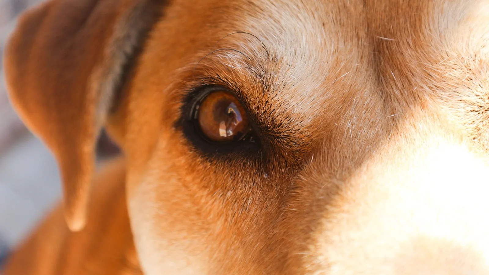
A dog that holds one eye shut is almost always in pain. The single most likely cause is a corneal ulcer, followed by a foreign body trapped under the eyelid or in the conjunctival sac. The first thing to check is whether the eye is suddenly swollen and rock-hard with a wide, unresponsive pupil, because that pattern points to glaucoma and needs same-day veterinary care. This guide is a triage tool. It helps you decide how fast your dog needs to be seen and what the veterinarian will likely do, not what you should do at home.
Squinting in dogs has a specific name. Veterinarians call it blepharospasm, and it is a reflex contraction of the orbicularis oculi muscle that closes the eyelids in response to pain [1][2]. That distinction matters. A dog that keeps an eye closed because it is tired, or because of mild discharge, behaves differently from a dog in true ocular pain. Painful squinting is usually accompanied by tearing, redness of the white of the eye, and reluctance to have the face touched. When you see that combination, treat it as a medical problem, not a cosmetic one.
This article is educational and is not a substitute for veterinary diagnosis or treatment.
Why a Closed Eye Means Pain, Not Just Irritation
Owners often describe a dog closed eye as "just a little weepy" or "maybe he got something in it." The clinical reality is narrower. Blepharospasm is a pain response, and it is one of the most reliable signs a veterinarian uses to localize disease to the surface of the eye [1][2]. In a controlled study of dogs with experimentally created corneal ulcers, squinting and corneal sensitivity were the two measures that changed most consistently with ulceration, and both improved when pain was treated [2].
That is why the presence of a dog eye closed should push you toward a veterinary visit rather than a wait-and-see approach. A dog cannot tell you the eye hurts, so the closed eyelid is the message. Mild conjunctivitis from allergies can cause some squinting, but it rarely produces a fully clenched eye that the dog refuses to open. A fully shut eye is a stronger signal.
There is also a mechanical reason squinting worsens the problem. When the eyelids stay closed, the cornea loses the thin tear film that normally spreads across it with every blink. A dry, unprotected cornea is more vulnerable to infection and to deeper ulceration. Squinting protects the eye from light and touch, but it also removes part of the eye's natural defense.
The Top Causes, From Most Common to Rarest
The list below runs from the conditions a veterinarian sees most often to the ones that are uncommon but serious. Each entry explains the mechanism, because understanding why the eye closes helps you judge urgency.
Corneal Ulcer
A corneal ulcer is a defect in the clear surface of the eye. It is the most common cause of a dog eye closed, and the one most likely to be missed by owners who assume the problem is minor. A large retrospective study of 347 dogs referred for corneal ulceration found that complex ulcers predominated, making up 38.5 percent of cases [3]. Complex ulcers included deep ulcers, ulcers with keratomalacia (melting), descemetoceles (where the ulcer reaches the deepest corneal layer), and corneal lacerations or foreign bodies [3].
The mechanism is straightforward. The cornea is densely supplied with pain receptors. When the surface epithelium is disrupted, those nerve endings are exposed, and every blink scrapes across them. The dog responds by clamping the eye shut. Tearing increases, the white of the eye turns red, and the dog may paw at the face or rub it along furniture.
Breed and age matter. The same study found that brachycephalic breeds had 2.757 times greater odds of presenting for corneal ulceration and 2.695 times greater odds of having a complex ulcer [3]. Shih Tzus were the most common breed for every ulcer type except spontaneous chronic corneal epithelial defects, where Boxers predominated [3]. Each 1 kg decrease in body weight increased the odds of an ulcer diagnosis, and each yearly increase in age raised the odds by 8.9 percent [3]. Older dogs were more likely to develop spontaneous chronic corneal epithelial defects and keratomalacia [3]. Dogs with diabetes mellitus also had increased odds of ulceration [3].
Foreign Body
A foreign body is any material trapped on or in the eye. Plant material is the classic culprit. Grass awns, seeds, and plant debris are the most common objects retrieved from the nasolacrimal sac in dogs with dacryocystitis, a painful inflammation of the tear drainage system [4]. In a large case series of 791 animals with confirmed foxtail foreign bodies, ocular foxtails were the most common location in cats, and foxtails were a recurring cause of ocular disease in dogs as well [5].
Foreign bodies cause a dog eye closed through direct mechanical irritation. A grass awn has a sharp tip and backward-pointing barbs, so it migrates forward and does not back out. It can lodge under the third eyelid, in the conjunctival fornix, in the tear sac, or even penetrate the sclera. One case report described a French Bulldog with a three-day history of conjunctival swelling that progressed to panuveitis and suspected scleral perforation, with a 12 mm grass awn eventually retrieved surgically [6]. Another report described a dog that ran into a thorned succulent plant and developed uveitis, with a thorn tip later found in the inferior iridocorneal angle [7].
The takeaway for owners is that a foreign body can be invisible on the surface. A dog eye closed after running through tall grass, especially in summer, should raise suspicion for a plant foreign body even if you cannot see anything [5].
Uveitis
Uveitis is inflammation of the uvea, the vascular middle layer of the eye. It is painful, and it causes squinting along with a red eye, a cloudy cornea, and often a constricted pupil. Uveitis is important because it can be a sign of disease elsewhere in the body rather than a primary eye problem.
One case report described a 10-month-old dog with sudden vision loss and severe breathing difficulty. Ocular examination found bilateral panuveitis, lens subluxation, secondary glaucoma, and retinal detachment. An intraocular parasite was visible on ultrasound, and the dog was ultimately diagnosed with Angiostrongylus vasorum infection [8]. The authors concluded that this parasite should be on the differential list for ocular uveitis to avoid serious complications from a delayed diagnosis [8].
Uveitis also follows trauma. In the French Bulldog with the migrating grass awn, panuveitis developed within 24 hours of initial medical management [6]. Any penetrating injury to the eye can trigger uveitis, and the inflammation can persist or recur. One case report noted recrudescence of anterior uveitis after medications were stopped, which eventually led to discovery of a retained intra-lenticular foreign body [9].
Glaucoma
Glaucoma is increased pressure inside the eye. It is one of the few true ocular emergencies in veterinary medicine, because sustained high pressure destroys the optic nerve and causes irreversible blindness. The classic presentation is a dog eye closed with a hard, swollen globe, a dilated pupil that does not respond to light, and a cloudy or blue-tinged cornea.
Glaucoma can be primary or secondary. Secondary glaucoma develops after another eye disease, including uveitis and lens instability. In the dog with Angiostrongylus vasorum infection, secondary glaucoma was part of a severe ocular picture that also included panuveitis and retinal detachment [8]. In a study of 33 canine eyes that had intraocular lens fixation surgery, glaucoma was the second most common cause of blindness after retinal detachment [10].
The urgency is different from other causes. A corneal ulcer can usually be managed over days with treatment. Glaucoma can blind an eye in hours. If the eye is suddenly hard, the pupil is wide, and the cornea looks cloudy, that is a same-day emergency.
Entropion and Eyelid Abnormalities
Entropion is a rolling inward of the eyelid margin, so the eyelashes and skin rub directly against the cornea. The constant friction causes pain, tearing, and squinting, and over time it can create corneal ulcers. Entropion is common in breeds with loose facial skin, and it often appears in young dogs as the head matures.
The mechanism is purely mechanical. Every blink drags hair across the corneal surface. The dog responds by squinting, which temporarily reduces contact but does not solve the underlying anatomy. Entropion is usually corrected surgically, and the corneal damage it causes is treated in the meantime.
Less Common Causes
Several other conditions can produce a dog eye closed. Calcareous corneal degeneration, in which mineral deposits form in the cornea, can be associated with corneal ulceration and is treated by chelation with an EDTA solution [11]. Ocular leech infestation, though rare, has been reported and presents as a suspected foreign body, with the leech attached to the conjunctiva near the limbus [12]. Intra-lenticular foreign bodies, embedded in the lens itself, are rare but documented, and one report noted that roughly 50 percent of perforating ocular injuries in dogs involve the lens [9].
Triage Table: Sign, Likely Cause, and Urgency
Use this table to match what you see with how fast your dog needs care. It does not replace an examination, but it helps you decide whether to call for an appointment today or go to an emergency clinic.
| Sign | Likely cause | How it is confirmed | Urgency |
|---|---|---|---|
| Eye shut with tearing and redness, no cloudiness | Corneal ulcer or foreign body | Fluorescein stain, eyelid eversion | Same day |
| Eye shut after running in tall grass or brush | Plant foreign body under eyelid or in tear sac | Eyelid eversion, ultrasound of tear sac | Same day |
| Eye shut with cloudy cornea and red eye | Uveitis | Slit lamp exam, intraocular pressure | Same day |
| Eye shut with hard, swollen globe and wide pupil | Glaucoma | Intraocular pressure measurement | Emergency, same day |
| Eye shut with a visible white or gray defect on the cornea | Deep ulcer or descemetocele | Fluorescein stain, depth assessment | Emergency, same day |
| Eye shut with a melting, gelatinous cornea | Keratomalacia | Fluorescein stain, clinical appearance | Emergency, same day |
| Eye shut with eyelid rolling inward | Entropion | Visual inspection of eyelid margin | Within 24 to 48 hours |
| Eye shut with no redness, no tearing, normal cornea | Possible non-ocular cause | Full ophthalmic and neurologic exam | Within 24 to 48 hours |
| Eye shut with sudden vision loss and breathing difficulty | Systemic disease with ocular involvement | Ocular exam plus systemic workup | Emergency, same day |
The Diagnostic Sequence a Veterinarian Follows
When your dog arrives with a closed eye, the veterinarian works through a logical sequence. Knowing the steps helps you understand why the visit takes time and why certain tests are not optional.
Step 1: History and Visual Inspection
The first step is a detailed history. When did the eye close? Was there trauma, a walk through tall grass, a recent grooming, or a fight with another animal? Has the dog been squinting on and off, or is this the first time? The veterinarian then inspects the eye from a distance before touching it, noting the degree of squinting, the presence of discharge, and whether the eye looks cloudy or swollen.
Step 2: Fluorescein Staining
Fluorescein stain is the single most important test for a dog eye closed. A drop of orange dye is placed on the eye, and the veterinarian examines it under a blue light. Healthy corneal epithelium repels the dye. Where the epithelium is missing, the dye penetrates the underlying stroma and glows bright green. This is how ulcers are detected, and it is how the veterinarian distinguishes a simple ulcer from a complex one.
The test is quick and non-invasive, and it should be done before any medication is placed on the eye. This is also why owners should never apply leftover eye drops before a visit. A steroid-containing drop can worsen an ulcer, and it can also mask the staining pattern.
Step 3: Schirmer Tear Test
The Schirmer tear test measures tear production. A small strip of filter paper is placed in the lower eyelid for one minute, and the distance the tears travel is measured. Low tear production means the cornea is not being adequately lubricated, which predisposes the dog to ulcers and makes healing slower. The test is especially relevant in older dogs and in breeds prone to dry eye.
Step 4: Intraocular Pressure
A tonometer measures the pressure inside the eye. This is the test that identifies glaucoma. Normal pressure is maintained by a balance of fluid production and drainage. When drainage fails, pressure rises, the globe becomes hard, and the optic nerve is damaged. Measuring pressure takes seconds and is essential in any dog with a cloudy cornea, a wide pupil, or a suddenly painful eye.
Step 5: Eyelid Eversion and Foreign Body Search
The veterinarian rolls the upper and lower eyelids to look for foreign material trapped against the conjunctiva. This is where grass awns and seeds hide. If nothing is found on the surface, the tear drainage system may be examined. Ultrasound has been used successfully to identify and guide removal of plant foreign bodies from the lacrimal sac [13]. In one series, ultrasound was positive in four of five affected lacrimal sacs, and all identified foreign bodies were removed successfully [13].
Step 6: Advanced Imaging When Needed
Some foreign bodies cannot be seen on surface examination. High-resolution MRI with a microscopy coil has been used to localize a perforating and migrating grass awn in a dog when ultrasound and conventional MRI failed [6]. This is a referral-level tool, but it shows how far imaging may need to go when a foreign body is suspected but not found.
Decision Flow for a Dog With a Closed Eye
The flowchart below walks through the main decision path, from the moment you notice the eye is shut to the point of veterinary care. It is a triage aid, not a diagnosis.
flowchart TD
    A[Dog eye is shut] --> B{Is the globe hard and swollen}
    B -->|Yes| C[Emergency visit now]
    B -->|No| D{Is the cornea cloudy or blue}
    D -->|Yes| C
    D -->|No| E{Did the dog run through grass or brush}
    E -->|Yes| F[Same day vet visit for foreign body check]
    E -->|No| G{Is there tearing and redness}
    G -->|Yes| H[Same day vet visit for stain and exam]
    G -->|No| I{Is the eyelid rolling inward}
    I -->|Yes| J[Vet visit within 24 to 48 hours]
    I -->|No| K[Vet visit within 24 to 48 hours for full exam]
Why You Should Never Use Steroid Drops Before an Exam
This point deserves its own section because it is the most common and most dangerous mistake owners make. Steroid eye drops reduce inflammation, so they can make a red, squinting eye look better for a day or two. But if the cornea has an ulcer, steroids suppress the immune response that keeps the defect from deepening. They also inhibit corneal healing. An ulcer that might have healed with appropriate treatment can progress to a descemetocele or a perforation when steroids are applied.
The rule is simple. Do not put any eye medication in your dog's eye before a veterinarian has examined it and performed a fluorescein stain. That includes leftover drops from a previous prescription, drops prescribed for another pet, and any human eye preparation. The risk of turning a manageable ulcer into a surgical emergency is too high.
What Treatment Usually Looks Like
Treatment depends entirely on the diagnosis, so this section describes categories rather than specific drugs. Your veterinarian will choose the exact medication and schedule.
For a simple corneal ulcer, the standard approach is a topical antibiotic to prevent infection, sometimes combined with a medication that dilates the pupil to relieve spasm. A study of bacteria isolated from canine corneal ulcers found that Staphylococcus species were most common, with Streptococcus and Pseudomonas also frequent, and that most isolates were sensitive to ciprofloxacin, followed by tobramycin, sulfa drugs, gentamicin, and tetracycline [14]. A separate European study found that 79.5 percent of corneal ulcer samples had positive bacterial growth, with Staphylococcus pseudintermedius and beta-hemolytic Streptococcus as the main organisms [15]. That study reported that 59 percent of isolates were resistant to penicillins, while 71 percent were susceptible to aminoglycosides and fluoroquinolones [15]. Multidrug resistance occurred in 27 percent of isolates [15]. This is why culture and sensitivity testing matters, especially in ulcers that are not healing.
For infected ulcers caused by Staphylococcus pseudintermedius, one study compared multidrug-resistant and non-multidrug-resistant cases. Median time to re-epithelialization was longer in the multidrug-resistant group at 29 days versus 22 days, though the difference was not statistically significant [16]. Follow-up time was significantly longer in dogs with multidrug-resistant keratitis [16].
For deep or melting ulcers, surgery is often required. A study of nine dogs with extensive keratomalacia affecting at least 50 percent of the corneal diameter used a 360-degree conjunctival graft combined with a biomaterial. Successful corneal healing and globe retention was achieved in 8 of 9 eyes, and 7 of 8 preserved globes were visual at the last follow-up [17]. A newer approach using a gelatinized cornea-derived extracellular matrix sealant for stromal ulcers less than 50 percent of corneal thickness showed complete re-epithelialization in 3.9 days versus 5.3 days with conventional therapy alone [18].
For calcareous corneal degeneration with associated ulceration, a chelation procedure using 13.8 percent EDTA solution has been described, with resolution in 26.3 percent of eyes, improvement in 57.9 percent, and recurrence in 15.8 percent at last follow-up [11].
Topical morphine has been studied for corneal pain in dogs. One study found that dogs treated with 1 percent morphine sulfate had significantly less blepharospasm and lower corneal sensitivity readings than controls, and the treatment did not interfere with wound healing [2]. A later study of topical 1 percent morphine in dogs and cats found no measurable analgesic effect against corneal pain [1]. The two studies disagree, so pain management should be discussed with your veterinarian rather than assumed.
Follow-Up and Recheck Timing
Recheck timing depends on the diagnosis. Simple ulcers are typically rechecked in a few days to confirm healing is progressing. Complex ulcers, ulcers that required surgery, and ulcers with multidrug-resistant infection need longer and more frequent follow-up. In the study of multidrug-resistant Staphylococcus pseudintermedius keratitis, follow-up time was significantly longer in the resistant group [16].
If your dog's eye is still shut after 48 hours of appropriate treatment, or if it is more painful than it was at the first visit, call your veterinarian. That pattern suggests the diagnosis was incomplete, the infection is resistant, or a foreign body was missed.
Prevention Practices
Prevention focuses on the causes that can be reduced. After walks in tall grass or brush, check your dog's eyes for seeds, awns, and plant debris. This is especially important in summer, when foxtail foreign bodies are most common [5]. If your dog has a known eye condition such as dry eye or entropion, keep scheduled rechecks so problems are caught before they become painful.
For breeds with a higher risk of corneal ulceration, including brachycephalic dogs and Shih Tzus, be alert to any squinting and seek care early [3]. If your dog has diabetes mellitus, which increases the odds of ulceration, monitor the eyes as part of routine diabetes care [3]. Avoid letting your dog ride with its head out of a car window, and keep thorny plants out of areas your dog explores.
Limitations and When to Contact a Veterinarian
This article cannot diagnose your dog's eye problem. Individual cases vary, and the same sign can have different causes in different dogs. Contact a veterinarian promptly if any of the following apply.
- The eye is suddenly hard, swollen, or cloudy, or the pupil is wide and does not react to light. This suggests glaucoma and needs same-day emergency care.
- The cornea has a visible white, gray, or gelatinous defect. This suggests a deep ulcer, a descemetocele, or keratomalacia, all of which are sight-threatening.
- The eye is bleeding, the globe looks ruptured, or there is a visible wound.
- The dog is pawing at the eye, crying, or reluctant to eat.
- The eye has been shut for more than 24 hours without improvement.
- The dog has a known foreign body that has not been removed.
- The dog has sudden vision loss, especially with breathing difficulty, which may indicate systemic disease with ocular involvement [8].
- The dog has a history of recent eye surgery, including intraocular lens placement, since glaucoma and retinal detachment are recognized complications [10].
Frequently Asked Questions
Should I wait to see if my dog's eye opens on its own?
No. A dog eye closed is a pain sign, and most causes do not resolve without treatment. Waiting risks progression of an ulcer or a missed foreign body.
What is the most common cause of a dog holding one eye shut?
A corneal ulcer is the most common cause. Foreign bodies and uveitis are also common, and glaucoma is the most urgent.
Can I use human eye drops on my dog?
No. Some human drops contain steroids, which can worsen a corneal ulcer. Do not apply any eye medication before a veterinarian has examined the eye.
How does a veterinarian tell if it is an ulcer?
Fluorescein stain is the standard test. The dye glows green where the corneal surface is missing.
What does a hard, cloudy eye with a wide pupil mean?
This pattern suggests glaucoma, which is an emergency. High pressure can damage the optic nerve within hours.
Can a grass awn cause a dog's eye to stay closed?
Yes. Grass awns and other plant material can lodge under the eyelid or in the tear sac and cause significant pain and inflammation [4][13].
Is entropion painful for dogs?
Yes. Entropion rolls the eyelid inward so hair rubs the cornea with every blink, causing pain and sometimes ulcers.
When should I go to an emergency clinic instead of my regular vet?
Go to an emergency clinic if the eye is hard and swollen, the cornea is cloudy, the pupil is wide, or there is a visible deep defect. These signs suggest glaucoma or a perforating ulcer, both of which are sight-threatening.
Related Articles
- Cat Eye Discharge: Causes and When to See a Vet
- Dog Eye Redness: When to See a Vet and When to Monitor
- Cat Eye Injury: When to See a Vet Immediately
- Cat Itchy Skin: Causes and When to See the Vet
- Cat Scooting: Causes and When to Call the Vet
- Dog Head Tilt: Causes and When to See a Vet
- Dog Pink Eye: Causes, Symptoms, and Treatment
- Dog Eye Ulcer: Symptoms, Causes and Treatment
- Bloodshot Eyes in Dogs: Causes and Hyphema Care
Sources
- Preliminary investigations into the analgesic effects of topical ocular 1% morphine solution in dogs and cats.
- Effect of topical administration of 1% morphine sulfate solution on signs of pain and corneal wound healing in dogs.
- Evaluation of corneal ulcer type, skull conformation, and other risk factors in dogs: A retrospective study of 347 cases.
- Nasolacrimal sac foreign body extraction using vitreoretinal forceps in 28 dogs.
- Clinical and clinicopathological characteristics, treatment, and outcome for dogs and cats with confirmed foxtail foreign body lesions: 791 cases (2009-2018).
- The localization of a conjunctivoscleral foreign body via high-resolution microscopy coil magnetic resonance imaging in a dog.
- Removal of an anterior chamber foreign body using hydropulsion and ophthalmic viscoelastic device capture in a dog.
- Irreversible Ocular Lesions in a Dog With Angiostrongylus Vasorum Infection.
- Diagnosis, management, and outcome of an intralenticular foreign body in a dog: A case report.
- The clinical outcomes of 33 canine eyes following the novel sulcus fixation technique of an injectable acrylic lens through a 3-mm corneal incision.
- Dissolution of presumed calcareous corneal degeneration with 13.8% ethylenediaminetetraacetic acid solution in 17 dogs.
- Topical hypertonic saline as a treatment for ocular and nasal hirudiniasis in a dog.
- Ultrasonography-guided removal of plant-based foreign bodies from the lacrimal sac in four dogs.
- Identification and antimicrobial susceptibility of bacteria isolated from corneal ulcers and healthy eyes of canines in Ibagué-Tolima, Colombia.
- Patterns of Bacterial Infection and Antibiotic Resistance in Canine Corneal Ulcers: A Retrospective Analysis (Berlin, Germany, 2021-2024).
- Impact of multi-drug resistance on clinical outcomes of dogs with corneal ulcers infected with Staphylococcus pseudintermedius.
- The Use of 360-Degree Conjunctival Graft and Biomaterials in the Treatment of Extensive Corneal Ulceration and Keratomalacia in Dogs: Nine Cases.
- Novel Non-Invasive Treatment for Canine Corneal Ulcers: Application of New Extracellular Matrix-Based Sticky Sealants.