Epizootic Hemorrhagic Disease in Deer: Signs and Spread
By Dr. Zubair Khalid, DVM, MS, PhD ·
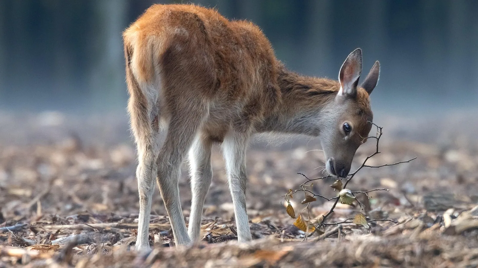
Epizootic hemorrhagic disease (EHD) in deer is a noncontagious, insect-borne viral illness caused by epizootic hemorrhagic disease virus (EHDV), an Orbivirus spread by Culicoides biting midges rather than by deer-to-deer contact [1][2]. White-tailed deer are the most severely affected species, and outbreaks cluster in late summer and early fall, ending with the first hard frost that kills adult midge activity [3][2].
This article explains the biology of EHDV, the clinical signs owners and wildlife managers see in the field, the seasonal and geographic patterns of spread, and how to tell EHD apart from bluetongue virus (BTV), a separate Orbivirus with overlapping vectors and similar lesions [3][4]. This article is educational and is not a substitute for veterinary diagnosis or treatment.
What Causes Epizootic Hemorrhagic Disease in Deer?
EHDV is an arthropod-borne RNA virus in the genus Orbivirus, family Sedoreoviridae [1]. Globally, seven known serotypes circulate among ruminant hosts and Culicoides vectors [1]. In North America, three serotypes (1, 2, and 6) are considered endemic, and all three were detected in both animals and midge pools during a single zoo outbreak in Minnesota [5].
The virus is not spread by direct contact between deer. It requires a biological vector: a female Culicoides midge takes a blood meal from an infected ruminant, the virus replicates in the midge, and a later bite inoculates a new host [6][2]. In North America, the only confirmed vector of EHDV is Culicoides sonorensis, although other Culicoides species are suspected vectors [6]. Field surveillance during the Minnesota Zoo outbreak detected EHDV RNA in pools of C. sonorensis, C. variipennis, and degraded C. variipennis complex midges [5]. Culicoides stellifer is a suspected vector in the southeastern United States, with field evidence from Florida [7].
Transmission efficiency is high. Experimental work showed that the bite of as few as five EHDV-infected C. sonorensis can infect a white-tailed deer, and all inoculated midges tested positive by virus isolation except one that likely failed intrathoracic inoculation [6]. This means a relatively small number of infectious midges in the right place at the right time can seed an outbreak.
The Vector and the Deer Host
Culicoides midges are small, blood-feeding flies sometimes called no-see-ums. They breed in moist soil, mud, and decaying organic matter, and their activity is tied to warm, humid conditions [4]. A study at a Brazilian zoological park found higher Culicoides prevalence during summer months, especially February, with elevated temperature, high humidity, fecal accumulation, and proximity to large animals associated with increased captures [4]. Culicoides insignis made up 75% of collected midges and C. pusillus 6%, both described as competent Orbivirus vectors, and C. insignis was shown to feed on deer and red deer [4].
White-tailed deer are highly susceptible to severe and often fatal EHD, while domestic cattle are important hosts in which inapparent infection is the most common outcome [1]. Fatal infection of cattle is rare [1]. This difference in species susceptibility is central to how EHD behaves at the wildlife-livestock interface [1].
Host genetics also matter. In Illinois wild white-tailed deer, researchers sequenced the entire coding region of the Toll-like receptor 3 (TLR3) gene, which helps the immune system detect double-stranded RNA viruses like EHDV [8]. They identified 85 haplotypes with 77 single nucleotide polymorphisms, and two non-synonymous SNPs differed significantly in frequency between EHD-positive and EHD-negative deer [8]. Both amino acid substitutions were predicted to affect protein structure or function, suggesting that some deer carry genetic variants that influence outbreak severity [8].
How Epizootic Hemorrhagic Disease Spreads


EHDV spreads through a vector-host-vector cycle, not through deer-to-deer contact, contaminated feed, or carcass handling [1][2]. The virus moves between ruminant hosts when infected Culicoides midges feed. Because midges are weak fliers that depend on wind, temperature, and humidity, the geographic footprint of an outbreak follows midge habitat and weather rather than deer movement alone.
The seasonal pattern is consistent. Outbreaks occur from late summer into early fall and stop with the first hard frost, when adult midge activity declines [3][2]. This is why EHD is often called a "seasonal hemorrhagic disease" and why carcasses tend to be found near water, where sick deer go to cool down and where midges breed.
The following flowchart summarizes the transmission cycle and the key decision points for a suspected case.
flowchart TD
    A[Infected ruminant] --> B[Midge takes blood meal]
    B --> C[Virus replicates in midge]
    C --> D[Infectious midge bites deer]
    D --> E[Incubation period]
    E --> F{Susceptible host}
    F --> G[White tailed deer severe disease]
    F --> H[Cattle mild or no disease]
    G --> I[Sudden death or acute signs]
    I --> J[Report and collect samples]
    J --> K[Test for EHDV and BTV]
    K --> L[Confirm diagnosis]
Geographic Spread and Emerging Patterns
EHDV has been detected globally across tropical and temperate regions, largely corresponding to the presence of Culicoides biting midges [2]. In North America, a 38-year survey of hemorrhagic disease reports in the Great Plains (North Dakota, South Dakota, Nebraska, Kansas, and Oklahoma) found that reports expanded northeast across latitude and longitude between 1982 and 2020 [3]. Reporting intensity increased significantly in three of the five states (North Dakota, South Dakota, and Kansas), and reporting frequency increased in all five states [3]. The authors noted that such changes in northern latitudes might lead to increased deer mortality in regions where epizootics have historically been less frequent [3].
EHDV-8 emerged in Europe in autumn 2022, first detected in Spain and later in Italy and Portugal [9][10][11]. In Spain, EHDV-8 caused severe disease in European red deer, with hyperacute to acute clinical signs and lesions related to vascular changes leading to death [9]. A cross-sectional serological study of 592 wild ruminants, mainly red deer, in southwestern Spain found anti-EHDV antibodies in 37 of 592 samples (6.3%), all from red deer and all from localities where clinical cases were confirmed [9]. This indicated limited spread in the first year of detection [9].
Phylodynamic analysis of Mediterranean EHDV suggests the region's strains share an ancestral root with North American strains that circulated in the 17th century, with the first incursions in France and Tunisia during the 1800s [12]. Significant transition routes were detected between North African and European countries [12].
Clinical Signs of Epizootic Hemorrhagic Disease in Deer
EHD can present as peracute, acute, or chronic disease, and the signs differ by species and individual susceptibility [1]. In white-tailed deer, the classic field presentation is sudden death, sometimes with no preceding signs, or an acute illness lasting days.
Common clinical signs in white-tailed deer include fever, swollen tongue and head, oral ulcers, and hoof sloughing [1]. The swollen tongue and head reflect vascular damage and edema. Oral ulcers and hoof lesions develop as the disease progresses. In the field, affected deer are often found near water, weak, depressed, and reluctant to move.
In European red deer infected with EHDV-8, frequent clinical signs included neurological signs, ptyalism (excessive salivation), lameness, and dyspnoea [11]. A 2023 outbreak in Sierras de Cazorla, Segura y Las Villas Natural Park in Spain described a consistent acute neuro-respiratory syndrome characterized by weakness, ataxia, nystagmus, and severe dyspnoea with frothy oral discharge [13]. During that outbreak, average morbidity and mortality in captive red deer were 6.9% and 4.9%, respectively, with a case fatality rate of 70.3%, higher in males and almost exclusively in adults [11].
In captive reindeer, an outbreak in September 2020 was associated with neurological signs and mortality. Four reindeer died or were euthanized after acute illness over a 12-day period, displaying abnormal behavior, neurologic signs, lethargy, and lameness [14].
In cattle, EHD is traditionally considered less severe, but clinical disease has increased. A study of 29 cattle farms in northwestern Spain during the 2022 emergence estimated an incidence of farms with clinical signs of 6.2% (n=467), with mean incidence of clinical signs, mortality, and lethality rates of 8.9%, 1.7%, and 23.0%, respectively [15]. Oral lesions, dysphagia, and hoof problems were common, and tongue edema, dehydration, and dyspnoea were associated with higher lethality [15]. Mortality and lethality were significantly higher in dairy cattle than in beef [15]. In a separate study of Spanish Holstein dairy cattle during the 2023 outbreak, about 66% of animals showed EHDV antibodies, only 25% showed severe symptoms, and the death rate was 2% to 3%. Age was the main risk factor for severe EHD, with older cows more susceptible, and production losses reached up to -9 kg/d in cows with moderate to severe symptoms, especially if the outbreak occurred mid-lactation [16].
Lesions and Necropsy Findings
Gross and microscopic lesions of EHD reflect widespread vascular injury. In white-tailed deer, field investigations commonly show hemorrhages and pulmonary edema [1]. A basic necropsy examination is an essential part of confirming an outbreak, and diagnostic sample collection should target spleen, lung, and affected tissues [1].
In European red deer infected with EHDV-8, gross examination revealed extensive vascular and epithelial lesions with generalized congestion, focal to multifocal hemorrhages, and marked pulmonary edema [11]. In the Spanish outbreak involving red deer, fallow deer, and mouflon, gross lesions included marked pulmonary edema, tracheal foam, and widespread congestion, while histopathology revealed lymphoid depletion, pulmonary hemorrhage, vascular injury, and renal tubular necrosis [13]. All animals tested positive for EHDV-8 with low RT-qPCR cycle threshold values, indicating high viral loads [13].
In captive reindeer, the most consistent gross finding was dark red streaks throughout the adrenal gland cortices (4/4). One animal had acute hemorrhage involving the subcutis and skeletal muscles over the ventrolateral body wall and back, and abomasal serosa. Histologically, the most common lesions were adrenal gland cortical hemorrhage (4/4) with necrosis (3/4) and lymphoplasmacytic meningoencephalitis with gliosis, glial nodules, satellitosis, and nonsuppurative perivascular cuffing (4/4). Brain lesions were most frequent in the gray matter of the cerebrum, hippocampus, and thalamus but also involved the cerebellum and brainstem [14].
EHDV vs Bluetongue Virus: Key Differences
Hemorrhagic disease of deer can be caused by either EHDV or bluetongue virus (BTV), and the two are often grouped together in field surveillance because they produce overlapping signs and share vectors [3]. Both are Orbiviruses transmitted primarily by Culicoides spp. [4]. Distinguishing them matters for diagnosis, reporting, and understanding outbreak dynamics.
The table below compares the two viruses by vector, hosts, and lesions.
| Feature | Epizootic Hemorrhagic Disease Virus (EHDV) | Bluetongue Virus (BTV) |
|---|---|---|
| Genus and family | Orbivirus, Sedoreoviridae [1] | Orbivirus, Reoviridae [4] |
| Primary vectors | Culicoides sonorensis confirmed in North America, other Culicoides suspected [6] | Culicoides spp., including C. insignis and C. pusillus in some regions [4] |
| Highly susceptible wildlife host | White-tailed deer, often severe and fatal [1] | White-tailed deer, also susceptible [3] |
| Cattle | Important host, usually inapparent, fatal infection rare [1] | Cattle can be affected, severity varies |
| Key lesions | Hemorrhages, pulmonary edema, vascular injury, lymphoid depletion [11][13] | Overlapping hemorrhagic and vascular lesions [3] |
| Seasonal pattern | Late summer to first frost, tied to midge activity [3][2] | Similar seasonal pattern, tied to midge activity [4] |
Both viruses circulate in the same ecosystems and can co-infect hosts. A bontebok at the Nashville Zoo was found to have coinfection of West Nile virus and EHDV, with meningoencephalitis and spinal myelitis [17]. This case illustrates that exotic zoo animals can be susceptible to endemic arboviruses and reinforces the value of cooperative surveillance among human, wildlife, and domestic animal health agencies [17].
Diagnosis and Reporting
Diagnosis of EHD relies on a combination of field signs, necropsy findings, and laboratory testing. Routine EHD diagnostics do not accurately quantify infectious virus, which would allow prediction of onward transmission risk, and are typically qualitative (virus isolation) or quantify viral genome copies (real-time PCR) that can remain detectable long after infectious virus is cleared [18]. Infectious EHDV titers are usually quantified through visible cytopathic effect in susceptible mammalian cell cultures, though not all susceptible cell lines show visible CPE, including KC cells derived from the EHDV biological insect vector, Culicoides sonorensis [18].
For field investigations, the necropsy examination is essential, and diagnostic sample collection should include spleen and affected tissues [1]. PCR and sequencing can confirm the serotype, as was done in the reindeer outbreak where EHDV-6 was detected through PCR and sequencing of the spleen in all cases [14]. In the Spanish red deer outbreak, partial sequences of VP2 and NS1 genes had greater than 99% nucleotide identity with EHDV-8 sequences from Tunisia and Italy [9].
EHD is a notifiable disease to the World Organisation for Animal Health [11]. In the United States, state wildlife agencies and veterinary diagnostic laboratories typically handle EHDV testing and reporting. The AVMA and state animal health officials can provide guidance on reporting requirements when an outbreak is suspected.
Treatment and Management
There is no specific antiviral treatment for EHD in deer. Management focuses on supportive care for valuable captive animals, vector control, and reducing exposure. The review of EHDV control notes a pressing need to identify new methods to prevent and mitigate outbreaks and reduce impacts on livestock and wildlife, including development of new investigative tools and progress in virus detection, disease mitigation, and vector control [2].
For captive deer, reindeer, and zoo ruminants, veterinarians may provide supportive care such as fluids, anti-inflammatory drugs, and wound care for hoof lesions, but outcomes depend on the severity of vascular damage. In the reindeer outbreak, affected animals died or were euthanized after acute illness [14]. In red deer, the case fatality rate was 70.3% [11].
Vector control is the primary preventive strategy. Reducing standing water, managing organic matter, and using approved insecticides in captive settings can lower midge populations. The Brazilian study found that fecal accumulation and proximity to large animals were associated with increased Culicoides capture, suggesting that sanitation and animal placement matter [4]. In Florida, midge abundance was positively associated with sites closer to large-animal feeders, and C. stellifer preferred mixed bottomland hardwood and agricultural, sand, and water habitats [7].
Vaccination is not widely available for wildlife. The control review discusses progress in basic and applied research focused on virus detection, disease mitigation, and vector control, but no licensed vaccine for wild deer is described [2].
Outbreak Patterns and Surveillance
EHD outbreaks are episodic and weather-dependent. The Great Plains survey documented that reports expanded northeast and increased in intensity and frequency over 38 years, with implications for increased deer mortality in northern regions where epizootics were historically less frequent [3]. This pattern is consistent with changing midge habitat and climate.
Zoos can serve as sentinels for re-emerging arboviruses. During a fall 2020 EHD outbreak at the Minnesota Zoo, insect surveillance detected EHDV in six pools of Culicoides biting midges, including three pools of C. sonorensis, two pools of C. variipennis, and a pool of degraded C. variipennis complex midges. All three endemic serotypes (1, 2, and 6) were detected in both animals and midge pools from the premises, yet no EHDV cases had been reported in wild animals near the zoo [5]. This highlights the value of animal holding facilities as sentinels for spatio-temporal pathogen dynamics [5].
In Europe, the emergence of EHDV-8 in 2022 and 2023 led to thousands of clinical outbreaks in cattle herds in mainland France, with a strong south-north seroprevalence gradient. The highest animal-level seroprevalence was in the southernmost zone at 82.6%, compared with 11.6% and 0.3% in more northern areas where clinical outbreaks were less frequent [19]. This gradient reflects the dependence of transmission on vector abundance and climate.
Limitations and When to Contact a Veterinarian
This article is educational and is not a substitute for veterinary diagnosis or treatment. Individual cases require a veterinarian, and wildlife cases should be reported to the appropriate state or provincial wildlife agency.
Contact a veterinarian promptly if you observe any of the following in captive deer, reindeer, or other ruminants:
- Sudden death in one or more animals, especially in late summer or early fall
- Fever, swollen tongue, swollen head, or oral ulcers
- Lameness, hoof swelling, or hoof sloughing
- Neurological signs such as ataxia, nystagmus, abnormal behavior, or weakness
- Severe respiratory distress, frothy oral discharge, or dyspnoea
- Excessive salivation or difficulty swallowing
For free-ranging deer, contact your state wildlife agency rather than a private veterinarian. Do not handle sick or dead wild deer without appropriate personal protective equipment, and follow agency guidance on carcass disposal and sample collection. EHDV is not known to infect humans, but other pathogens can be present, and safe handling practices protect you and your animals.
Frequently Asked Questions
What causes epizootic hemorrhagic disease in deer?
EHD in deer is caused by epizootic hemorrhagic disease virus, an Orbivirus in the family Sedoreoviridae, transmitted by Culicoides biting midges [1][2]. It is not spread by deer-to-deer contact.
Is epizootic hemorrhagic disease contagious to other deer?
No. EHDV requires a Culicoides midge vector and does not spread directly between deer [1][2]. Outbreaks end with the first hard frost when midge activity stops [3].
What are the first signs of EHD in deer?
The first signs are often sudden death or an acute illness with fever, swollen tongue and head, oral ulcers, and reluctance to move [1]. Some deer are found dead near water with no observed preceding signs.
Can cattle get epizootic hemorrhagic disease?
Yes, cattle can be infected, but inapparent infection is the most common outcome and fatal infection is rare [1]. Clinical disease has increased in some regions, with oral lesions, dysphagia, and hoof problems reported [15].
How is EHD different from bluetongue?
EHDV and BTV are separate Orbiviruses with overlapping Culicoides vectors and similar hemorrhagic lesions [3][4]. Laboratory testing is needed to tell them apart because field signs alone cannot distinguish them.
Does EHD affect humans?
EHDV is not a human pathogen. The virus infects ruminants and is transmitted by midges, and there is no evidence of human infection from deer or midges.
What should I do if I find a dead deer in late summer?
Contact your state wildlife agency. Do not handle the carcass without protective equipment. Agency staff can collect samples and determine whether EHDV or another cause is responsible.
Is there a vaccine for epizootic hemorrhagic disease?
No licensed vaccine for wild deer is described in the current literature. Control relies on vector management, surveillance, and supportive care for captive animals [2].
Related Articles
- Rabbit Hemorrhagic Disease (RHDV2): Prevention and Outbreak Management
- Chronic Wasting Disease Deer Prion Diagnostics Surveillance
- Rabbit Biosecurity for Housing: Preventing Disease Introduction and Spread
- Epizootic Haematopoietic Necrosis Virus (EHNV)
- Chronic Wasting Disease in Deer: Prion Biology and Diagnostic Challenges in Wildlife
- Model Validation and Calibration for Animal Disease Spread
- Bullseye Rash: Tick Bite and Lyme Disease Signs
Sources
- Recognition of Field Signs, Necropsy Procedures, and Evaluation of Macroscopic Lesions of Cervids Infected with Epizootic Hemorrhagic Disease Virus.
- Perspectives on the Changing Landscape of Epizootic Hemorrhagic Disease Virus Control.
- Patterns of Hemorrhagic Disease in White-Tailed Deer (Odocoileus virginianus) in the Great Plains of the USA, 1982-2020.
- The Study of Bluetongue Virus (BTV) and Epizootic Hemorrhagic Disease Virus (EHDV) Circulation and Vectors at the Municipal Parks and Zoobotanical Foundation of Belo Horizonte, Minas Gerais, Brazil (FPMZB-BH).
- Using Zoos as Sentinels for Re-Emerging Arboviruses: Vector Surveillance during an Outbreak of Epizootic Hemorrhagic Disease at the Minnesota Zoo.
- Apparent transmission efficiency of Culicoides sonorensis (Diptera: Ceratopogonidae) for epizootic hemorrhagic disease virus serotype 2 in white-tailed deer (Odocoileus virginianus).
- Modeling Abundance of Culicoides stellifer, a Candidate Orbivirus Vector, Indicates Nonrandom Hemorrhagic Disease Risk for White-Tailed Deer (Odocoileus virginianus).
- The Impact of Variation in the Toll-like Receptor 3 Gene on Epizootic Hemorrhagic Disease in Illinois Wild White-Tailed Deer (Odocoileus virginianus).
- Emergence of epizootic hemorrhagic disease in red deer (Cervus elaphus), Spain, 2022.
- Emergence of epizootic hemorrhagic disease in Europe.
- Unveiling the clinical signs and pathology in red deer (Cervus elaphus) naturally infected with epizootic haemorrhagic disease virus serotype 8.
- Emergence and evolution of epizootic hemorrhagic disease virus in the Mediterranean region: spatio-temporal dynamics and epidemiological insights.
- Epizootic Haemorrhagic Disease Virus (EHDV) Infection in Red Deer (Cervus elaphus), Fallow Deer (Dama dama) and Mouflon (Ovis orientalis musimon) in South-Eastern Spain: Implications for Wildlife Health and Ruminant Disease Ecology.
- Outbreak of epizootic hemorrhagic disease in captive reindeer (Rangifer tarandus).
- Description of the Clinical Findings Associated With the Epizootic Hemorrhagic Disease in Cattle From Northwestern Spain During the Emergence.
- Epidemiological and genetic factors affecting severe epizootic hemorrhagic disease in Spanish Holstein cattle during the Southern Europe outbreak of 2023.
- West Nile Virus and Epizootic Hemorrhagic Disease Virus Co-Infection in a Novel Host at the Nashville Zoo.
- Epizootic Hemorrhagic Disease Virus Titration.
- Heterogenous within-herd seroprevalence against epizootic hemorrhagic disease virus type 8 (EHDV-8) after massive virus circulation in cattle in France, 2023.