
Chronic Wasting Disease in Deer: Prion Biology and Diagnostic Challenges in Wildlife
Introduction
Chronic wasting disease (CWD) is a progressive, fatal neurodegenerative disorder affecting members of the family Cervidae, including white-tailed deer (Odocoileus virginianus), mule deer (Odocoileus hemionus), elk (Cervus canadensis), and moose (Alces alces). The disease is caused by the accumulation of a misfolded isoform (PrPSc) of the host-encoded cellular prion protein (PrPC). Unlike conventional infectious agents such as bacteria or viruses, prions lack nucleic acids and propagate solely through conformational templating. This unique biology presents profound challenges for ante-mortem diagnosis, surveillance in free-ranging populations, and management of captive herds. Although CWD is classified under wildlife-bacteria surveillance frameworks in some diagnostic portals due to its transmissible nature and reportable status, it is essential to clarify that prions are proteinaceous infectious particles and not bacterial organisms. This review provides an exhaustive examination of prion biology, the biophysical basis of misfolding, current diagnostic platforms, and the specific obstacles encountered when applying these technologies to wildlife populations.
Prion Biology and Molecular Mechanisms
Structure of the Cellular Prion Protein
The cellular prion protein (PrPC) is a glycosylphosphatidylinositol (GPI)-anchored glycoprotein expressed predominantly on the surface of neurons and cells of the lymphoid and reticuloendothelial systems. In cervids, the PRNP gene encodes a protein of approximately 253 amino acids. The mature PrPC contains a flexible N-terminal domain with octapeptide repeats and a globular C-terminal domain composed primarily of alpha-helices. Under physiological conditions, PrPC is soluble, sensitive to proteolytic digestion, and has a high content of alpha-helical secondary structure (approximately 42%) with minimal beta-sheet content (approximately 3%) [1, 2].
Conformational Conversion to PrPSc
The pathogenic isoform, PrPSc, is derived from PrPC through a post-translational conformational rearrangement. In PrPSc, the alpha-helical content is substantially reduced, and the beta-sheet content increases to approximately 43% [3]. This conversion is autocatalytic: PrPSc acts as a template that induces the misfolding of additional PrPC molecules. The process follows a nucleation-polymerization model in which small oligomeric seeds of PrPSc elongate by recruiting monomeric PrPC [4]. The resulting aggregates are insoluble, partially resistant to proteinase K digestion, and accumulate in the central nervous system (CNS) and lymphoid tissues.
Strain Diversity and Species Barriers
Prion strains are defined by distinct conformational variants of PrPSc that produce characteristic patterns of neuropathology, incubation periods, and biochemical properties (e.g., glycosylation profile, protease resistance, and conformational stability). In CWD, multiple strains have been identified in both captive and free-ranging cervids [5, 6]. Strain diversity complicates diagnostic detection because different conformers may exhibit variable affinity for antibodies used in immunoassays or differential amplification efficiency in in vitro conversion assays. The species barrier, which limits cross-species transmission, is influenced by the degree of sequence homology between the donor PrPSc and the recipient PrPC. CWD has been experimentally transmitted to non-cervid species including voles, hamsters, and transgenic mice expressing cervid PRNP, but natural transmission to livestock or humans has not been conclusively demonstrated [7, 8].
Pathogenesis and Tissue Distribution
Routes of Infection and Peripheral Pathogenesis
Natural transmission of CWD occurs horizontally through direct contact with infected animals or indirectly through environmental contamination. PrPSc is shed in saliva, urine, feces, and placental tissues [9, 10]. The oral route is considered the primary mode of transmission. Following oral exposure, PrPSc is taken up by gut-associated lymphoid tissues, including Peyer's patches, and subsequently disseminates to peripheral lymph nodes, tonsils, and the spleen [11]. This extraneural replication phase is critical for diagnostic sampling because lymphoid tissues become positive months to years before the onset of clinical signs.
Neuroinvasion and CNS Pathology
Neuroinvasion occurs via peripheral nerves, primarily the vagus and splanchnic nerves, which transport PrPSc from the enteric nervous system to the dorsal motor nucleus of the vagus in the medulla oblongata [12]. From the brainstem, the infection spreads rostrally to the thalamus, hypothalamus, and cerebral cortex. Histopathologic hallmarks include spongiform vacuolation, neuronal loss, astrogliosis, and microglial activation. The accumulation of PrPSc in the CNS correlates with the severity of clinical signs, which include progressive weight loss, behavioral changes, ataxia, and excessive salivation.
Prion Shedding and Environmental Persistence
A distinguishing feature of CWD is the remarkable persistence of PrPSc in the environment. Prions bind tightly to soil particles, particularly clay minerals such as montmorillonite, and retain infectivity for years [13, 14]. This environmental reservoir complicates eradication efforts because contaminated premises remain infectious even after depopulation. The mechanisms of prion-soil interaction involve electrostatic and hydrophobic forces, and bound PrPSc may exhibit enhanced resistance to proteolytic degradation [15].
Diagnostic Platforms for CWD
Immunohistochemistry (IHC)
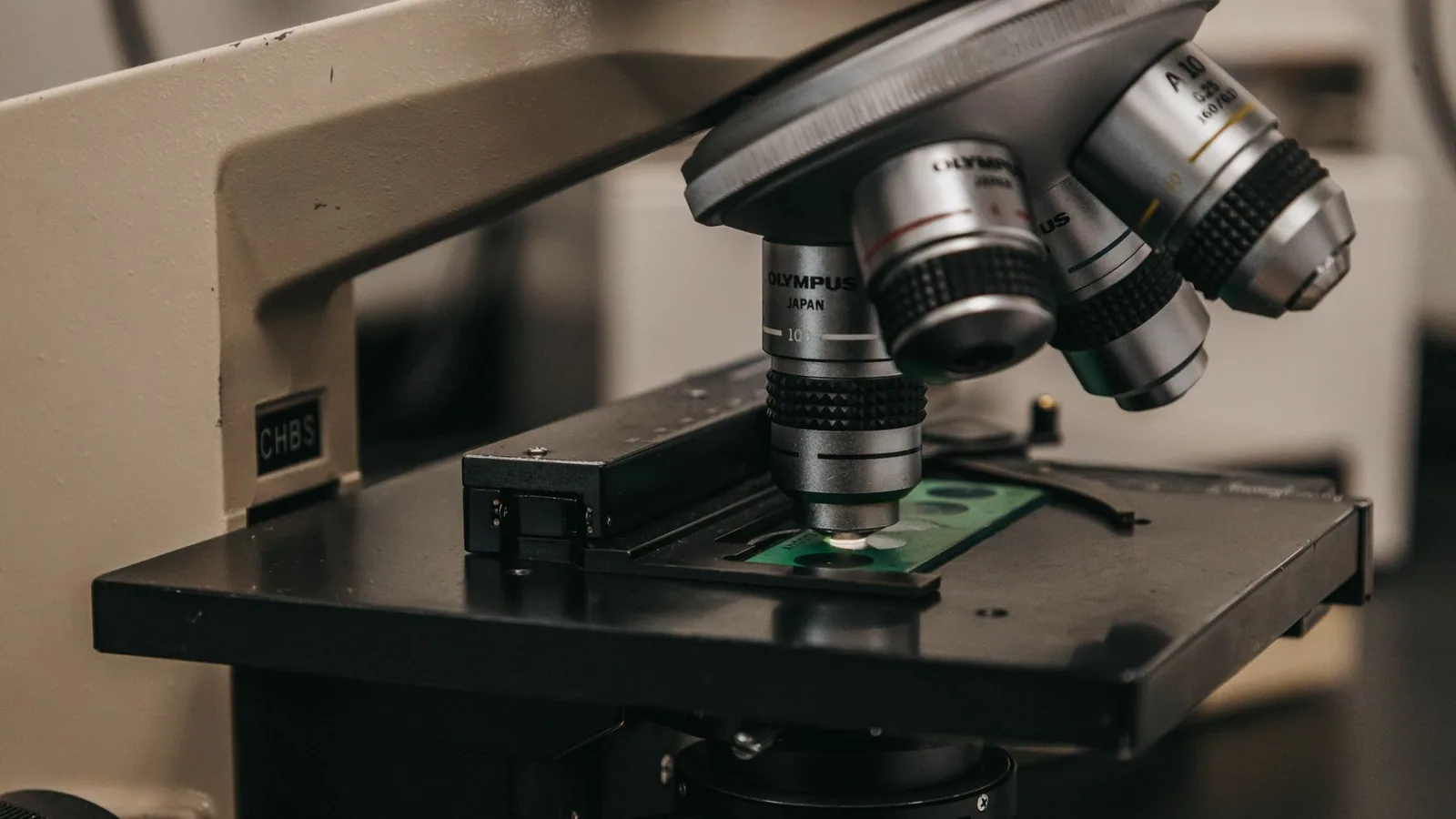
Immunohistochemistry on formalin-fixed, paraffin-embedded tissues remains the gold standard for confirmatory diagnosis of CWD. The recommended target tissues are the obex (medulla oblongata at the level of the obex) and retropharyngeal lymph nodes [16]. Monoclonal antibodies directed against prion protein epitopes (e.g., F99/97.6.1, BAR224) are used to detect PrPSc after antigen retrieval and proteinase K digestion. IHC provides high specificity and allows visualization of the anatomical distribution of PrPSc deposition. However, the technique is labor-intensive, requires specialized training, and is not suitable for high-throughput screening of large numbers of samples.
Enzyme-Linked Immunosorbent Assay (ELISA)
Several commercial ELISA kits have been developed for CWD detection in cervid tissues. These assays typically use a combination of capture and detection antibodies that recognize epitopes exposed only in the misfolded conformation of PrPSc. The ELISA format offers higher throughput and shorter turnaround times compared to IHC. Sensitivity and specificity estimates for ELISA on retropharyngeal lymph node samples exceed 95% in experimentally infected animals [17, 18]. However, performance may decline in free-ranging populations with low prevalence or early-stage infections. False positives can occur due to cross-reactivity with PrPC if the proteinase K digestion step is incomplete. The diagnostic workflow for ELISA is analogous to that described for Enzyme-Linked Immunosorbent Assay (ELISA) for Feline Leukemia Virus, although the target analyte differs fundamentally.
Western Blotting
Western blotting is used as a confirmatory method to detect proteinase K-resistant PrPSc. Following electrophoresis and transfer to a membrane, PrPSc is detected using anti-prion antibodies. The characteristic banding pattern reflects the di-, mono-, and unglycosylated isoforms of PrPSc. Western blotting can differentiate CWD from other transmissible spongiform encephalopathies based on molecular mass and glycoform ratios [19]. The technique is semi-quantitative but is not practical for large-scale surveillance due to low throughput and technical complexity.
Real-Time Quaking-Induced Conversion (RT-QuIC)
RT-QuIC is a highly sensitive in vitro amplification assay that exploits the autocatalytic conversion of recombinant PrPC substrate by PrPSc seeds present in the test sample. The reaction is performed in a microtiter plate format, and amyloid formation is monitored in real time using the fluorescent dye thioflavin T [20, 21]. RT-QuIC has been applied to many sample types including brain tissue, lymph nodes, rectal mucosa, nasal brushings, saliva, urine, and feces [22, 23]. The assay can detect as little as femtogram quantities of PrPSc, making it the most sensitive ante-mortem diagnostic tool currently available.
The RT-QuIC protocol for CWD typically uses recombinant cervid PrPC substrate (amino acids 23-231) expressed in Escherichia coli. Reaction conditions include incubation at 50 degrees Celsius with intermittent shaking (double-orbital shaking at 700 rpm for 60 seconds, followed by 60 seconds of rest). Positive samples produce a sigmoidal increase in fluorescence, with the lag phase inversely proportional to the seed concentration [24]. The specificity of RT-QuIC approaches 100% when performed on brain tissue, but false positives due to spontaneous substrate aggregation can occur, particularly with prolonged incubation times.
Protein Misfolding Cyclic Amplification (PMCA)
PMCA is an alternative amplification technique that uses sonication to break PrPSc aggregates into smaller seeds, thereby accelerating the conversion process. The assay uses brain homogenate from transgenic mice expressing cervid PrPC as a substrate. PMCA has been used to detect PrPSc in blood, urine, and feces from experimentally infected deer [25, 26]. The sensitivity of PMCA is comparable to or exceeds that of RT-QuIC for certain sample types, but the requirement for animal-derived substrate limits standardization and scalability.
Ante-Mortem Sampling Strategies
Ante-mortem diagnosis of CWD in live animals is essential for surveillance and management of captive herds. Rectal mucosa-associated lymphoid tissue (RAMALT) biopsy is a minimally invasive procedure that collects lymphoid follicles from the rectal wall. PrPSc can be detected in RAMALT samples by IHC or RT-QuIC months before clinical onset [27]. Nasal brushings have also been evaluated as a sampling method, with RT-QuIC achieving high sensitivity in experimentally infected deer [28]. Third eyelid (nictitating membrane) biopsies are another option, although sensitivity is lower than for RAMALT.
Table 1 summarizes the diagnostic platforms, sample types, and performance characteristics for CWD detection.
| Diagnostic Platform | Sample Type | Sensitivity (Late-Stage) | Specificity | Turnaround Time | Throughput |
|---|---|---|---|---|---|
| Immunohistochemistry | Obex, lymph node | >99% | >99% | 2-3 days | Low |
| ELISA | Lymph node, brain | 95-98% | 98-99% | 4-6 hours | High |
| Western Blotting | Brain | >99% | >99% | 1-2 days | Low |
| RT-QuIC | Brain, lymph node, RAMALT, nasal brush, saliva, urine, feces | >95% (brain), 70-90% (RAMALT) | >99% | 24-48 hours | Moderate |
| PMCA | Blood, urine, feces | 80-95% | >95% | 3-7 days | Low |
Diagnostic Challenges in Wildlife Populations
Sample Quality and Collection Logistics
Free-ranging wildlife present unique challenges for diagnostic sampling. Carcasses may be autolyzed or scavenged before collection, compromising tissue integrity and antigen stability. PrPSc is relatively resistant to proteolysis, but prolonged autolysis can degrade PrPC and reduce the sensitivity of detection assays [29]. In remote field settings, maintaining the cold chain for sample transport is often impractical. Fixation in formalin is standard for IHC but precludes the use of RT-QuIC or ELISA, which require fresh or frozen tissue.
Subclinical and Preclinical Infections
A substantial proportion of infected cervids harbor PrPSc in lymphoid tissues without detectable CNS involvement. These subclinical carriers shed prions into the environment and contribute to transmission but may test negative on brain-based diagnostic assays [30]. Surveillance programs that rely solely on examination of the obex will underestimate true prevalence. The inclusion of lymphoid tissue testing (retropharyngeal lymph node or RAMALT) is essential for accurate prevalence estimation.
Strain Diversity and Diagnostic Sensitivity
The existence of multiple CWD strains with distinct conformational properties can affect diagnostic assay performance. Some strains may exhibit reduced affinity for certain monoclonal antibodies used in ELISA or IHC, leading to false-negative results [31]. Similarly, RT-QuIC amplification efficiency varies among strains, and the choice of recombinant substrate can influence detection limits. The use of a pan-strain antibody cocktail or multiple RT-QuIC substrates may mitigate this issue but adds complexity to the diagnostic workflow.
Environmental Contamination and Surveillance
Environmental sampling for CWD is an emerging area of research. Soil, water, and plant material can harbor infectious PrPSc, but detection in environmental matrices is challenging due to low concentrations and the presence of inhibitors that interfere with amplification assays [32]. Methods for extracting prions from soil using sodium dodecyl sulfate and proteinase K have been described, but sensitivity remains suboptimal [33]. The development of robust environmental surveillance tools would enable early detection of CWD introduction into new geographic areas.
Population-Level Surveillance Strategies
Surveillance for CWD in free-ranging cervids typically relies on hunter-harvested samples, road-kill collections, and targeted culling of clinically suspect animals. Each sampling strategy has inherent biases. Hunter-harvested samples are biased toward healthy animals because hunters preferentially select mature males in good body condition. Road-kill samples may overrepresent animals with neurologic impairment that are more likely to be struck by vehicles. Targeted culling of clinically suspect animals is resource-intensive and may not detect early-stage infections [34].
The following Mermaid diagram illustrates a decision tree for CWD diagnostic workflow in wildlife surveillance.
flowchart TD
 A[Sample Collection] --> B{Sample Type}
 B --> C[Brain / Obex]
 B --> D[Lymph Node]
 B --> E[RAMALT / Nasal Brush]
 B --> F[Environmental Sample]
 C --> G[IHC or ELISA]
 D --> G
 E --> H[RT-QuIC]
 F --> I[PMCA or RT-QuIC with extraction]
 G --> J{Result}
 J --> K[Positive]
 J --> L[Negative]
 K --> M[Confirmatory IHC or Western Blot]
 L --> N[Consider repeat testing or alternative sample]
 M --> O[Report to wildlife agency]
 N --> P[Surveillance classification]
Management and Control Implications
Captive Herd Management
In captive cervid facilities, control of CWD relies on a combination of testing, movement restrictions, and depopulation. Ante-mortem testing using RAMALT biopsy and RT-QuIC enables identification of infected animals before they become clinically ill. However, the sensitivity of ante-mortem testing is not 100%, and a negative result does not guarantee freedom from infection. Repeated testing at intervals of 6 to 12 months is recommended for high-risk herds [35]. Depopulation of infected herds remains the most effective measure for eliminating CWD from a facility, but the economic and genetic consequences are substantial.
Free-Ranging Population Management
Management of CWD in free-ranging populations is considerably more difficult. Strategies include reducing population density through increased harvest, banning supplemental feeding (which concentrates animals and facilitates transmission), and establishing surveillance zones around detected cases [36]. Culling of infected animals has been attempted in some regions but has not consistently reduced prevalence. Mathematical modeling suggests that sustained, aggressive culling combined with harvest restrictions may slow the spread of CWD, but eradication from free-ranging populations is unlikely once the disease becomes established.
One Health Considerations
Although no cases of natural CWD transmission to humans have been documented, the potential for zoonotic spillover remains a concern. Experimental studies have shown that CWD prions can infect cynomolgus macaques, a non-human primate model, under certain conditions. The species barrier between cervids and humans is thought to be substantial, but the emergence of new strains or the accumulation of mutations in the PRNP gene could alter this barrier. Continued surveillance of human populations with high exposure risk, such as hunters and venison consumers, is warranted.
Future Directions
Advances in Amplification Assays
Second-generation RT-QuIC assays using engineered recombinant substrates with enhanced conversion efficiency are under development. These substrates may incorporate stabilizing mutations or chimeric sequences that broaden the range of detectable strains. Digital RT-QuIC, in which the reaction is partitioned into thousands of nanoliter droplets, offers the potential for absolute quantification of PrPSc seeds and improved sensitivity.
Proteomics and Biomarker Discovery
Proteomic analysis of cerebrospinal fluid and blood from CWD-infected cervids has identified candidate biomarkers, including glial fibrillary acidic protein (GFAP), neurofilament light chain (NfL), and total tau protein [41, 42]. These biomarkers reflect neuropathological changes and may complement prion-specific assays for ante-mortem diagnosis. Multiplexed biomarker panels could improve diagnostic accuracy and provide prognostic information.
Genomic Surveillance and Host Genetics
Polymorphisms in the cervid PRNP gene influence susceptibility to CWD. In white-tailed deer, the Q95H and G96S polymorphisms are associated with reduced susceptibility and prolonged incubation periods [43, 44]. In elk, the M132L polymorphism has a similar effect. Genotyping of PRNP alleles in captive and free-ranging populations can inform selective breeding programs and predict the trajectory of CWD outbreaks. The integration of genomic data with diagnostic testing represents a powerful approach for disease management, analogous to strategies used for Porcine Reproductive and Respiratory Syndrome Coinfections with Bacterial Pathogens in Swine where host genetics influence disease outcome.
Computational Modeling and Bioinformatics
Computational models of prion propagation, including molecular dynamics simulations of PrPC to PrPSc conversion, are providing insights into the structural determinants of strain diversity and species barriers. Machine learning algorithms trained on RT-QuIC kinetic data can classify samples with high accuracy and distinguish between different prion strains. These bioinformatic tools are increasingly integrated into diagnostic workflows and may eventually enable real-time classification of CWD strains in the field.
Conclusion
Chronic wasting disease represents one of the most challenging infectious diseases facing wildlife management and veterinary diagnostics. The unique biology of prions, including their conformational replication, strain diversity, and environmental persistence, necessitates specialized diagnostic approaches that differ fundamentally from those used for conventional pathogens. RT-QuIC and PMCA have revolutionized ante-mortem detection, but their application in free-ranging populations is constrained by sample quality, logistical barriers, and the need for standardized protocols. Continued investment in assay development, biomarker discovery, and computational modeling is essential for effective surveillance and control. The integration of these tools into comprehensive management programs will determine the trajectory of CWD in cervid populations worldwide.
References
[1] Prusiner SB. Prions. Proceedings of the National Academy of Sciences of the United States of America. 1998;95(23):13363-13383.
[2] Wopfner F, Weidenhofer G, Schneider R, et al. Analysis of 27 mammalian and 9 avian PrPs reveals high conservation of flexible regions of the prion protein. Journal of Molecular Biology. 1999;289(5):1163-1178.
[3] Pan KM, Baldwin M, Nguyen J, et al. Conversion of alpha-helices into beta-sheets features in the formation of the scrapie prion protein. Proceedings of the National Academy of Sciences of the United States of America. 1993;90(23):10962-10966.
[4] Jarrett JT, Lansbury PT. Seeding "one-dimensional crystallization" of amyloid: a pathogenic mechanism in Alzheimer's disease and scrapie? Cell. 1993;73(6):1055-1058.
[5] Angers RC, Kang HE, Napier D, et al. Prion strain mutation determined by prion protein conformational compatibility and primary structure. Science. 2010;328(5982):1154-1158.
[6] Perrott MR, Sigurdson CJ, Mason GL, Hoover EA. Evidence for distinct chronic wasting disease (CWD) strains in elk and deer. Journal of General Virology. 2012;93(Pt 1):203-212.
[7] Raymond GJ, Bossers A, Raymond LD, et al. Evidence of a molecular barrier limiting susceptibility of humans, cattle and sheep to chronic wasting disease. EMBO Journal. 2000;19(17):4425-4430.
[8] Sigurdson CJ, Manco G, Schwarz P, et al. Strain fidelity of chronic wasting disease upon murine adaptation. Journal of Virology. 2006;80(24):12303-12311.
[9] Mathiason CK, Powers JG, Dahmes SJ, et al. Infectious prions in the saliva and blood of deer with chronic wasting disease. Science. 2006;314(5796):133-136.
[10] Haley NJ, Seelig DM, Zabel MD, Telling GC, Hoover EA. Detection of CWD prions in urine and saliva of deer by transgenic mouse bioassay. PLoS One. 2009;4(3):e4848.
[11] Sigurdson CJ, Williams ES, Miller MW, Spraker TR, O'Rourke KI, Hoover EA. Oral transmission and early lymphoid tropism of chronic wasting disease PrPres in mule deer fawns (Odocoileus hemionus). Journal of General Virology. 1999;80(Pt 10):2757-2764.
[12] Kimberlin RH, Walker CA. Pathogenesis of scrapie in mice after intragastric infection. Virus Research. 1989;12(3):213-220.
[13] Johnson CJ, Phillips KE, Schramm PT, McKenzie D, Aiken JM, Pedersen JA. Prions adhere to soil minerals and remain infectious. PLoS Pathogens. 2006;2(4):e32.
[14] Saunders SE, Bartelt-Hunt SL, Bartz JC. Occurrence, transmission, and zoonotic potential of chronic wasting disease. Emerging Infectious Diseases. 2012;18(3):369-376.
[15] Johnson CJ, Pedersen JA, Chappell RJ, McKenzie D, Aiken JM. Oral transmissibility of prion disease is enhanced by binding to soil particles. PLoS Pathogens. 2007;3(7):e93.
[16] Spraker TR, Zink RR, Cummings BA, Sigurdson CJ, Miller MW, O'Rourke KI. Comparison of histological lesions and immunohistochemical staining of proteinase-resistant prion protein in a naturally occurring spongiform encephalopathy of free-ranging mule deer (Odocoileus hemionus) with those of chronic wasting disease of captive mule deer. Veterinary Pathology. 2002;39(1):110-119.
[17] Hibler CP, Wilson KL, Spraker TR, et al. Field validation and assessment of an enzyme-linked immunosorbent assay for detecting chronic wasting disease in mule deer. Journal of Veterinary Diagnostic Investigation. 2003;15(4):321-328.
[18] O'Rourke KI, Zhuang D, Lyda A, et al. Abundant PrPCWD in tonsil from mule deer with preclinical chronic wasting disease. Journal of Veterinary Diagnostic Investigation. 2003;15(4):320-323.
[19] Baron TG, Biacabe AG, Bencsik A, Langeveld JP. Transmission of the bovine spongiform encephalopathy agent to mice in the absence of detectable abnormal prion protein. Science. 2007;315(5815):1118-1121.
[20] Atarashi R, Moore RA, Sim VL, et al. Ultrasensitive detection of scrapie prion protein using seeded conversion of recombinant prion protein. Nature Methods. 2007;4(8):645-650.
[21] Wilham JM, Orru CD, Bessen RA, et al. Rapid end-point quantitation of prion seeding activity with sensitivity comparable to bioassay. PLoS Pathogens. 2010;6(12):e1001217.
[22] Haley NJ, Carver S, Hoon-Hanks LL, et al. Detection of chronic wasting disease in the lymph nodes of free-ranging cervids by real-time quaking-induced conversion. Journal of Clinical Microbiology. 2014;52(9):3237-3243.
[23] Henderson DM, Davenport KA, Haley NJ, et al. Quantitative assessment of prion infectivity in tissues and body fluids by real-time quaking-induced conversion. Journal of General Virology. 2015;96(Pt 1):210-219.
[24] Orru CD, Groveman BR, Raymond LD, et al. Bank vole prion protein as a broadly reactive substrate for RT-QuIC detection of diverse prion strains. mBio. 2015;6(3):e00530-15.
[25] Saborio GP, Permanne B, Soto C. Sensitive detection of pathological prion protein by cyclic amplification of protein misfolding. Nature. 2001;411(6839):810-813.
[26] Castilla J, Saa P, Soto C. Detection of prions in blood. Nature Medicine. 2005;11(9):982-985.
[27] Thomsen BV, Schneider DA, O'Rourke KI, et al. Diagnostic accuracy of rectal mucosa biopsy testing for chronic wasting disease in white-tailed deer (Odocoileus virginianus) and mule deer (Odocoileus hemionus). Journal of Veterinary Diagnostic Investigation. 2012;24(5):878-887.
[28] Haley NJ, Siepker C, Hoon-Hanks LL, et al. Seeded amplification of chronic wasting disease prions in nasal brushings and rectal biopsies from experimentally infected deer. Journal of Clinical Microbiology. 2016;54(4):1101-1108.
[29] Jewell JE, Brown J, Kreeger T, Williams ES. Prion protein in cardiac muscle of elk (Cervus elaphus nelsoni) with chronic wasting disease. Journal of General Virology. 2006;87(Pt 11):3443-3450.
[30] Fox KA, Jewell JE, Williams ES, Miller MW. Patterns of PrPCWD accumulation in the brains of mule deer (Odocoileus hemionus) with chronic wasting disease. Journal of General Virology. 2006;87(Pt 5):1391-1399.
[31] Bian J, Khaychuk V, Angers RC, et al. Prion replication without host adaptation during interspecies transmissions. Proceedings of the National Academy of Sciences of the United States of America. 2017;114(5):1141-1146.
[32] Nichols TA, Pulford B, Wyckoff AC, et al. Detection of protease-resistant cervid prion protein in water from a CWD-endemic area. Prion. 2009;3(3):171-183.
[33] Saunders SE, Bartz JC, Bartelt-Hunt SL. Soil-mediated prion transmission: is local soil-type a key determinant of prion disease incidence? Chemosphere. 2012;87(7):661-667.
[34] Miller MW, Williams ES, McCarty CW, et al. Epizootiology of chronic wasting disease in free-ranging cervids in Colorado and Wyoming. Journal of Wildlife Diseases. 2000;36(4):676-690.
[35] Wolfe LL, Conner MM, Baker TH, et al. Evaluation of antemortem sampling to estimate chronic wasting disease prevalence in free-ranging mule deer. Journal of Wildlife Management. 2014;78(7):1159-1168.
[36] Williams ES, Miller MW, Kreeger TJ, Kahn RH, Thorne ET. Chronic wasting disease of deer and elk: a review with recommendations for management. Journal of Wildlife Management.
Disclaimer: This article is for educational and informational purposes only. It is not intended to substitute for professional veterinary advice, diagnosis, treatment, or regulatory guidance. Always consult a licensed veterinarian or qualified specialist regarding animal health, disease diagnosis, and therapeutic decisions.