Tooth Root Abscess in Dogs
By Dr. Zubair Khalid, DVM, MS, PhD ·
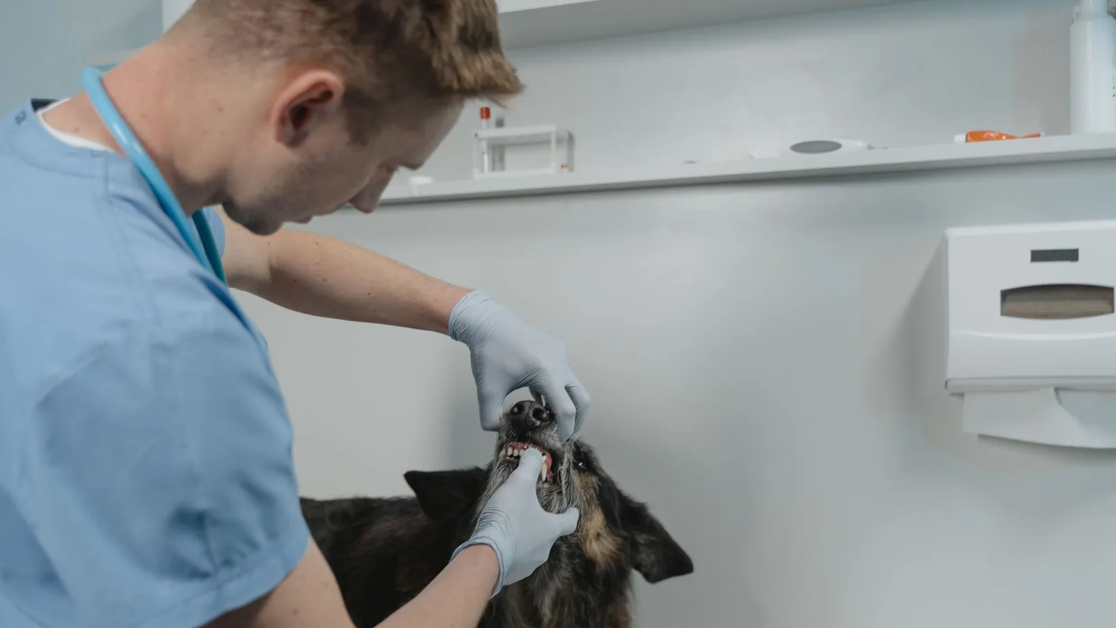
A tooth root abscess in dogs is a pocket of pus and infection at the tip of a tooth root. It develops when the pulp inside the tooth dies and bacteria move through the root canal into the bone around the apex. The infection causes inflammation, bone destruction, and swelling. Without treatment, it can spread to the eye, the nasal cavity, or the soft tissues of the face.
The signs can be obvious or subtle. One dog may have a swollen area under the eye and a fractured canine tooth. Another may have only chronic discharge from one eye. Facial swelling is one of the most recognizable features of a tooth root abscess, and it should prompt a veterinary visit rather than watchful waiting.
This article is educational and is not a substitute for veterinary diagnosis or treatment.
At a Glance: Tooth Root Abscess in Dogs
| Presentation | What it may mean | Urgency | First step |
|---|---|---|---|
| Swelling under the eye or over the upper canine root | Periapical abscess of a maxillary tooth [1][2] | Prompt | Oral examination and dental radiographs |
| Fractured canine tooth with a visible pulp cavity | Pulp exposure and risk of root infection [1][2] | Prompt | Dental radiographs and a treatment plan |
| Chronic discharge from one eye with no obvious ocular cause | Possible nasolacrimal compression from a root abscess [2] | Prompt | Thorough oral examination plus eye examination |
| Eye pushed forward, pain on opening the mouth, or inability to close the eye | Orbital or retrobulbar extension [3][4] | Emergency | Immediate veterinary assessment and imaging |
| Swelling or eye signs after a dental extraction | Possible globe injury, retrobulbar abscess, or sialadenitis [5][4] | Emergency | Immediate recheck |
What Is a Tooth Root Abscess?
Tooth Anatomy and the Root Apex
A dog's tooth has a crown above the gum line and a root below it. The pulp in the center contains blood vessels, nerves, and connective tissue. It extends through the root canal and exits at the apical foramen, where the root sits in the alveolar bone.
The maxillary canine tooth has a long root that sits close to important structures. In a cat, a root abscess of the upper canine compressed the nasolacrimal duct and caused chronic ocular discharge [2]. In dogs, periapical abscess of a maxillary canine can lead to an oronasal fistula when the bone between the root and the nasal cavity is lost [6]. These relationships explain why a dental infection can produce eye or nose signs.
How the Abscess Forms
The pulp is normally sterile. A fracture, a deep cavity, or advanced periodontal disease can breach the barrier and let bacteria in. Trauma to the upper canine that exposed the pulp cavity was the suspected route of infection in a colony of marmosets with facial abscesses [1]. In a cat with a fractured upper canine and pulpal exposure, dental radiographs showed a severe periapical lucency at the root apex [2].
Once bacteria enter the pulp, the tissue becomes inflamed. If the pulp does not recover, it dies. An experimental study in cats tracked what happens after pulp exposure. Within the first week, changes were limited to acute inflammation in the coronal pulp. By 30 days, some teeth had necrosis and abscess spread to the end of the root. By 90 days, complete pulp necrosis, internal resorption, and a large periapical lesion were present in 54.5% of samples [7]. The timeline supports early treatment of any tooth with exposed pulp.
When the pulp dies, bacteria and inflammatory debris leave the root canal and enter the periapical bone. The body walls off the infection, and pus collects. Experimental abscess formation in dog alveolar bone and cementum produced a clear tissue reaction [8]. The abscess can then expand through the bone and into soft tissue. The direction of spread determines whether the dog develops facial swelling, eye signs, or nasal signs.
Causes and Risk Factors
Pulp Exposure From Fracture or Trauma
A fracture that exposes the pulp creates a direct path for bacteria. Trauma to the upper canine was the presumed cause of root abscessation in the marmoset colony [1]. A fractured upper canine with pulpal exposure was the dental finding in a cat with a root abscess and facial swelling [2].
A tooth can fracture without causing immediate collapse. The infection develops later. That delay is why a broken tooth needs a dental examination even if the dog seems comfortable.
Periodontal Disease
Periodontal disease is one of the main oral cavity diseases in dogs. Its main complication is the formation of a periapical abscess [6]. When periodontal disease affects a maxillary canine, the infection and bone loss can lead to an oronasal fistula [6]. Severe periodontal disease was also present in the cat with an upper canine root abscess [2].
Tooth Anatomy and Immature Teeth
Immature teeth with open apices are a special risk group. In a two-year-old Chow Chow, a necrotic immature mandibular canine had an open apex, a wide root canal, thin dentinal walls, and periapical bone resorption [9]. The open apex allows infection to spread more easily into the periapical bone. It also makes treatment more complex, because the root has not finished forming.
Which Teeth Are Most Often Involved
The maxillary canine is a common focus in reported cases of root abscess with facial or ocular signs [1][2][6]. Its long root and its proximity to the nasal cavity, nasolacrimal duct, and orbit make complications more likely when infection spreads. The mandibular canine can also be affected, as the Chow Chow case shows [9].
Reported cases span young and older dogs, including a two-year-old Chow Chow [9] and a ten-year-old Cavalier King Charles Spaniel [4]. The common factor is dental disease or tooth injury.
Dog Tooth Abscess Symptoms
Signs vary with the tooth involved and the direction the infection takes.
Facial Swelling
Facial swelling is the most visible sign. In a cat with an upper canine root abscess, the left side of the face was swollen [2]. In the marmoset colony, facial abscesses were traced to abscessation of the upper canine root [1]. The swelling may be painful when touched or when the mouth is opened [4].
Swelling alone does not confirm an abscess. A veterinarian needs to examine the mouth and take dental radiographs to find the tooth and the periapical lesion.
Ocular Signs
Eye signs can be the first thing an owner notices. A ten-year-old cat presented with chronic ocular discharge from the left eye. Examination showed epiphora and mucopurulent discharge. The cause was traced to a root abscess of the upper canine that narrowed the nasolacrimal duct [2]. The ocular discharge and facial swelling resolved after the tooth was removed [2].
More severe orbital signs are possible. Exophthalmos, lagophthalmos, pain on opening the mouth, and a corneal ulcer occurred in a dog with retrobulbar disease after extensive dental extractions [4]. In a series of dogs with retrobulbar cellulitis or abscessation, six cases presented with vision loss [3]. These signs indicate that infection or inflammation has reached the space behind the eye.
Oral Signs
Oral examination may reveal a fractured tooth, severe periodontal disease, or pain when the mouth is opened [2][4].
Dental radiographs are needed to confirm periapical disease. A visual examination of the tooth does not show the root apex [1][2].
Signs Owners May Miss
Chronic discharge from one eye, mild facial asymmetry, or a small swelling that comes and goes can be the only signs. In the cat case, the owner's complaint was chronic ocular discharge, and the dental problem was found during a thorough oral examination [2]. A thorough oral examination is especially important when nasolacrimal obstruction is evident [2].
How Veterinarians Diagnose a Tooth Root Abscess
Oral Examination
The examination starts with the mouth. A veterinarian looks for fractures, periodontal pockets, fistulas, and swelling. Pain on opening the mouth is recorded [4]. In a dog with eye signs, the eye examination and the oral examination are linked, because a dental cause can explain the eye signs [2].
Dental Radiographs
Dental radiographs are the most useful diagnostic aid for tooth root infection [1]. They show the root, the periapical bone, and the extent of the lesion. In the cat case, radiographs showed a severe periapical lucency at the apex of the upper left canine [2]. In the marmoset work, radiography was the most useful diagnostic aid in establishing the nature and extent of the lesion [1].
Radiographs also help the veterinarian decide whether the tooth can be saved. Your veterinarian will make that judgment.
Advanced Imaging
When the infection has spread beyond the tooth, computed tomography (CT) and ultrasound can help. In a review of 38 dogs with retrobulbar cellulitis or abscessation, CT was the most common form of orbital imaging, used in 48.5% of cases, followed by ocular ultrasound in 18.2% [3]. CT revealed a tubular fluid-filled structure consistent with a retrobulbar abscess and zygomatic sialadenitis in a dog after extensive dental extractions [4]. Ultrasound was consistent with a penetrating globe injury in a dog with periorbital swelling after a maxillary tooth extraction [5].
Culture, Cytology, and Susceptibility
Samples can be taken for cytology and culture when the infection is deep or has spread. In the dog with retrobulbar disease after extractions, cytology showed septic suppurative inflammation and culture grew Bacteroides pyogenes [4]. In the 38-dog series, 15 of 18 cultures grew aerobic bacteria, anaerobic bacteria, or both [3].
Culture and susceptibility testing matter because antibiotic choice should match the organisms present. In that series, the antibiotics with the highest susceptibility patterns included gentamicin, amoxicillin/clavulanic acid, cephalothin, chloramphenicol, and imipenem. No bacteria were susceptible to cefovecin [3]. Your veterinarian will interpret the culture from your dog.
What a Diagnosis Needs
A confirmed diagnosis usually requires three things: a clinical sign, an oral examination finding, and a dental radiograph showing periapical disease. When the eye or orbit is involved, advanced imaging and cultures may be added [3][4].
What Else Can Cause Facial Swelling?
Facial swelling in a dog has several possible causes. A tooth root abscess is one of them, but not the only one.
Retrobulbar cellulitis or abscessation can cause swelling behind the eye, exophthalmos, and pain. A review of 38 dogs described the diagnostic imaging, bacterial isolates, and susceptibility patterns for these lesions [3]. Zygomatic sialadenitis can also cause exophthalmos and retrobulbar disease. It was diagnosed in a dog after extensive dental extractions [4].
A dog can also develop periorbital swelling after a dental extraction because of a complication. In one dog, periorbital swelling and blepharospasm developed four days after an ipsilateral maxillary tooth extraction. The differential diagnoses included iatrogenic globe penetration and peribulbar abscess or cellulitis. Ultrasound was consistent with a penetrating wound to the globe [5].
These conditions overlap with tooth root abscess in their signs. That overlap is why imaging and a full oral examination are needed. A swelling that looks dental may be orbital, and an orbital swelling may have a dental source.
Tooth Root Abscess Dog Treatment
The main goal is to remove the source of infection. That means treating or removing the affected tooth. Antibiotics support healing but do not replace source control.
Source Control: Extraction or Endodontic Treatment
In the marmoset cases, antibiotic therapy alone was inadequate. Providing drainage by extracting the tooth, in conjunction with antibiotics, was the most effective treatment [1]. In the cat with an upper canine root abscess, removal of the fractured canine resolved the ocular discharge and facial swelling. The signs did not recur over two years.
Endodontic treatment is an alternative when the tooth can be saved. In a two-year-old Chow Chow with a necrotic immature mandibular canine, endodontic treatment included apexification. The procedure induced apical closure by calcified tissue formation and resolved the periapical inflammation [9]. Root canal therapy has also been studied in dogs in the context of replanted teeth [10].
The choice between extraction and endodontic treatment depends on the tooth, the root, the surrounding bone, and the dog's health. Your veterinarian will discuss whether the tooth can be saved.
Tooth Root Abscess Dog Surgery
Extraction is a common surgical treatment. The tooth, the infected root, and the periapical debris are removed. Draining the abscess is part of the procedure.
Maxillary canine extraction can leave a communication between the mouth and the nasal cavity. This is called an oronasal fistula. In a study of dogs with oronasal fistulas after extraction of maxillary canines compromised by severe periodontal disease, fistulas were repaired with a double-mucoperiosteal flap 15 days after extraction or with a single-flap technique immediately after extraction [6].
Extraction is not risk-free. In one five-year-old dog, periorbital swelling and blepharospasm developed four days after an ipsilateral maxillary tooth extraction. The dog had exophthalmos, pain on retropulsion, and absent menace and pupillary light reflexes. Ultrasound was consistent with a penetrating wound to the globe [5]. The dog was treated medically and was visual at ten months [5]. This is a rare complication, but it shows why extraction of a maxillary tooth is a surgical procedure that needs careful planning.
Retrobulbar disease can also follow extensive dental extractions. A ten-year-old Cavalier King Charles Spaniel developed acute right-sided exophthalmos after a dental procedure involving 15 extractions. CT showed a probable retrobulbar abscess and zygomatic sialadenitis. The dog required transpalpebral exenteration and zygomatic sialoadenectomy after clinical deterioration and vision loss [4]. Extensive dental work in a small dog is a reminder that post-operative monitoring matters.
Dog Tooth Root Abscess Antibiotics
Antibiotics are used to control bacterial infection, but they do not cure an abscess on their own. In the marmoset cases, antibiotic therapy alone was inadequate [1]. An abscess is a walled-off collection of pus and infected tissue, and antibiotics alone do not remove it. Drainage and removal of the infected tooth are needed.
When antibiotics are used, the choice should be based on culture and susceptibility when possible. In the 38-dog series of retrobulbar infections, amoxicillin/clavulanic acid was among the antibiotics with the highest susceptibility patterns, and cefovecin was not effective against any isolate [3]. In a survey of pet owners, amoxicillin/clavulanic acid was the most commonly used systemic antibiotic in dogs and cats, followed by amoxicillin, metronidazole, and doxycycline [11].
Antibiotics for dogs are prescription medications in the United States. A veterinarian must examine the dog and decide whether antibiotics are needed, which antibiotic to use, and for how long.
Pain Control and Supportive Care
Pain control is part of treatment. In the dog with retrobulbar disease after extractions, initial medical management included intravenous fluids, broad-spectrum systemic antibiotics, analgesics, topical antibiotics, and ocular antihypertensive medications [4]. The exact drugs and doses are chosen by the veterinarian.
Follow-Up
Follow-up confirms that the infection has resolved and the surgical site is healing. In the cat case, the ocular discharge and facial swelling resolved after extraction, and there was no recurrence after two years [2]. In the dog with globe penetration, re-examination at ten months showed vision and a chorioretinal scar [5]. Your veterinarian will set a recheck schedule based on the procedure and the tissues involved.
Dog Tooth Root Abscess Home Treatment: What Not to Do
Home treatment is not a substitute for veterinary care. The infection starts inside the tooth and the bone. It cannot be cured by cleaning the outside of the face or by giving antibiotics from a previous prescription.
Do not rely on antibiotics alone. In the marmoset cases, antibiotic therapy alone was inadequate, and drainage plus extraction was more effective [1]. The same principle applies to dogs. Antibiotics may reduce the bacterial load, but the abscess and the dead tooth remain.
Do not try to open or drain a swelling at home. In reported cases, drainage was provided by a veterinarian during extraction or surgery [1][6].
Do not give human pain relievers or leftover antibiotics without veterinary advice. Ask your veterinarian before giving any medication.
Do not wait for the swelling to resolve on its own. The experimental pulp exposure study showed that infection can progress from inflammation to complete pulp necrosis and a large periapical lesion over 30 to 90 days [7]. Waiting allows the infection to spread.
Complications and Prognosis
Oronasal Fistula
A maxillary canine root abscess can destroy the bone between the root and the nasal cavity. When the tooth is extracted, a hole can remain. This is an oronasal fistula. In dogs with severe periodontal disease, extraction of a compromised maxillary canine was followed by an oronasal fistula that required surgical repair [6].
Orbital and Retrobulbar Complications
The orbit is close to the maxillary tooth roots. Infection can spread into the retrobulbar space. Retrobulbar cellulitis or abscessation can cause exophthalmos, pain, and vision loss. In a series of 38 dogs, six presented with vision loss [3]. Most cases in that series were managed medically, and 18.4% were managed surgically [3].
A tooth root abscess can also cause ocular signs without direct orbital infection. In the cat case, the root abscess compressed the nasolacrimal duct and caused chronic ocular discharge [2]. That sign resolved after extraction [2].
Complications of Extraction
Extraction of a maxillary tooth can injure the eye. A dog developed globe penetration after an ipsilateral maxillary tooth extraction [5]. The dog was treated medically and remained visual at ten months [5]. This is a rare but serious complication.
Extensive dental extractions can also be followed by retrobulbar disease and sialadenitis [4]. A dog needed exenteration after progressive deterioration and vision loss [4].
Prognosis
The prognosis is good when the source of infection is removed and the surrounding tissues are healthy. In the cat case, signs resolved and did not recur over two years [2]. In the dog with a necrotic immature canine, endodontic treatment resolved the periapical inflammation [9].
The prognosis is more guarded when the infection has reached the orbit or when vision is already lost. In the 38-dog series, vision loss was present in six cases [3]. In the dog with retrobulbar disease after extensive extractions, vision loss and clinical deterioration led to exenteration [4].
Prevention
Prevention starts with regular oral examinations. A thorough oral examination is important when nasolacrimal obstruction or one-sided ocular discharge is present, because a dental cause may be found [2]. Dental radiographs are the most useful diagnostic aid when root infection is suspected [1].
Treat fractured teeth early. A tooth with pulp exposure is at risk of pulp death and periapical abscess [1][2][7]. If a dog breaks a canine tooth, a dental examination and radiographs should follow. Waiting for pain or swelling allows the infection to progress.
Control periodontal disease. Periodontal disease is one of the main oral cavity diseases in dogs, and periapical abscess is its main complication [6]. Home dental care and regular professional dental assessment are the foundation of periodontal control. The AAHA Dental Care Guidelines recommend home dental care and routine professional evaluation.
Emergency Red Flags
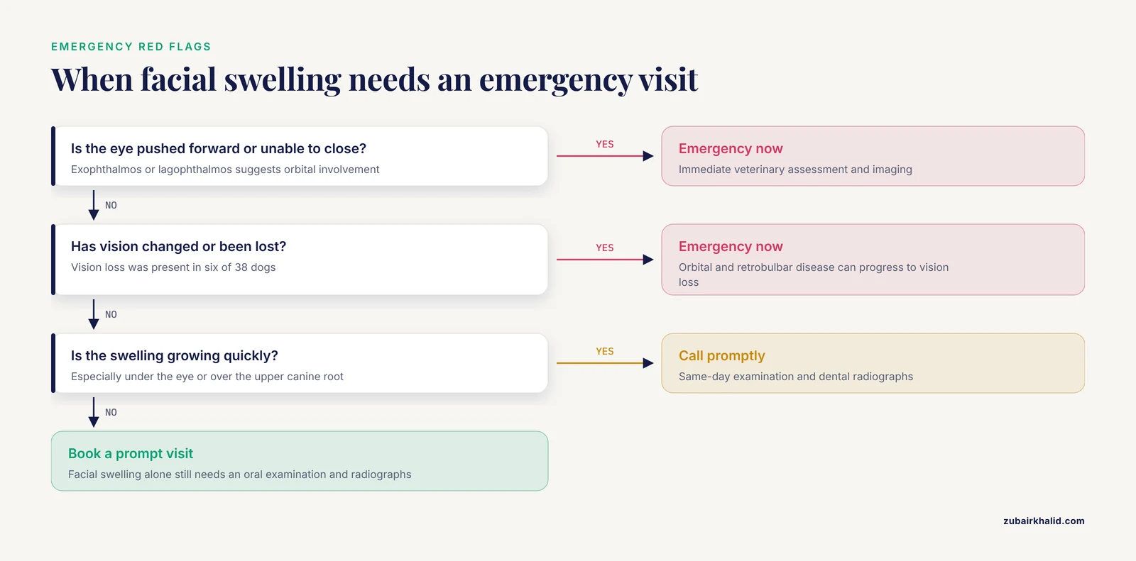
Call your veterinarian immediately if you see any of the following:
- Facial swelling that is growing quickly, especially under the eye or over the upper canine root [1][2].
- The eye is pushed forward, the dog cannot close the eye, or the eye looks different.
- Pain when the mouth is opened [4].
- Vision loss or a change in vision [3].
- Discharge from one eye that persists or worsens [2].
- Swelling, eye signs, or pain after a dental extraction [5][4].
- A fractured canine tooth with a visible pulp cavity [1][2].
These signs do not prove an abscess. They do mean the dog needs an examination. Orbital and retrobulbar disease can progress to vision loss [3][4].
Limitations and When to Contact a Veterinarian
This article is educational and is not a substitute for veterinary diagnosis or treatment. Individual dogs differ in anatomy, dental disease, and response to treatment. A veterinarian who can examine the mouth, take radiographs, and assess the eye needs to make the plan for your dog.
Contact a veterinarian promptly if your dog has facial swelling, a fractured tooth, one-sided eye discharge, pain when opening the mouth, or a change in vision. Contact an emergency service if the eye is pushed forward, the dog cannot close the eye, or swelling is rapidly worsening. Do not wait for a tooth root abscess to drain on its own.
How Much Does Treating a Tooth Root Abscess Cost?
Treatment costs vary widely, but most owners can expect a total bill between $800 and $2,500 for a single affected tooth. The largest components are anesthesia, dental radiographs, and the procedure itself.
Anesthesia adds expense because it requires an intravenous catheter, monitoring equipment, and trained staff. Dental radiographs are not optional for a root abscess; they are needed to see the root tip and surrounding bone, and each tooth requires multiple views.
The procedure type drives the bill. Extraction of a single-rooted tooth costs less than extraction of a multi-rooted carnassial tooth. Root canal therapy is more expensive than extraction because it takes longer and requires specialized equipment. Tooth location matters too; a maxillary canine with a long root is more complex to extract than a small incisor.
Regional pricing also varies. Specialty hospitals and urban practices generally charge more than general practices in rural areas.
Ask for a written, itemized estimate before treatment. It should list examination, radiographs, anesthesia, monitoring, medications, and the procedure, and it should state what happens if complications arise.
What to Expect During Recovery and Aftercare
The first 24 to 48 hours focus on protecting the surgical site and keeping your dog comfortable.
Food and Water
Offer soft food for 7 to 14 days after the procedure. Canned food or soaked kibble is easier to eat. Provide fresh water. Call your veterinarian if your dog refuses food for more than 24 hours.
Pain Relief and Activity
Give all pain medications as prescribed. Keep your dog quiet for the first week. No hard chew toys, bones, or rough play. Use a cone or collar if your dog paws at the mouth or rubs the face.
Oral Care
Do not brush the surgical area until your veterinarian says it is safe. Gentle rinsing may be recommended. Follow the recheck schedule to assess healing.
When to Call Your Veterinarian
Call if you see bleeding that does not stop, swelling that increases after the first day, refusal to eat, vomiting, or discharge from the incision. Call if your dog seems painful despite medication.
Root Canal Therapy vs. Extraction: How the Decision Is Made
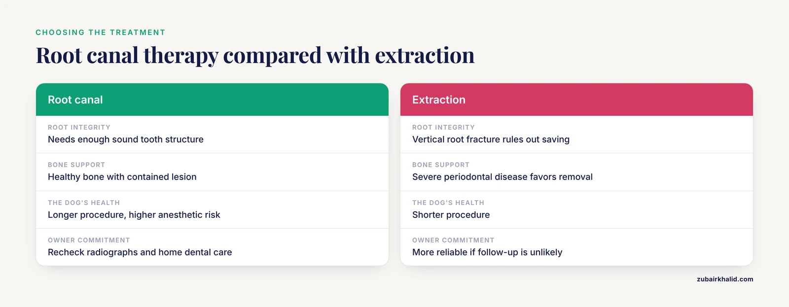
Dental radiographs guide this decision because they show the root, the periapical lesion, and the bone around the tooth [1].
Root Integrity
Root canal therapy requires enough sound tooth structure. A vertical root fracture, a canal that cannot be sealed, or a fracture below the gum line usually rules out saving the tooth.
Bone Support
The tooth needs enough periodontal support to remain functional. Severe periodontal disease with advanced bone loss favors extraction [6]. A tooth with healthy surrounding bone and a contained periapical lesion can often be treated endodontically [9].
The Dog's Health
Root canal therapy takes longer than extraction. A dog with heart, kidney, or other systemic disease may face higher anesthetic risk with a longer procedure.
Owner Commitment
Saving a tooth requires follow-up. The owner must return for recheck radiographs, provide home dental care, and accept that the tooth may still fail. If follow-up is unlikely, extraction is more reliable.
The final recommendation is based on which option gives the dog the best long-term outcome with the least risk.
Related Guides
- Cat Tooth Abscess and Infection: Home Care Limits: Feline counterpart with similar root abscess signs and home care limits
- Split Tooth in Dogs: Signs, Pain, and Treatment: Another tooth fracture type that can expose pulp and lead to abscess
- Tooth Resorption in Dogs: Causes, Stages, and Symptoms: Related dental disease; resorption can accompany pulp necrosis and infection
- Broken Tooth in a Cat: Treatment for Fractures and Lesions: Cat-specific fracture treatment; article cites feline cases with root abscess
- Cat Abscess: Home Care and When to See a Vet: General cat abscess home care guidance parallels the limits described for dogs
- Dog Conjunctivitis Treatment: What You Can Use at Home: Eye discharge can be a sign of tooth root abscess; useful differential
Frequently Asked Questions
Can a tooth root abscess in dogs heal on its own?
No. A tooth root abscess needs source control, which means treating or removing the affected tooth. Antibiotics alone do not cure it.
What are the most common dog tooth abscess symptoms?
Facial swelling, one-sided eye discharge, a fractured tooth, and pain when the mouth is opened are common signs. Some dogs show only subtle asymmetry or chronic discharge.
How is a tooth root abscess in dogs treated?
Treatment removes the source of infection. The tooth is extracted or treated endodontically, with antibiotics and pain control as needed.
Will my dog need surgery for a tooth root abscess?
Many dogs need extraction. Some teeth can be saved with endodontic treatment. Your veterinarian will decide based on the tooth and the surrounding bone.
Are dog tooth root abscess antibiotics enough?
No. Antibiotics alone do not cure a tooth root abscess. They are used with drainage and extraction or endodontic treatment.
Can I use dog tooth root abscess home treatment?
No home treatment can remove the infected tooth or drain the abscess safely. Do not open the swelling at home, and do not give medications without veterinary advice.
Why does a tooth root abscess cause facial swelling?
The abscess expands through the bone and into the soft tissues of the face. The swelling location depends on which tooth is affected and where the infection spreads.
When is a tooth root abscess an emergency?
It is an emergency when the eye is pushed forward, the dog cannot close the eye, vision changes, or swelling is rapidly worsening. These signs suggest orbital or retrobulbar involvement.
Sources
- Canine tooth root infection as a cause of facial abscess in the common marmoset (Callithrix jacchus).
- Nasolacrimal obstruction caused by root abscess of the upper canine in a cat.
- Canine Retrobulbar Cellulitis and Abscessation in the Southeastern United States: A review of case management, diagnostic imaging, bacterial isolates, and susceptibility patterns.
- Zygomatic Sialadenitis as a Rare Cause of Exophthalmos in a Dog.
- Successful medical treatment for globe penetration following tooth extraction in a dog.
- Self-Curing Glass Ionomer Cement Covered by Photopolymerizable Adhesive for Protection of Mucoperiosteal or Gingival Flap Sutures in Canine Oral Surgery.
- Dental pulp reaction to exposure at different time intervals in open apex canine teeth of cats.
- Reaction of alveolar bone and cementum to experimental abscess formation in the dog.
- Endodontic treatment including apexification in a chow chow with a necrotic immature mandibular canine tooth.
- THE EFFECT OF ROOT CANAL THERAPY ON REPLANTED TEETH OF DOGS; A GROSS, ROENTGENOGRAPHIC, AND HISTOLOGIC STUDY.
- Online-Assisted Survey on Antibiotic Use by Pet Owners in Dogs and Cats.