Turtle Scutes: Anatomy, Shedding, and Shell Health
By Dr. Zubair Khalid, DVM, MS, PhD ·
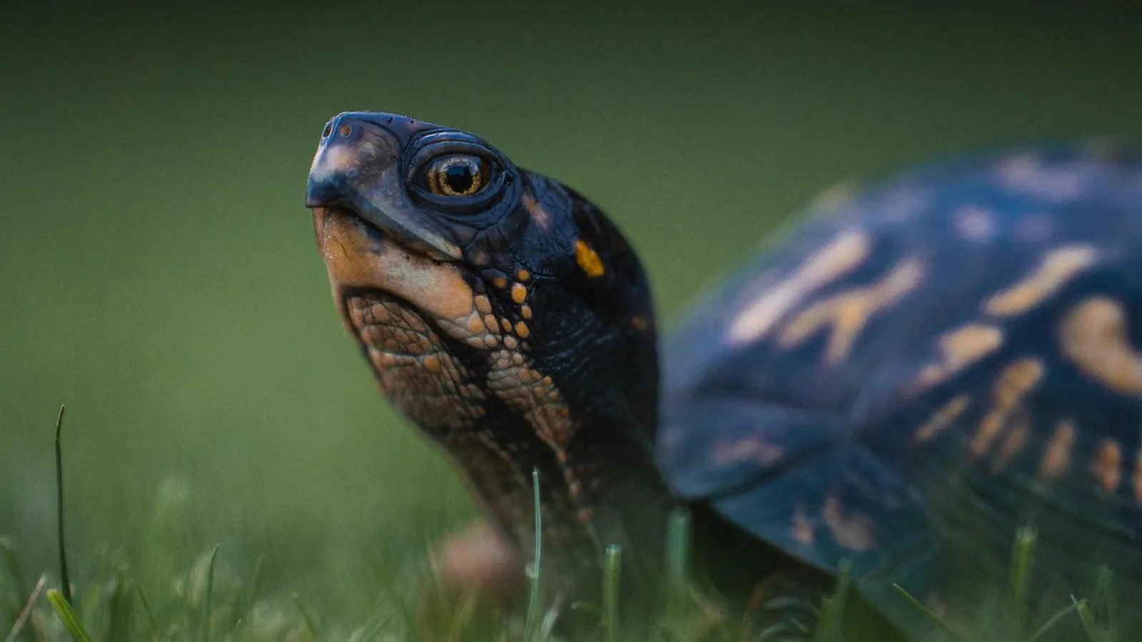
Turtle scutes are the large, keratinized epidermal plates that cover the outer surface of the carapace (the dorsal shell) and the plastron (the ventral shell), separated from one another by grooves called furrows. They are not bones. They are the horny, cornified outer layer of the shell, produced by the epidermis and sitting directly over the bony plates of the underlying skeleton.
Scutes matter because they are the part of the shell an owner actually sees and touches, and because their condition is one of the most reliable outward indicators of husbandry quality. A shell that sheds cleanly, sits flat, and stays smooth usually reflects correct diet, lighting, humidity, and basking. A shell with stacked, retained scutes, a peaked or pyramid-shaped profile, or soft, discolored, foul-smelling patches points to a problem that needs correction and often veterinary care.
This article is educational and is not a substitute for veterinary diagnosis or treatment.
What Scutes Are and Where They Come From
The turtle shell is a composite structure. It is built from basal endochondral axial skeletal elements (ribs and vertebrae) plus plates of bone, all overlain by keratinous ectodermal scutes [1]. In other words, the shell has a bony foundation and a keratin covering, and the two develop as interacting modules rather than as a single tissue.
Scutes are an evolutionary novelty unique to turtles. They are autapomorphic for the group, meaning they define turtles and are not shared with other reptiles in the same form, and their tessellation pattern is stable enough across species to be used for taxonomic identification [2]. The small epidermal bumps called tubercles found on the shells of some species are a separate feature and are considered homologous to ordinary reptilian scales, whereas the scutes themselves are not [2].
At the molecular level, scutes are built from beta-keratins, which are glycine-proline-tyrosine rich proteins similar to those of crocodilians and birds [3]. Turtle beta-keratins belong to a well-conserved multigene family, and their mRNAs are expressed in the differentiating beta-layers of the scute [3]. Later immunocytochemical work showed that two beta-protein subtypes, designated Tu2 and Tu17, co-accumulate in the thick corneous layer of hard-shelled turtle epidermis, and the relative amounts of these subtypes in relation to the alpha-keratin meshwork are thought to determine how hard and inflexible the finished scute becomes [4]. An antibody raised against a turtle scute beta-keratin of roughly 13 to 16 kDa localized the protein to the corneous layer of both carapace and plastron across multiple chelonian species [5].
Not every turtle builds scutes. Soft-shelled turtles such as Trionyx spiniferus have a smooth, unscaled, pliable shell. Their epidermis has a thick corneous layer of alpha-corneocytes with only some beta-keratin present, and their mechanical protection comes largely from a very thick, plywood-patterned dermal collagen layer rather than from cornified scutes and dermal bones [6]. This contrast is useful because it shows that the scute is a specialization, not an inevitable feature of having a shell.
How the Pattern Is Laid Down
Scutes develop from an array of patterned placodes, which are localized signaling centers in the embryonic epidermis. Experimentally inhibiting Shh, Bmp, or Fgf signaling disrupts the placodal pattern, and a computational model using two coupled reaction-diffusion systems reproduces both natural and abnormal variation in turtle scutes [1]. The placodes are topographically associated with the somites of the embryo, and one model proposes that horny scutes develop from a mosaic of placodes corresponding to the paths of myosepta, with vertebral and pleural scutes developing staggered in adjacent segments and marginal scutes developing in every segment [7]. That scheme predicts little variation in the marginals and identifies intercalary supernumerary scutes as a likely variation in the vertebral and pleural rows [7].
The pattern is stable phylogenetically but labile in the individual [2]. An examination of 655 newly hatched olive ridley sea turtles (Lepidochelys olivacea), a species known for a highly variable horny shell, found 120 distinct carapacial scute patterns and 10 plastral patterns. Vertebral scute number ranged from 4 to 10, with five, six, and seven occurring at nearly equal frequency (about 31.5% on average), and the typical symmetric sea turtle pattern of five pairs of pleurals appeared in only about 12% of animals [7].
Environment pushes the pattern around during incubation. High temperatures are sufficient to produce anomalous scute patterns, and the correlation is even stronger when conditions are dry [8]. The variation is not random: more anomalies appear in the midline vertebral scutes and during a critical window of development [8]. These anomalies are largely cosmetic in survivors. A 25-year photo database of loggerhead (Caretta caretta) and green (Chelonia mydas) turtles on Florida's east coast found that anomalous scute forms and patterns are stable throughout growth, with limited evidence of selection against them in the size classes examined, and their frequency stayed stable in juvenile cohorts from 1994 onward [9].
Naming the Scutes: A Working Map


Scute names are positional. Each name tells you where on the shell the plate sits, which is why the same vocabulary works across species even when counts differ.
| Scute name | Location | Notes |
|---|---|---|
| Nuchal | Single scute at the front midline of the carapace, over the neck | Also called the cervical scute. Its upturned wall can change shape with growth in some species [10] |
| Vertebral | Midline row down the carapace, over the vertebral column | The row most prone to anomalous variation [8] |
| Costal (pleural) | Paired rows flanking the vertebrals, over the ribs | Sea turtles typically show five pairs, but this pattern appeared in only about 12% of olive ridleys sampled [7] |
| Marginal | Peripheral rim of the carapace | Predicted to vary least because they develop in every segment [7] |
| Supracaudal | Single scute at the rear midline of the carapace, over the tail | Often counted with the marginals |
| Gular | Anterior midline pair on the plastron | Paired in most species |
| Humeral | Anterior plastron, lateral to the gulars | Paired |
| Pectoral | Mid-anterior plastron | Paired |
| Abdominal | Mid-plastron | Paired |
| Femoral | Posterior plastron | Paired |
| Anal | Rear midline of the plastron | Paired |
The vertebral and costal scutes are the ones most often used as external radiographic landmarks. In loggerhead sea turtles, the vertebral and lateral scutes serve as important external reference points for identifying internal structures such as the bronchi, coracoid bones, the caudal border of the pulmonary fields, and the acetabulum on dorsoventral radiographs [11]. That is a practical point for clinicians: the scute pattern is not just a shell feature, it is a coordinate system for the whole body.
Scutes as a Record of Growth
Because scutes are deposited incrementally, they carry a chronological record. Sectioned and polished posterior marginal scutes from 36 hawksbill turtles (Eretmochelys imbricata) were used for bomb radiocarbon dating, and the authors found that Hawaii hawksbills deposit eight growth lines annually, with a range of 5 to 14 [12]. Fitting von Bertalanffy growth models to those data produced a somatic growth parameter (k) of 0.13, range 0.1 to 0.2, and an estimate of first breeding at 29 years, range 23 to 36 [12]. Growth-line counts are therefore not a simple one-line-per-year tally, and species-specific deposition rates matter.
Mechanical Behavior
Scutes are not just inert armor plating. Compression testing of a Terrapene carolina box turtle shell, combined with finite element analysis, identified three constitutive regimes: linear elastic, perfectly inelastic, and densification, in which hardening occurs [13]. These responses arise as deformation proceeds through three distinctive layers of the carapace, and the overall stress-strain behavior resembles that of metallic foams [13]. The practical takeaway is that the shell is a layered, energy-absorbing structure, and its performance depends on all layers remaining intact.
Normal Shedding: What Species-Appropriate Shedding Looks Like
Shedding, or ecdysis, is the normal replacement of the outer keratin layer. The pattern differs by species and by habitat, and knowing the normal pattern for your species is the single best way to judge whether something is wrong.
Aquatic turtles typically shed scutes individually. A scute lifts at the edges, separates along the furrows, and comes away as a whole plate or a few large pieces. This is the pattern most owners see in sliders, cooters, painted turtles, and similar species.
Tortoises typically shed in small pieces. Rather than losing whole plates, a tortoise flakes off small fragments of keratin from the surface of each scute over time. A tortoise that drops an entire scute in one piece is more likely to have a problem than a tortoise that sheds fine flakes.
Some species retain scutes. In these animals, the outer keratin layer is worn down gradually rather than shed in identifiable pieces, and old scutes may remain visibly stacked. This can be normal for the species, which is why species identification comes before any judgment about shedding.
Shedding is driven by growth and by the mechanical and thermal environment. Basking provides the heat and drying that help the outer keratin layer separate cleanly. Humidity and soaking soften the seams. A turtle with no access to a proper basking site, or one kept too cool, tends to shed poorly regardless of diet.
When Shedding Goes Wrong
Retained Scutes
Retained scutes are scutes that should have separated but did not. They appear as stacked, raised, or overlapping plates, sometimes with trapped debris or a musty smell underneath. The usual causes are environmental: inadequate basking temperature or duration, low humidity for species that need it, poor water quality in aquatic setups, and insufficient dietary support for normal keratin turnover. A retained scute that is not loose should never be peeled off by hand, because pulling can tear the underlying epidermis and open a route for infection.
Pyramiding
Pyramiding is abnormal upward growth of the scutes, producing a peaked or cone-shaped carapace instead of a smooth domed profile. It is a growth deformity rather than a shedding problem, and it reflects a mismatch between growth rate and shell development. In practice it is associated with excessive dietary protein and energy, inadequate calcium and vitamin D support, and insufficient ultraviolet B exposure or inappropriate basking temperatures. Once pyramiding has developed, the shape change is permanent, so the goal of management is to stop progression and prevent it in animals still growing.
Shell Rot (Ulcerative Shell Disease)
Shell rot, more precisely called ulcerative shell disease, is a progressive destructive lesion of the shell. It typically presents as pitting, softening, discoloration, a foul odor, or areas of exposed underlying tissue, and it can involve the scutes, the bone, or both. It is a clinical diagnosis, not a cosmetic one, and it requires veterinary evaluation.
A study of 19 map turtles (Graptemys spp.) kept under natural conditions and presented for chronic shell abnormalities used a shell scoring system that divided the 54 scutes into six regions, scoring each region for lesion extent and severity and summing the results into a total shell disease score (TSDS) [14]. Scores in that series were low, ranging from 4 to 22 out of a possible 54, with a median of 9 [14]. Investigators collected cytology tape strips and full-thickness shell biopsies aseptically under ketamine-medetomidine-morphine anesthesia for microbiologic, histologic, and ultrastructural evaluation [14]. Small clefts and pitting lesions were noted in 8 of 19 sections, and although there was no evidence of erosion, ulceration, inflammation, or infectious agents, algae and diatoms were observed [14]. Six biopsies yielded aerobic isolates including Chryseobacterium indologenes, Aeromonas hydrophila, Ralstonia pickettii, and Morganella morganii [14].
That study is instructive for two reasons. First, it shows that visible shell surface change does not automatically equal infection, which is exactly why culture and biopsy matter. Second, it shows that a standardized scoring system lets a clinician track lesion extent and severity over time rather than relying on a subjective impression. No hematologic or biochemistry parameter correlated with the shell disease score in that series [14], so bloodwork alone will not tell you how bad a shell is.
Because the organisms involved vary and because superficial contamination is common, the choice of antimicrobial therapy has to follow culture and sensitivity results from a veterinarian. Dosing is not something an owner can extrapolate from a textbook or a forum post.
How Shell Health Is Assessed
Assessment starts with the hands and the eyes, then moves to imaging and laboratory work when indicated.
Visual and tactile examination. Note the number and position of scutes, look for asymmetry, and check whether any scute is loose, stacked, or missing. Press gently along the margins and over any discolored area to detect softening. Photograph the shell from above, below, and both sides at each visit so changes can be compared over time rather than recalled.
Furrow and seam inspection. Clean furrows are a good sign. Dark, wet, or debris-filled furrows suggest that scutes are not separating normally or that the animal is spending too much time in unsanitary conditions.
Radiography. Dorsoventral radiographs of the neck and body are useful for internal assessment, and the vertebral and lateral scutes serve as external landmarks for identifying the bronchi, coracoid bones, the caudal border of the pulmonary fields, and the acetabulum [11]. Distortion or superimposition from the natural curvature of the shell can limit interpretation in some views [11].
Cytology, culture, and biopsy. For suspected shell rot, tape-strip cytology and full-thickness shell biopsy collected aseptically provide the most reliable information [14]. Histology distinguishes true tissue destruction from surface staining or algal colonization.
Husbandry audit. Because most shell problems are husbandry problems, the history matters as much as the physical exam. Record basking surface temperature, ambient temperature, humidity, UVB source and its replacement interval, water quality parameters, and the full diet including any supplements.
Comparative Notes Across Species
The scute pattern is conservative across species but the shedding behavior and the clinical priorities are not.
Hard-shelled aquatic turtles (sliders, cooters, painted turtles, map turtles) shed individual scutes and are prone to retained scutes and shell rot when water quality, basking, or diet is poor. The map turtle series above is a reminder that chronic shell abnormalities in this group can be multifactorial and sometimes non-infectious [14].
Tortoises shed in small flakes and are the group most associated with pyramiding when fed high-protein, high-energy diets with inadequate calcium or UVB. They are also sensitive to humidity, and low humidity during growth contributes to shell deformities.
Sea turtles show the most variable scute patterns, with the classic five-pair pleural arrangement appearing in only a minority of olive ridleys [7]. Anomalous patterns are common, environmentally influenced, and largely stable and non-costly once animals reach juvenile size [8][9]. Nest shading in a green turtle conservation experiment reduced the production of nonmodal scute patterns and produced hatchlings with a larger carapace size and a better self-righting response [15].
Soft-shelled turtles lack cornified scutes entirely and rely on a thick collagenous dermis for protection [6]. Their shell problems look different and cannot be assessed with the same scute-by-scute framework.
Box turtles have a rigid shell structure that makes them convenient subjects for pattern analysis, and their scute color patterns have been quantified using digital image algorithms that measure symmetry and other pattern features across 19 variables [16]. Symmetry of scute pattern is a recognized component of developmental and evolutionary study [16].
Clinical Relevance, Limitations and Common Mistakes
The clinical relevance of scutes is that they are the most accessible window into shell health, and shell health is usually a readout of husbandry. A careful scute-by-scute examination, combined with a husbandry history, will identify most problems before they become advanced.
Common mistakes owners make:
- Peeling retained scutes off by hand. This tears the epidermis underneath and creates an entry point for infection. Let loose scutes fall off on their own.
- Assuming all shedding is the same. Aquatic turtles shed whole scutes, tortoises shed flakes, and some species retain scutes. Judging a tortoise by aquatic turtle standards leads to unnecessary worry, and the reverse leads to missed problems.
- Treating shell discoloration as infection without diagnostics. Algae, diatoms, and staining can mimic early shell rot, and the map turtle series found pitting and clefts without evidence of erosion, ulceration, inflammation, or infectious agents [14].
- Relying on bloodwork to grade shell disease. No hematologic or biochemistry parameter correlated with the shell disease score in that series [14].
- Assuming a deformed scute pattern means a sick animal. Anomalous patterns are common, environmentally induced, and stable through growth with limited evidence of a survival cost in the size classes examined [8][9].
- Ignoring the environment because the shell "looks fine." Pyramiding and retained scutes develop slowly. By the time the shape change is obvious, it is often permanent.
Limitations: individual animals vary, species requirements differ substantially, and a definitive diagnosis of shell disease requires veterinary examination with appropriate sampling. Any turtle with a soft, foul-smelling, bleeding, or rapidly enlarging shell lesion should be seen by a veterinarian promptly.
Quick Review
- Scutes are keratinized epidermal plates over the carapace and plastron, separated by furrows. They are not bone.
- The main carapacial scutes are nuchal, vertebral, costal (pleural), marginal, and supracaudal. The main plastral scutes are gular, humeral, pectoral, abdominal, femoral, and anal.
- Scutes develop from epidermal placodes patterned by Shh, Bmp, and Fgf signaling, and the pattern is stable across species but variable within individuals [1][2].
- Normal shedding differs by species. Aquatic turtles shed individual scutes, tortoises shed small flakes, and some species retain scutes.
- Retained scutes, pyramiding, and shell rot point to husbandry problems such as poor diet, low humidity, or inadequate basking.
- Shell rot is a clinical diagnosis. It requires veterinary evaluation with culture and often biopsy, and treatment follows those results [14].
- Scutes double as radiographic landmarks for internal anatomy [11].
Frequently Asked Questions
Should I pull off a loose scute?
No. Let it separate on its own. Pulling a scute that is not fully detached can tear the epidermis beneath it and create an opening for infection. If a scute has been stuck for a long time or the shell underneath looks abnormal, have a veterinarian examine it.
Why is my turtle's shell peeling in big pieces instead of flakes?
Large pieces are normal for many aquatic turtles, which shed scutes individually. Flaking in small fragments is more typical of tortoises. The pattern that matters is whether shedding is progressing cleanly and whether the new surface underneath looks smooth and intact.
What causes pyramiding?
Pyramiding is abnormal upward growth of the scutes, usually linked to excessive dietary protein and energy, inadequate calcium or vitamin D, and insufficient UVB or inappropriate basking temperatures. The shape change is permanent once it develops, so the focus shifts to stopping progression.
Is shell rot contagious to my other turtles?
Shell rot reflects an interaction between the animal, its environment, and opportunistic organisms rather than a single contagious disease. Because affected animals often share the same inadequate setup, separate them and correct the environment while a veterinarian evaluates the primary case.
Can a shell heal after shell rot?
Keratin and bone can repair to varying degrees depending on how deep the lesion went and whether the underlying bone was involved. Healing is slow in reptiles. Follow your veterinarian's plan and keep photographing the lesion so progress can be measured objectively.
Do all turtles shed their scutes?
No. Shedding pattern varies by species. Aquatic turtles typically shed individual scutes, tortoises shed small flakes, and some species retain scutes and wear them down gradually. Soft-shelled turtles lack cornified scutes altogether [6].
Related Articles
- Red-Eared Slider Turtle Care: Pyramiding & Shell Health Management
- Turtle Shell Diseases: Identification and Treatment
- Pet Turtle Shell Rot Signs
- Hamster Care: Pyramiding & Shell Health Management
- Chinchilla Care: Pyramiding & Shell Health Management
- Rabbit Care: Pyramiding & Shell Health Management
- Bantam Silkies: Breed Care and Health Guide
- Bully Pit Mix: Breed Traits, Health, and Care
Sources
- The origin and loss of periodic patterning in the turtle shell.
- The integumental appendages of the turtle shell: an evo-devo perspective.
- Beta-keratins of turtle shell are glycine-proline-tyrosine rich proteins similar to those of crocodilians and birds.
- Immunocytochemistry suggests that the prevalence of a sub-type of beta-proteins determines the hardness in the epidermis of the hard-shelled turtle.
- Immunological characterization of a newly developed antibody for localization of a beta-keratin in turtle epidermis.
- Skin structure and cornification proteins in the soft-shelled turtle Trionyx spiniferus.
- Supernumerary scutes verify a segment-dependent model of the horny shell development in turtles.
- Environmental Causation of Turtle Scute Anomalies in ovo and in silico.
- No evidence of selection against anomalous scute arrangements between juvenile and adult sea turtles in Florida.
- New insights on the anatomy and ontogeny of the largest extinct freshwater turtles.
- Cervical and coelomic radiologic features of the loggerhead sea turtle, Caretta caretta.
- Time in tortoiseshell: a bomb radiocarbon-validated chronology in sea turtle scutes.
- Compressive behavior of a turtle's shell: experiment, modeling, and simulation.
- Investigation of shell disease in map turtles (Graptemys spp.).
- Investigating the effects of nest shading on the green turtle (Chelonia mydas) hatchling phenotype in the Ogasawara islands using a field-based split clutch experiment.
- New approaches for capturing and estimating variation in complex animal color patterns from digital photographs: application to the Eastern Box Turtle (Terrapene carolina).