Teeth Diagram: Types, Numbers and Animal Comparison
By Dr. Zubair Khalid, DVM, MS, PhD ·
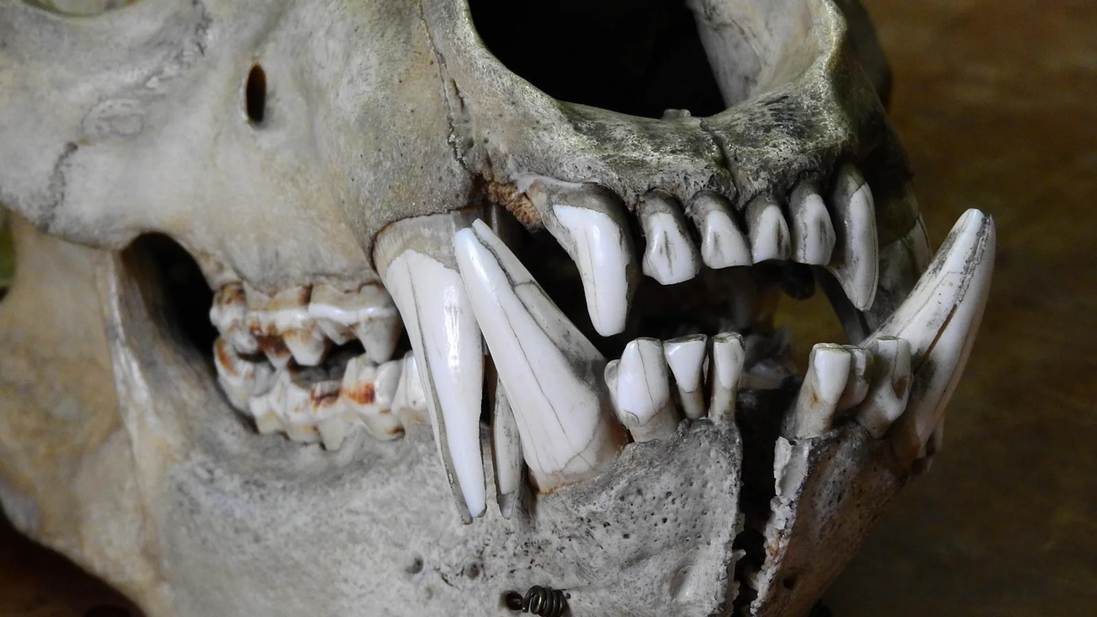
A teeth diagram labels four tooth types (incisors, canines, premolars and molars) and counts them in each jaw quadrant using a dental formula. The human adult formula is 2-1-2-3 over 2-1-2-3, the dog is 3-1-4-2 over 3-1-4-3, the cat is 3-1-3-1 over 3-1-2-1, and the horse is 3-1-3-3 over 3-1-3-3, with the canine often absent in mares.
This article is educational and is not a substitute for veterinary diagnosis or treatment.
What a Teeth Diagram Shows
A teeth diagram is a map of the mouth. It divides each jaw into a left and a right side, giving four quadrants: upper right, upper left, lower right and lower left. Every tooth sits in one quadrant, and every quadrant holds a fixed set of tooth types in a fixed order from front to back.
The order never changes in mammals. Incisors sit at the front. Canines come next. Premolars follow, then molars at the back. A tooth diagram therefore reads like a sentence written from the midline outward, and the dental formula is simply the count of each tooth type in one quadrant.
Veterinarians and dentists use several numbering systems to point at a single tooth. The most common in veterinary practice is the modified Triadan system, in which the mouth is split into four quadrants numbered 1 through 4 for the permanent upper right, upper left, lower left and lower right, and each tooth gets a two-digit number. The first digit is the quadrant and the second is the tooth position from the midline. A permanent upper right canine in a dog is tooth 104. A permanent lower left fourth premolar in a dog is tooth 308. Deciduous teeth use quadrants 5 through 8, so the deciduous upper right canine is 504.
Labeled teeth diagrams in textbooks and clinical charts usually show both the anatomical name and the Triadan number, because the number is what gets written in the medical record. An oral examination note that says "fractured 108" is unambiguous, while "broken upper tooth" is not.
Anatomy Terms You Need First
Crown. The part of the tooth visible above the gum line.
Root. The part anchored in the alveolar bone of the jaw.
Enamel. The hard outer layer of the crown. It is the hardest tissue in the body.
Dentin. The layer beneath enamel and cementum. It forms most of the tooth body.
Pulp. The soft tissue at the center of the tooth containing nerves, blood vessels and connective tissue. The dental pulp is a vital tissue integral to tooth vitality [1].
Cementum. The bone-like layer covering the root surface.
Periodontium. The supporting apparatus: gingiva, periodontal ligament, cementum and alveolar bone.
Apex. The tip of the root.
These terms matter because they explain why species differ. A tooth diagram that shows only crowns tells you what you can see. A tooth diagram that shows roots and the pulp chamber tells you what can hurt.
The Four Tooth Types and What They Do
Incisors
Incisors are the front teeth. They cut, nibble and groom. In dogs and cats they are small and arranged in a row between the two canines. In horses they are large, curved and used to shear grass. In humans they are flat-edged and used for biting.
Canines
Canines are the long, pointed teeth behind the incisors. In dogs they are the largest teeth in the mouth and are used for grasping and holding prey. In cats they are similarly prominent and slightly recurved. In horses, canine teeth (also called tushes) are small, often only in males, and sit close to the incisors.
Premolars
Premolars sit behind the canines. In dogs and cats they are the main shearing teeth, working against each other like scissor blades. In horses they are large grinding teeth that form the front part of the cheek tooth row. In humans they are transitional teeth used for tearing and crushing.
Molars
Molars are the rearmost teeth. In dogs and cats they crush and grind. In horses they are the largest and most important grinding teeth. In humans they are the main grinding teeth.
Dental Formulae Stated Precisely
A dental formula is written as a fraction. The top line is one upper quadrant. The bottom line is one lower quadrant. Multiply by two for the whole mouth.
The order is always incisors, canines, premolars, molars.
Human adult
2-1-2-3 over 2-1-2-3
That is 2 incisors, 1 canine, 2 premolars and 3 molars in each quadrant. Total permanent teeth: 32.
Dog
3-1-4-2 over 3-1-4-3
Upper quadrant: 3 incisors, 1 canine, 4 premolars, 2 molars. Lower quadrant: 3 incisors, 1 canine, 4 premolars, 3 molars. Total permanent teeth: 42.
The dog has one more lower molar than upper molar in each quadrant. That is why the dog formula is asymmetric between jaws.
Cat
3-1-3-1 over 3-1-2-1
Upper quadrant: 3 incisors, 1 canine, 3 premolars, 1 molar. Lower quadrant: 3 incisors, 1 canine, 2 premolars, 1 molar. Total permanent teeth: 30.
Cats lack the upper first premolar and the lower first premolar. This is a defining feature of the feline formula and a common exam point. A cat with 30 permanent teeth is normal. A cat with 32 is not.
Horse
3-1-3-3 over 3-1-3-3
Upper quadrant: 3 incisors, 1 canine, 3 premolars, 3 molars. Lower quadrant: same. Total permanent teeth: 36 to 44 depending on canine and wolf tooth presence.
The canine is often absent in mares. When present in mares it is usually small. The wolf tooth is a vestigial first premolar, not counted in the standard formula, and may be present or absent.
Brachydont Versus Hypsodont Teeth
This distinction explains most of the practical differences between species.
Brachydont teeth have a short crown, a distinct neck and a fully formed root. They erupt once, reach their full length, and then stop. The crown is covered in enamel and the root is covered in cementum. Dogs, cats and humans are brachydont.
Hypsodont teeth have a long crown and short roots, or in the extreme case no true root at all. The crown continues to erupt as it wears. Horse cheek teeth are hypsodont and open-rooted, meaning the root canal stays open and the tooth keeps erupting throughout life. This is why a horse can grind abrasive forage for decades without wearing its teeth to the gum line.
The horse's incisors are also hypsodont and continue to erupt. A horse's teeth therefore change shape with age, which is the basis of aging horses by their incisor appearance.
Brachydont teeth do not regrow. A fractured dog canine will not repair itself. A worn horse cheek tooth will be replaced by continued eruption, but only until the reserve crown is exhausted.
Open-Rooted Versus Closed-Rooted
Open-rooted teeth keep an open apical foramen, the opening at the root tip where vessels and nerves enter. The pulp chamber stays large and the tooth continues to erupt. Horse cheek teeth are open-rooted.
Closed-rooted teeth seal the apex after eruption. The pulp chamber narrows with age. Dog, cat and human teeth are closed-rooted.
This matters clinically. Root canal treatment in a dog is a standard procedure on a closed-rooted tooth. Root canal treatment in a horse cheek tooth is a different problem entirely, because the tooth is designed to keep erupting and the pulp is not meant to be permanently sealed.
Comparison Table: Tooth Type Counts Per Species
The table below gives the count per quadrant. Multiply by two for the whole mouth. The formula is written upper over lower.
| Species | Incisors | Canines | Premolars | Molars | Total permanent teeth | Tooth type |
|---|---|---|---|---|---|---|
| Human adult | 2 over 2 | 1 over 1 | 2 over 2 | 3 over 3 | 32 | Brachydont |
| Dog | 3 over 3 | 1 over 1 | 4 over 4 | 2 over 3 | 42 | Brachydont |
| Cat | 3 over 3 | 1 over 1 | 3 over 2 | 1 over 1 | 30 | Brachydont |
| Horse | 3 over 3 | 1 over 1 (often absent in mares) | 3 over 3 | 3 over 3 | 36 to 44 | Hypsodont, open-rooted cheek teeth |
The dog formula has 4 upper premolars and 4 lower premolars per quadrant. The cat has 3 upper premolars and 2 lower premolars per quadrant, reflecting the missing first premolars.
Deciduous (Baby) Teeth
All four species have a deciduous set. The deciduous formula differs from the permanent formula because premolars are present but molars are not.
Dog deciduous formula: 3-1-3 over 3-1-3. Total 28 deciduous teeth.
Cat deciduous formula: 3-1-3 over 3-1-2. Total 26 deciduous teeth.
Human deciduous formula: 2-1-2 over 2-1-2. Total 20 deciduous teeth.
Horse deciduous formula: 3-1-3 over 3-1-3. Total 24 to 28 deciduous teeth, with the deciduous premolars later replaced by permanent premolars and the molars erupting behind them without deciduous predecessors.
The rule to remember: deciduous teeth have no molars. Molars are permanent-only teeth in these species.
How Teeth Diagrams Are Used in Veterinary Practice
A teeth diagram is not decoration. It is a clinical record. During a dental examination under anesthesia, the veterinarian or technician charts every tooth by Triadan number and records findings: missing, fractured, mobile, discolored, resorbed, or covered in calculus.
Charting drives treatment planning. A dog with a fractured 108 (upper right canine) with pulp exposure needs a decision: extraction or root canal. A cat with a resorptive lesion on 307 needs that tooth addressed. A horse with a sharp enamel point on 109 needs floating.
Dental radiography complements the chart. Intraoral radiographs show roots, pulp chambers and bone that the eye cannot see. Cone beam computed tomography (CBCT) gives three-dimensional detail and is used in both human and veterinary dentistry to map root canal anatomy. A CBCT study of permanent canines found that all examined teeth were single-rooted, and that mandibular canines showed significantly greater morphological complexity in the middle third than maxillary canines, including an 8.5% prevalence of Class III configurations [2]. That level of variation is invisible on a plain radiograph.
Three-dimensional visualization is changing how dental anatomy is taught. A randomized study of 79 dental students compared classification accuracy using holograms versus conventional periapical radiographs for identifying endodontic structures, with holograms created from CBCT data and annotated by calibrated examiners [3]. The takeaway for anatomy teaching is that flat images have limits. A tooth diagram on paper is a starting point, not the whole story.
Magnification helps too. A study of extracted single-rooted mandibular teeth found lingual canals in 36% of the teeth, and none of the techniques tested detected all of them. Sensitivity was 0.40 for direct visual inspection, 0.59 for dental loupes at 6x magnification, and 0.68 for a dental operating microscope [4]. The practical lesson is that hidden anatomy is common and magnification improves detection, but does not guarantee it.
Common Myths About Teeth Diagrams
Myth: All mammals have the same number of teeth. False. The range across common domestic species runs from 26 deciduous teeth in a kitten to 44 permanent teeth in a horse.
Myth: A cat with 30 teeth has missing teeth. False. Thirty is the normal permanent count. Cats lack the upper first premolar and the lower first premolar by design.
Myth: Horse teeth stop growing. False. Horse cheek teeth are open-rooted and continuously erupting. They wear down and erupt to compensate.
Myth: A dog's teeth are just smaller versions of human teeth. False. The dog has 42 teeth, not 32, and the dental formula differs in every quadrant. The dog also has carnassial teeth, the upper fourth premolar and lower first molar, which shear against each other.
Myth: Tooth roots are simple cones. False. Root canal anatomy varies. Canine teeth are usually single-rooted, but canal shape changes along the root, and a CBCT study found the most frequent transition was from oval at the cervical level to circular at the apical third [2].
Myth: You can age any animal by tooth wear alone. Partly true for horses, unreliable for dogs and cats. Diet, chewing behavior and dental disease all affect wear.
Practical Implications for Owners and Keepers
For dog and cat owners, the dental formula tells you how many teeth to expect and where problems hide. The carnassial teeth at the back of the mouth collect tartar and periodontal disease early. The small incisors fracture easily. The canine teeth are the ones most often broken by chewing on hard objects.
For horse owners, the hypsodont design means dental care is a lifelong commitment. Sharp enamel points form on the outside of the upper cheek teeth and the inside of the lower cheek teeth because of the sideways grinding motion. These points cut the cheek and tongue. Routine floating smooths them.
For all species, the teeth diagram is the shared language between owner and veterinarian. When your veterinarian says "tooth 208," you can look at a chart and know it is the upper left first molar in a dog.
Dental disease is common across species, and the chart is how it gets tracked over time. Comparing charts from year to year shows which teeth are deteriorating and which are stable.
Clinical Relevance, Limitations and Common Mistakes
The dental formula is a species-level rule, not an individual guarantee. Congenital absence, extra teeth (supernumerary teeth), retained deciduous teeth and tooth resorption all change the count in a real patient. A dog with 41 teeth may be perfectly normal if one tooth never formed.
Common mistakes in reading a teeth diagram:
Confusing quadrant numbering. The modified Triadan system numbers quadrants 1 to 4 clockwise from the patient's upper right. Getting the quadrant wrong puts the finding on the wrong side of the mouth.
Forgetting that deciduous teeth use quadrants 5 to 8. A deciduous tooth charted as 604 is not the same as 104.
Assuming the horse formula always totals 40. Canine presence varies, and wolf teeth are extra.
Reading the formula as a whole-mouth count. The formula is per quadrant. Multiply by two.
Treating brachydont and hypsodont teeth the same way. Extraction technique, anesthesia planning and aftercare differ.
Mistaking normal feline tooth count for pathology. Cats have 30 permanent teeth.
Individual animals vary, and only a veterinarian who examines your animal can interpret its dental chart. Radiographs and, where indicated, advanced imaging give the full picture.
Frequently Asked Questions
What is the dental formula for a dog?
3-1-4-2 over 3-1-4-3, for a total of 42 permanent teeth.
What is the dental formula for a cat?
3-1-3-1 over 3-1-2-1, for a total of 30 permanent teeth.
Why do cats have fewer teeth than dogs?
Cats lack the upper first premolar and the lower first premolar, which removes two teeth per side compared with the dog pattern.
What is the dental formula for a horse?
3-1-3-3 over 3-1-3-3, with the canine often absent in mares.
What does brachydont mean?
Brachydont describes a tooth with a short crown and a fully formed closed root that stops erupting after it reaches position. Dogs and cats are brachydont.
What does hypsodont mean?
Hypsodont describes a tooth with a long crown and short or absent roots that continues to erupt as it wears. Horse cheek teeth are hypsodont.
Do horse teeth keep growing?
Horse cheek teeth keep erupting throughout life from a reserve crown. They do not grow new enamel, but the tooth continues to move into the mouth as the surface wears.
How many teeth does a puppy have?
A puppy has 28 deciduous teeth, which are gradually replaced by 42 permanent teeth.
Related Articles
- Diagram of DNA Damage: Types, Causes, and Repair Pathways
- How to Determine the Minimum Number of Animals for Statistical Power
- Cat Breeds: Health Comparison and Care Tips for Popular Types
- How To Brush a Dog's Teeth Safely
- How To Brush a Cat's Teeth Safely
- How to Brush Your Dog's Teeth: A Step-by-Step Guide
- Incisor Teeth: Anatomy, Function, and Species Comparison
- Lung Lobes Anatomy: Human and Animal Comparison
- Primary Dentition: Deciduous Teeth Explained
Further Reading
- Comparative histological characteristics of the nasal cavity of Beagle dogs and rabbits.
- Comparison of artificial intelligence and lecturers' grading in the evaluation of handwritten open-ended anatomy examinations.
Sources
- Preclinical dental students' awareness and integration of pulp biology knowledge: a cross-sectional study assessing the understanding of pulpal functions and implications for clinical endodontic competency and practice.
- Comparative cone beam computed tomography analysis of root canal morphology in permanent maxillary and mandibular canines within a Saudi Arabian subpopulation in the Albaha region.
- Assessment of classification accuracy in endodontics by dental students: a comparative study of holographic imaging and conventional radiography.
- Detection of lingual canal in mandibular single-rooted teeth using dental operating microscope and dental loupes: a comparative study.