Incisor Teeth: Anatomy, Function, and Species Comparison
By Dr. Zubair Khalid, DVM, MS, PhD ·
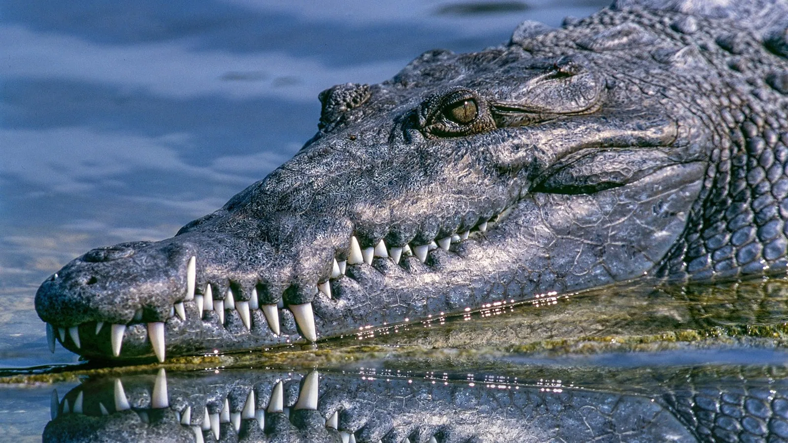
Incisor teeth are the most rostral or anterior teeth in the dental arch, positioned in the premaxilla of the upper jaw and the rostral mandible of the lower jaw. They are single-rooted (with one important exception in rabbits), have a simple, chisel-shaped crown, and act primarily to cut, crop, nip, or groom rather than to grind.
Incisors matter in veterinary practice because they are the teeth most often damaged by trauma, the teeth most affected by malocclusion in rabbits and rodents, and the teeth whose eruption patterns are used to estimate age in both animals and humans. A working knowledge of incisor anatomy and species variation is foundational for oral examination, dental charting, and recognizing when a patient needs intervention.
This article is educational and is not a substitute for veterinary diagnosis or treatment.
What Counts as an Incisor Tooth
An incisor tooth sits at the front of the mouth, immediately adjacent to its partner across the midline. In most mammals the incisors form a continuous row in each jaw, and the upper and lower rows oppose each other for shearing or grasping.
The term incisive teeth refers to the same structures and is often used in comparative anatomy texts. The adjective "incisive" also appears in the name of the incisive bone (the premaxilla), the bony segment that houses the upper incisors.
What Incisors Are Not
Students frequently confuse incisors with the teeth next to them. The table below separates the terms.
| Feature | Incisor | Canine | Premolar / Molar |
|---|---|---|---|
| Position | Most rostral | Immediately caudal to incisors | Caudal to canine |
| Crown shape | Chisel or shovel | Conical, pointed | Blocky, multi-cusped |
| Primary function | Cutting, cropping, grooming | Grasping, defense, holding | Grinding, crushing |
| Root number | One (rabbit upper has two) | One | Multiple in most species |
| Growth pattern | Brachyodont or continuously growing | Brachyodont | Brachyodont |
The canine tooth is a single-cusped tooth used for grasping and, in some species, as a weapon. Incisors are distinguished by their position, their flattened or spatulate crown, and their cutting edge.
Anatomy of an Incisor Tooth
Every incisor, regardless of species, is built from the same four hard and soft tissues arranged in concentric layers.
Crown and Root
The crown is the portion of the tooth covered by enamel and visible above the gingival margin. The root is the portion embedded in the alveolus (the bony socket) and covered by cementum. The boundary between crown and root is the cementoenamel junction, often abbreviated CEJ.
Incisors are single-rooted in humans, dogs, cats, horses, and most rodents. The upper incisor of the rabbit is the classic exception: it has a small secondary (peg) incisor sitting immediately behind the main incisor, so the upper jaw carries two incisor teeth per side rather than one.
Enamel
Enamel is the hardest tissue in the body. It is approximately 96 percent mineral by weight, mostly hydroxyapatite, with a small amount of organic matrix and water. Enamel is produced by ameloblasts, which are lost when the tooth erupts, so mature enamel cannot regenerate.
In brachyodont (short-crowned) teeth such as human, dog, and cat incisors, enamel forms a complete cap over the crown. In continuously growing (elodont) incisors such as those of rabbits and rodents, enamel covers only the labial (front) surface. The lingual (back) surface is dentin, and the differential wear between hard enamel in front and softer dentin behind is what maintains the sharp chisel edge.
Dentin
Dentin lies beneath the enamel and forms the bulk of the crown and root. It is roughly 70 percent mineral, 20 percent organic matrix (mostly type I collagen), and 10 percent water. Dentin is produced by odontoblasts that line the pulp chamber and remain active for life.
Dentin is softer than enamel but harder than bone. It contains microscopic tubules that radiate outward from the pulp, and these tubules transmit fluid and stimuli, which is why exposed dentin is sensitive.
Pulp
The pulp is the soft tissue core of the tooth. It contains blood vessels, nerves, connective tissue, and the odontoblasts that produce dentin. The pulp occupies the pulp chamber in the crown and continues into the root as the root canal, exiting at the apical foramen.
Pulp volume relative to total tooth volume is used in forensic age estimation. A cone-beam computed tomography study of maxillary anterior teeth in children aged 8 to 18 years found that the pulp-to-tooth ratio correlated strongly with chronological age, and that the maxillary central incisor gave a better age estimate than the canine [1].
Periodontium
The tooth is anchored by the periodontium, which includes the gingiva, periodontal ligament, cementum, and alveolar bone. The periodontal ligament is a thin layer of collagen fibers that suspends the root in its socket and absorbs occlusal force. The incisive canal, a bony channel in the anterior maxilla, runs near the roots of the upper incisors, and its proximity to those roots is relevant when teeth are moved or when implants are placed [2].
Dental Formula Notation
Veterinary and human dental anatomy both use the dental formula, a shorthand that records the number of each tooth type in one half of the upper jaw and one half of the lower jaw.
The formula is written as a fraction. The numerator is the upper jaw (maxilla) and the denominator is the lower jaw (mandible). Each number represents one side only, so the total tooth count is the sum of all numbers multiplied by two.
The four tooth types appear in order from rostral to caudal: incisor (I), canine (C), premolar (P), and molar (M).
Worked Examples
Human deciduous and permanent formulas differ. The permanent human formula is:
I 2/2, C 1/1, P 2/2, M 3/3
That means two incisors per side per jaw (eight incisors total), one canine per side per jaw, two premolars per side per jaw, and three molars per side per jaw, for a total of 32 permanent teeth.
The dog formula is:
I 3/3, C 1/1, P 4/4, M 2/3
That gives 12 incisors, 4 canines, 16 premolars, and 10 molars, for a total of 42 teeth in the adult dog.
The cat formula is:
I 3/3, C 1/1, P 3/2, M 1/1
That gives 12 incisors, 4 canines, 10 premolars, and 4 molars, for a total of 30 teeth in the adult cat.
The horse formula is:
I 3/3, C 0-1/0-1, P 3-4/3-4, M 3/3
Horses have 6 upper and 6 lower incisors. The canine teeth are small or absent in mares and are usually present as vestigial "wolf teeth" or canines in geldings and stallions.
Incisor Anatomy and Function by Species
The incisors of each species reflect its diet and its social behavior. The sections below compare humans, dogs, cats, horses, rabbits, and rodents.
Human Incisors
Humans have eight incisors: two central incisors and two lateral incisors in each jaw. They are brachyodont, single-rooted, and fully enamel-covered. The upper central incisor is the largest of the group and is the tooth most often used as a reference in dental anatomy and esthetic dentistry.
Human incisors erupt in a predictable sequence. A prospective study of 1,063 children in Buenos Aires City found that the lower central incisors were the first permanent teeth to erupt and the upper second molars were the last, with lower incisors and canines erupting significantly earlier than their upper counterparts [3]. Girls erupted earlier than boys for nearly all teeth except the upper central incisors [3].
Primary (deciduous) incisors erupt earlier still. A longitudinal cohort study of 100 infants measured crown eruption on serial maxillary casts and found that preterm infants had a mean eruption delay of about 2.7 months compared with full-term infants, with the largest delay (4.5 months) seen for canines [4].
Human incisors function mainly in cutting and incising food and in speech articulation. They are also the teeth most commonly affected by traumatic injury in children aged 7 to 12 years [5].
Dog Incisors
Dogs have 12 incisors: three per side in each jaw. The three upper incisors are named, from medial to lateral, the central incisor (I1), intermediate incisor (I2), and corner incisor (I3). The lower incisors follow the same naming.
Dog incisors are small, closely spaced, and used for grooming, nibbling, and grasping. They are not the primary biting teeth. The canines do the heavy holding, and the carnassial teeth (upper fourth premolar and lower first molar) do the shearing.
Incisor eruption in the dog begins with the deciduous incisors at roughly 3 to 4 weeks of age and the permanent incisors at roughly 3 to 5 months. The exact timing varies with breed size, and large-breed dogs tend to erupt slightly later than small-breed dogs.
Cat Incisors
Cats also have 12 incisors, arranged in the same 3/3 pattern as the dog. They are small and delicate and are used primarily for grooming and for nibbling meat from bone.
Cat incisors erupt as deciduous teeth at about 2 to 4 weeks and are replaced by permanent incisors at about 3 to 4 months. The upper and lower incisor rows are aligned so that the upper incisors slightly overlap the lower ones when the mouth is closed.
Horse Incisors
Horses have 6 upper and 6 lower incisors. They are used to crop grass and other forage, which is then passed back to the premolars and molars for grinding.
Horse incisors are brachyodont but have a long reserve crown, so they continue to erupt for much of the animal's life even though they are not continuously growing in the same sense as rodent incisors. The occlusal (biting) surface shows a characteristic pattern of enamel folds, and the shape of the incisal table changes with age. This change is the basis for estimating a horse's age by examining the incisors.
The deciduous incisors erupt shortly after birth, and the permanent incisors erupt in a sequence that is well documented in equine dentistry. The permanent central incisors erupt at roughly 2.5 years, the intermediates at roughly 3.5 years, and the corners at roughly 4.5 years.
Rabbit Incisors
Rabbits have a distinctive incisor arrangement. The upper jaw has two incisors per side: a large main incisor and a small peg incisor directly behind it. The lower jaw has one incisor per side. This gives the dental formula I 2/1, C 0/0, P 3/2, M 3/3, for a total of 28 teeth.
Rabbit incisors are open-rooted (elodont), meaning they grow continuously throughout life. The upper incisors grow at roughly 2 mm per week and the lower incisors slightly faster. Growth is balanced by wear from the opposing teeth and from abrasive food.
The peg incisor is easy to miss on a quick oral examination. A rabbit with a missing peg incisor or with abnormal wear on the main incisor should be evaluated for malocclusion, because the peg incisor helps guide the main incisor and contributes to normal wear.
Rodent Incisors
Rodents, including rats, mice, hamsters, guinea pigs, and chinchillas, have a reduced incisor count. Most rodents have one incisor per side in each jaw, giving the formula I 1/1, C 0/0, P 0/0, M 3/3 in many species.
Rodent incisors are open-rooted and grow continuously. The enamel is present only on the labial surface, and the softer dentin on the lingual surface wears faster, which keeps the incisal edge sharp. If the upper and lower incisors do not meet properly, the teeth can grow into the palate or through the jaw, a condition called malocclusion.
Guinea pigs and chinchillas are particularly prone to incisor malocclusion because their cheek teeth also grow continuously and can develop overgrowth that changes the occlusion of the incisors.
Summary Table: Incisor Comparison Across Species
| Species | Incisor count | Eruption time (permanent) | Primary function |
|---|---|---|---|
| Human | 8 | Lower central incisors first, around 6 to 7 years; upper central incisors around 7 to 8 years [3] | Cutting, speech |
| Dog | 12 | 3 to 5 months | Grooming, nibbling, grasping |
| Cat | 12 | 3 to 4 months | Grooming, nibbling |
| Horse | 12 (6 upper, 6 lower) | Central at ~2.5 years, intermediate at ~3.5 years, corner at ~4.5 years | Cropping grass |
| Rabbit | 6 (4 upper including peg incisors, 2 lower) | Continuous growth, no fixed eruption endpoint | Cropping, gnawing |
| Rodent | 4 (1 upper, 1 lower per side) | Continuous growth | Gnawing, opening seeds |
How Incisors Are Examined in Practice
A complete incisor examination in a veterinary patient follows a consistent sequence.
First, observe the patient from the front with the mouth closed. Note the position of the upper and lower incisors relative to each other, the presence of any obvious overgrowth, and any asymmetry between the left and right sides.
Second, lift the upper lip and examine the upper incisors. Count them, check for fractures, discoloration, and mobility, and inspect the gingival margin for inflammation.
Third, examine the lower incisors in the same way. In rabbits and rodents, use an otoscope cone or a dental mirror to look at the lingual surface of the upper incisors, because this is where early malocclusion shows up.
Fourth, assess occlusion by gently closing the mouth and observing how the incisal edges meet. In a normal rabbit, the upper incisors should overlap the lower incisors and the wear surfaces should be even.
Radiography is used when the roots or the reserve crowns need to be evaluated. Intraoral radiographs are standard in dogs and cats. In rabbits and rodents, skull radiographs or computed tomography are often needed because the reserve crowns extend deep into the skull.
Clinical Relevance, Limitations and Common Mistakes
Incisor problems are common in small animal and exotic practice, and several clinical patterns are worth knowing.
Traumatic injury to incisors is most frequent in young animals and in animals that chew on hard objects. A fractured incisor with pulp exposure requires treatment, because the exposed pulp can become infected and lead to periapical disease. Traumatic intrusion of a permanent incisor is a severe injury that can be complicated by pulp necrosis, root resorption, and ankylosis, and management often requires a multidisciplinary approach [6]. In some cases, spontaneous re-eruption can occur even after a long period of non-eruption, so conservative management with careful follow-up is sometimes appropriate [7].
Malocclusion in rabbits and rodents is one of the most common reasons for presentation. It can be congenital or acquired, and it can affect the incisors alone or both the incisors and the cheek teeth. Clinical signs include drooling, weight loss, difficulty eating, and visible overgrowth of the incisors. Treatment usually involves trimming or grinding the incisors under anesthesia and addressing any underlying cheek tooth disease.
Molar incisor hypomineralisation is a human condition in which the enamel of the first permanent molars and often the incisors is defective. It is relevant to veterinary students mainly as a comparative example of how enamel defects present, and it is mentioned here because it is a well-studied cause of incisor enamel abnormality in children [8][9].
Common mistakes students make when examining incisors include the following.
Counting only the visible teeth and missing the rabbit peg incisor. The peg incisor sits behind the main upper incisor and is easy to overlook.
Assuming that a discolored incisor is normal. Discoloration usually indicates pulp disease or previous trauma and should be investigated.
Forgetting that rodent and rabbit incisors grow continuously. A tooth that looks short today can overgrow within weeks if the opposing tooth is lost.
Treating incisor malocclusion without examining the cheek teeth. In rabbits and guinea pigs, incisor overgrowth is often secondary to cheek tooth disease, and trimming the incisors alone will not solve the problem.
Using human eruption times as a reference for animals. The timing and sequence of eruption differ substantially across species, and species-specific references should be used.
Quick Review
- Incisors are the most rostral teeth, single-rooted in most species, and used for cutting, cropping, or grooming.
- Every incisor is built from enamel, dentin, pulp, and cementum, with the periodontium anchoring it in the alveolus.
- Dental formula notation records tooth counts per side per jaw, with the upper jaw written above the lower jaw.
- Humans have 8 incisors, dogs and cats have 12, horses have 12, rabbits have 6 (including peg incisors), and rodents have 4.
- Rabbit and rodent incisors are open-rooted and grow continuously, so malocclusion is a lifelong risk.
- Human lower central incisors are typically the first permanent teeth to erupt [3].
- Incisor trauma is common in young animals and in children aged 7 to 12 years [5].
Frequently Asked Questions
How many incisors does a dog have?
A dog has 12 incisors, three per side in the upper jaw and three per side in the lower jaw.
Do rabbit incisors keep growing?
Yes. Rabbit incisors are open-rooted and grow continuously throughout life, which is why regular dental checks are important.
What is the dental formula for a cat?
The cat dental formula is I 3/3, C 1/1, P 3/2, M 1/1, for a total of 30 adult teeth.
Why do rodent incisors stay sharp?
Rodent incisors have enamel only on the front surface and softer dentin behind. The dentin wears faster, which maintains a sharp chisel edge.
Which permanent tooth erupts first in humans?
The lower central incisors are typically the first permanent teeth to erupt, followed by the upper central incisors [3].
Can a broken incisor heal on its own?
No. Enamel cannot regenerate, and a fractured incisor with pulp exposure needs veterinary or dental treatment to prevent infection.
Related Articles
- Snake Anatomy: Do Snakes Have Teeth, Spines, and Other Structures?
- Equine Larynx Anatomy and Function in Respiration
- Canine Lymphatic System: Anatomy and Function
- Equine Hoof Anatomy: Structures and Function
- Parrot Species Comparison: Choosing the Right Parrot for You
- Fish Gills: Anatomy, Function, and Common Health Issues
- Teeth Diagram: Types, Numbers and Animal Comparison
- Primary Dentition: Deciduous Teeth Explained
- Teratoma: Tumors with Hair and Teeth
Sources
- A Comparative Evaluation of Chronological Age with Dental Age Based on Volumetric Analysis of Pulp-Tooth Ratio of Maxillary Anterior Teeth: A Cone-beam Computed Tomography Study.
- 3D analysis of incisive canal morphological changes and proximity to maxillary incisor roots following anterior tooth movement: a comparison of aligner and fixed appliance therapy.
- Chronology and sequence of permanent tooth eruption in children in Buenos Aires City.
- Timing of primary maxillary tooth eruption in preterm compared to term infants: a retrospective longitudinal cohort study.
- AI in Learning Anatomy and Restoring Central Incisors: A Comparative Study.
- Management of a Severe Traumatic Intrusion of a Permanent Incisor With Spontaneous Re-Eruption After Maxillary Expansion: A Surgical-Orthodontic Case Report.
- Spontaneous eruption of a maxillary central incisor near the nasal floor following primary incisor avulsion: a 10-year follow-up case report.
- Tooth-level outcomes in molar incisor hypomineralisation affected molars with post-eruptive breakdown: a randomized clinical trial comparing glass ionomer and resin composite.
- Assessment of the effectiveness of intraosseous anaesthesia during restorative procedures on teeth affected by molar incisor hypomineralisation in children.