Lingual Tonsils: Anatomy, Location, and Function
By Dr. Zubair Khalid, DVM, MS, PhD ·
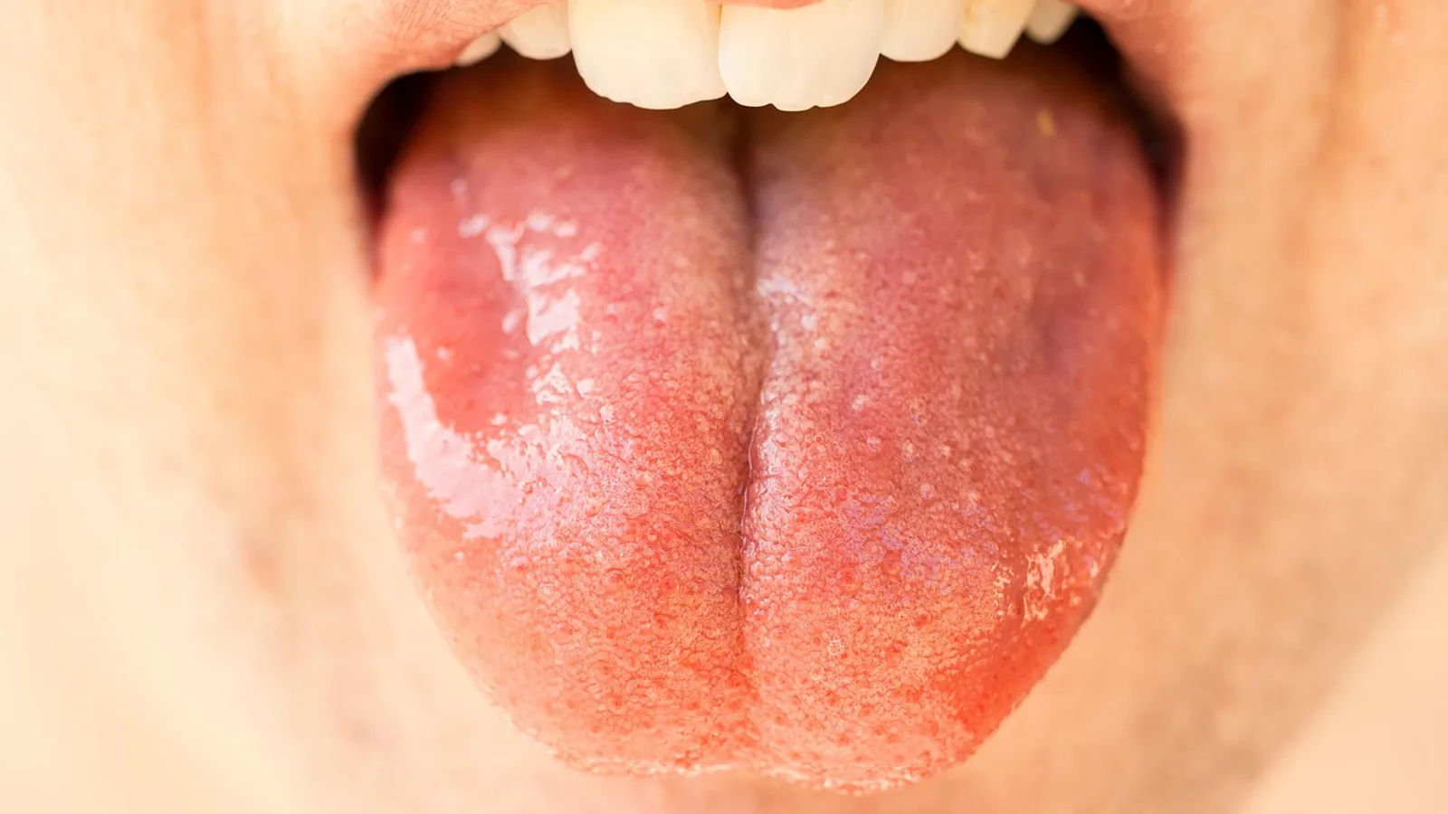
The lingual tonsils are unencapsulated aggregates of lymphoid tissue embedded in the submucosa at the root (base) of the tongue, sitting posterior to the circumvallate papillae and forming the oral component of Waldeyer's ring. They consist of stratified squamous epithelium invaginated into crypts, beneath which sit lymphoid follicles with germinal centers, all supported by connective tissue rather than a fibrous capsule.
Why the Lingual Tonsils Matter
Every swallow carries a sample of the outside world across the base of the tongue. Food, saliva, oral bacteria, inhaled dust, and viral particles all contact this surface within seconds of entering the pharynx. The lingual tonsils sit at that checkpoint and sample antigens before they reach the larynx and lower airway.
For veterinary students, the lingual tonsils matter for three practical reasons. First, they are a standard examinable component of the mucosal immune system, and confusing them with the palatine tonsils is a common error. Second, they are a recognized site of lymphoid accumulation for pathogens, including the agents of transmissible spongiform encephalopathies, which has direct regulatory consequences at slaughter [1][2]. Third, lingual tonsillitis and lingual tonsil hypertrophy are real clinical entities in dogs, cats, and other species, and they can produce dysphagia or upper airway obstruction [3][4].
This article is educational and is not a substitute for veterinary diagnosis or treatment.
Where Are the Lingual Tonsils Located?
The lingual tonsils occupy the root of the tongue, the caudal third of the dorsal lingual surface. The key landmark is the row of circumvallate (vallate) papillae, which sits just rostral to the tonsillar tissue. In the bovine tongue, organized lymphoid tissue extends up to 30 mm rostral of the most caudal vallate papilla, and this disseminate portion is not visible to the naked eye [5][2]. That single number explains why regulatory trimming of the tongue root is anatomically difficult: lymphoid tissue is present beyond what a gross inspection reveals.
The lingual tonsils are one of several tonsils arranged in a ring around the entrance to the pharynx. Collectively this ring is called Waldeyer's ring, and it includes the palatine tonsils, the pharyngeal tonsil, the tubal tonsils, the tonsil of the soft palate, and the paraepiglottic tonsil in species that have them [6][7]. In the camel, all six tonsil types are present and well developed, with the lingual, palatine, velar, and paraepiglottic tonsils arranged as closely assembled lymphoid follicles with numerous crypt openings into the oropharyngeal tube [7].
Position Relative to Neighboring Structures
Rostrally, the lingual tonsil borders the vallate papillae and the body of the tongue. Caudally, it transitions into the oropharynx and the tissues of the tongue root. Dorsally it faces the soft palate. Ventrally it sits above the intrinsic musculature of the tongue, separated from muscle by lamina propria and, in many species, by seromucous glands [8][9].
This position is clinically significant. Because the lingual tonsil sits at the base of the tongue, hypertrophy narrows the oropharyngeal airway at exactly the point where the tongue base, soft palate, and epiglottis converge. That is why lingual tonsil hypertrophy is studied as a contributor to obstructive sleep apnea in people and why base-of-tongue reduction surgery improves swallowing and quality of life in chronic lingual tonsillitis [3][4].
Structure of the Lingual Tonsils
The lingual tonsils are built from four repeating components: surface epithelium, crypts, lymphoid follicles, and supporting connective tissue. There is no capsule.
Epithelium and Crypts
The outer surface is lined by stratified squamous epithelium, keratinized in some species and non-keratinized in others. In the horse, the outer surface epithelium is stratified squamous non-keratinized and is modified by heavy lymphoid infiltration to form reticular epithelium within the crypts [8]. In the one-humped camel, each macroscopic nodule has a single central crypt covered by keratinized stratified squamous epithelium, and the crypt itself is lined by stratified squamous non-keratinized epithelium infiltrated with lymphocytes [6].
Reticular epithelium is the functional heart of the tonsil. It is a discontinuous, lymphocyte-filled epithelium that brings antigens into close contact with immune cells. The equine lingual tonsil crypt epithelium contains numerous CD4 and CD8 positive cells along with macrophages and IgGb and IgA positive cells [10]. This arrangement is what allows the tonsil to initiate and maintain immune responses to incoming infectious agents and antigens [8].
One structural detail is worth remembering for exams: no M cells have been identified in the equine lingual tonsil or the camel lingual tonsil [8][6]. M cells are specialized antigen-transporting cells found in gut-associated lymphoid tissue and in some tonsils, but the equine lingual tonsil instead relies on its reticular epithelium. Vimentin filaments were not detected in the horse, consistent with the absence of M cells [8].
Lymphoid Follicles and Germinal Centers
Beneath the epithelium, the lamina propria contains lymphoid follicles. In the horse, follicles consist of a parafollicular area, a corona, and a germinal center. The parafollicular area holds large numbers of CD4 and CD8 positive lymphocytes, macrophages, and interdigitating cells. The germinal center contains mainly IgGb and IgG(T) positive cells, plasma cells, and small numbers of follicular dendritic cells, macrophages, and CD4, CD8, and IgA positive cells [10].
Follicular dendritic cells are the marker that distinguishes true organized tonsillar tissue from nonspecific lymphocyte accumulations. In cattle, monoclonal antibodies CNA.42 and D46 detect bovine follicular dendritic cells in situ, and immunopositivity for these cells was used to confirm that lymphoid nodules up to 30 mm rostral of the caudal vallate papilla are genuinely part of the lingual tonsil [5].
High Endothelial Venules
Tonsils need a way to recruit lymphocytes from the bloodstream. They use high endothelial venules, which are postcapillary venules with tall, cuboidal endothelial cells. In the equine lingual tonsil, venules in the parafollicular and interfollicular areas show irregular nuclei and cytoplasmic processes on the luminal surface, features characteristic of high endothelial venules. These cells contain a vesiculo-vacuolar organelle whose stomata and diaphragms communicate with each other and with inter-endothelial clefts, forming a structural basis for enhanced permeability and extravasation of macromolecules [10].
Absence of a Capsule
The lingual tonsils are not encapsulated. They differ from the palatine tonsils in this respect, and the distinction is not trivial. A capsule allows a surgeon to dissect a tonsil along a defined plane. Without one, lingual tonsillar tissue blends into the surrounding lamina propria, submucosa, and muscle, and complete removal is difficult. This is precisely the problem described for bovine tongues at slaughter, where variations in distribution suggest that traces of tonsillar tissue may remain even after the most rigorous trimming of the tongue root [1].
Comparison of Tonsil Types
| Tonsil | Typical location | Surface epithelium | Capsule |
|---|---|---|---|
| Lingual | Root of tongue, posterior to vallate papillae | Stratified squamous, keratinized or non-keratinized, with crypts and reticular epithelium | Absent (incomplete in camel) |
| Palatine | Lateral wall of the oropharynx | Stratified squamous with crypts | Present |
| Pharyngeal | Roof of the nasopharynx | Pseudostratified columnar ciliated in yaks | Present |
| Tubal | Around the pharyngeal opening of the auditory tube | Pseudostratified columnar ciliated in yaks | Present |
| Soft palate | Oral surface of the soft palate | Stratified squamous non-keratinized with perforations or small crypts | Absent |
| Paraepiglottic | Base of the epiglottis | Stratified squamous | Present (species dependent) |
The camel lingual tonsil is a partial exception to the "no capsule" rule. Each nodule possesses a single central crypt and is surrounded by an incomplete capsule [6]. That detail is worth noting because it shows the capsule is a spectrum rather than a binary feature.
Species Differences in Lingual Tonsil Anatomy
The lingual tonsil is not equally developed across domestic species. This is one of the most examinable comparative points in veterinary anatomy.
Dogs and Cats
The lingual tonsils are consistently present and prominent in dogs and cats as part of the oropharyngeal lymphoid ring. They are located at the base of the tongue and share the general architecture described above: stratified squamous epithelium with crypts, underlying lymphoid follicles, and no capsule. In clinical practice, lingual tonsillar tissue in these species is encountered during oral examination, during evaluation of dysphagia, and as a differential for a base-of-tongue mass.
Horses
The equine lingual tonsil is well developed and has been studied in detail. It sits at the root of the tongue and presents an irregular surface with rounded elevations, numerous folds, and crypts. The crypt epithelium is reticular and heavily infiltrated with lymphoid cells. Lamellated structures resembling Hassall's corpuscles appear toward the outer surface epithelium, and microplicae are visible on the surface of both the outer and reticular epithelia [8]. Mucus glandular acini in the deeper lamina propria contain glycogen and acidic, neutral, and weakly sulphated mucopolysaccharides [8].
The horse also has a tonsil of the soft palate, an oval flat structure on the oral surface of the soft palate, but its epithelium is perforated by holes or small crypts rather than true crypts and reticular epithelium. Its lymphoid tissue is much less abundant than in the lingual and palatine tonsils [11]. Do not confuse the two.
Ruminants
Ruminant anatomy is the source of the most common misconception. In sheep and goats, lymphoid cells are scattered in the subepithelial propria-submucosa and in the connective tissue cores of the lingual papillae, with relatively more lymphocytes in the cores of fungiform and vallate papillae. Aggregations are especially abundant beneath the grooves surrounding the vallate papillae in small ruminants. However, CD21-positive lymphocytes are numerous in the lymphoid nodules of the bovine lingual tonsil but very scarce in ovine and caprine tongues. On that basis, the lymphoid aggregations in the tongues of small ruminants should not be called a lingual tonsil [12].
Cattle do have a well-organized lingual tonsil at the root of the tongue [12]. It has a macroscopically visible part and a disseminate part extending up to 30 mm rostral of the most caudal vallate papilla [5][2]. In a survey of cattle tongues from British abattoirs, macroscopically identifiable tonsillar tissue was present in more than 75 percent of tongues, and histological examination revealed lymphoid tissue in more than 90 percent of tongues even when no visible tonsillar tissue remained [1].
Yaks have a lingual tonsil as one of their oropharyngeal tonsils, along with the palatine tonsil and the tonsil of the soft palate. In yaks, IgA and IgG mRNA and protein expression from highest to lowest was pharyngeal tonsil, palatine tonsil, tonsil of the soft palate, lingual tonsil, and tubal tonsil. IgG expression was significantly higher than IgA across yak tonsils [9].
Pigs
The porcine lingual tonsil is small. In a stereological study, the lingual tonsil measured 9.4 ± 6.4 mm³ in conventionally reared piglets and 6.3 ± 2.6 mm³ in artificially reared piglets at day 21 postpartum. The tonsil of the soft palate was by far the largest tonsil, with a mean volume of 967.2 ± 122.4 mm³ in conventionally reared piglets [13]. Rearing strategy had no significant effect on the volume of the lingual tonsil or the tonsil of the soft palate [13].
Porcine tonsils also develop after birth. At birth, tonsils consist of diffuse lymphoid tissue without lymphoid follicle aggregations. Lymphoid follicles appear in the soft palate tonsil at 7 days, and by 21 days the lymphoid layer of the nasopharyngeal, soft palate, and lingual tonsils is thicker with clearly visible follicles in the lamina propria. Secondary lymphoid follicles appear in the nasopharyngeal tonsil at 50 days and in the soft palate tonsil at 120 days. Dendritic cells, CD3 positive T cells, and IgA positive B cells increase continuously, especially during the first 21 days [14].
Camels
The camel has all six tonsil types, and they are well developed compared with other domestic species. The lingual tonsils appear as several spherical macroscopic nodules protruding into the oropharynx at the root of the tongue in animals of all ages. Each nodule has a single central crypt, an incomplete capsule, secondary lymphoid nodules extending under the apical epithelium, and interfollicular areas with diffuse lymphocytes, high endothelial venules, macrophages, dendritic cells, and plasma cells [6]. The organization into isolated units with separate crypts increases the surface area available for antigen contact [6].
Species Without Lingual Tonsils
Not every mammal has one. In a study of three Australian marsupials, the koala and two possum species, oropharyngeal tonsils were consistently found in the dorsolateral wall of the caudal oropharynx and additionally in the ventral soft palate of the koala, but aggregated lymphoid tissue was not detected in the tongue, esophagus, or stomach of any of the three species [15].
Function of the Lingual Tonsils
The lingual tonsils are secondary lymphoid tissue and part of the mucosa-associated lymphoid tissue network. Their function is antigen sampling and initiation of local immune responses at the entrance to the digestive and respiratory tracts [6].
Antigen Sampling
Food and inhaled particles contact the lingual tonsil surface directly. The crypts increase surface area and trap material long enough for immune cells to encounter it. Reticular epithelium, with its infiltrating lymphocytes and macrophages, provides the cellular interface. In the horse, the crypt epithelium contains CD4 and CD8 positive cells, macrophages, and IgGb and IgA positive cells [10].
Lymphocyte Recruitment and Organization
High endothelial venules allow lymphocytes to leave the bloodstream and enter the tonsil. In the horse, lymphocytes are visible within the high endothelial venules, consistent with transmural migration [10]. Once inside, B cells organize into follicles and germinal centers while T cells populate the parafollicular and interfollicular regions [10][16].
Antibody Production
Tonsils produce local antibody. In the horse, germinal centers contain mainly IgGb and IgG(T) positive cells along with plasma cells [10]. In yaks, IgG expression is significantly higher than IgA across tonsils, and IgA and IgG antibody-secreting cells are distributed in the subepithelial areas of the non-reticular crypt epithelium, the reticular crypt epithelium, and lymphoid follicles [9]. In sheep, CD21 positive B lymphocytes localize within tonsillar lymphoid follicles, while CD2 and CD3 positive T lymphocytes are numerous in interfollicular regions and aligned underneath and within the epithelium [16].
Developmental Timing
Tonsils are not fully formed at birth. In pigs, the lingual tonsil develops from diffuse lymphoid tissue into organized follicles over the first three weeks of life, with dendritic cells, T cells, and IgA positive B cells increasing continuously during that window [14]. This timing matters for vaccination: oral and nasal vaccine responses depend on the maturation of these local immune tissues.
Clinical Relevance, Limitations and Common Mistakes
Lingual Tonsillitis and Lingual Tonsils Infection
Lingual tonsillitis is inflammation of the lingual tonsils. Causes include viral infection, bacterial infection, and laryngopharyngeal reflux. Chronic lingual tonsillitis presents with recurrent infections, throat discomfort, dyspnea, dysphagia, and lingual tonsil hypertrophy [3]. In people, chronic lingual tonsillitis is treated in selected cases with base-of-tongue reduction, and outcomes include improved swallowing and quality of life, reduced tonsillitis scores, and reduced Friedman grade of lingual tonsil hypertrophy [3].
Lingual tonsil hypertrophy is a recognized contributor to upper airway obstruction. In a study of 117 adults with obstructive sleep apnea, patients with a prior palatine tonsillectomy had significantly greater lingual tonsil hypertrophy grades, with a 57 percent increased risk of grade III to IV hypertrophy [4]. This is a human finding, but the anatomical principle transfers: when one component of Waldeyer's ring is removed, the remaining lymphoid tissue at the tongue base can enlarge. A veterinarian evaluating a dog or cat with stertor, dysphagia, or exercise intolerance should consider the base of the tongue as a site of obstruction, not only the palate and larynx.
Airway Obstruction and Dysphagia
Because the lingual tonsils sit at the base of the tongue, enlargement narrows the oropharyngeal airway. Clinical signs to watch for include noisy breathing, stertor, difficulty swallowing, gagging, and reduced exercise tolerance. Dysphagia in particular can reflect mechanical obstruction by hypertrophied tonsillar tissue or pain from active inflammation.
Lymphoid Tissue as a Pathogen Reservoir
Tonsillar lymphoid tissue can harbor pathogens. In cattle experimentally exposed orally to the bovine spongiform encephalopathy agent, traces of infectivity were found in the palatine tonsil of cattle killed 10 months after exposure, and because infectivity may be present throughout the tonsils, the anatomical distribution of the lingual tonsil was mapped in detail [1]. In sheep, tonsilar lymphoid tissue biopsy has been used to identify scrapie infection, and lateral transmission between sheep under natural conditions has been demonstrated using that approach [17]. These findings are the reason lingual tonsil tissue is treated as specified risk material in bovine carcasses and why adequate removal of the tongue root is regulated [2].
Common Mistakes
The most frequent student error is calling the scattered lingual lymphocytes of sheep and goats a lingual tonsil. They are not. CD21 positive lymphocytes are numerous in bovine lingual tonsil nodules but very scarce in ovine and caprine tongues, so the term should be reserved for the organized structure [12].
The second error is assuming the lingual tonsil has a capsule. It does not, and that is why surgical removal is difficult and why trimming at slaughter may leave residual tissue [1].
The third error is confusing the lingual tonsil with the tonsil of the soft palate. In the horse these are distinct structures with different epithelia, and the soft palate tonsil has far less lymphoid tissue [11].
The fourth error is expecting M cells. The equine and camel lingual tonsils lack M cells and rely on reticular epithelium instead [8][6].
Quick Review
- Lingual tonsils sit at the root of the tongue, posterior to the circumvallate papillae, and form part of Waldeyer's ring.
- They are unencapsulated, with stratified squamous epithelium, crypts, reticular epithelium, and lymphoid follicles.
- They differ from palatine tonsils mainly by the absence of a capsule.
- They are prominent in dogs and cats, well developed and well studied in horses, organized in cattle, and absent as a true tonsil in sheep and goats.
- Lingual tonsillitis has viral, bacterial, and reflux causes and can produce dysphagia or airway obstruction.
- Tonsillar lymphoid tissue can harbor pathogens, which drives regulatory removal of the bovine tongue root.
- No M cells have been identified in equine or camel lingual tonsils.
Frequently Asked Questions
What are the lingual tonsils?
The lingual tonsils are unencapsulated lymphoid aggregates at the base of the tongue, posterior to the circumvallate papillae, forming the oral part of Waldeyer's ring.
Are lingual tonsils encapsulated?
No. The lingual tonsils lack a capsule, which distinguishes them from the palatine tonsils and makes complete surgical removal difficult.
What causes lingual tonsillitis?
Lingual tonsillitis can be caused by viral infection, bacterial infection, or laryngopharyngeal reflux, and it presents with recurrent infection, throat discomfort, dyspnea, dysphagia, and tonsillar hypertrophy.
Do dogs and cats have lingual tonsils?
Yes. Lingual tonsils are prominent in dogs and cats and are part of the oropharyngeal lymphoid ring at the base of the tongue.
Do sheep and goats have a lingual tonsil?
No. Sheep and goats have scattered lingual lymphoid aggregations, but these lack the organized follicular structure of a true tonsil and should not be called a lingual tonsil.
Can lingual tonsils cause airway obstruction?
Yes. Lingual tonsil hypertrophy narrows the oropharyngeal airway and is a recognized contributor to upper airway obstruction and obstructive sleep apnea.
Related Articles
- RNA Location: Types, Functions, and Cellular Distribution
- Z-DNA Found in Biology: Location, Function, and Detection
- Prokaryotic DNA: Location, Structure, and Function
- Equine Larynx Anatomy and Function in Respiration
- Canine Lymphatic System: Anatomy and Function
- Equine Hoof Anatomy: Structures and Function
- Circumvallate Papillae: Structure and Function
- Proteus Mirabilis: Characteristics and Infections
- Dog Eyelid Infection: Causes, Signs, and Treatment
Sources
- Pathogenesis of experimental bovine spongiform encephalopathy: preclinical infectivity in tonsil and observations on the distribution of lingual tonsil in slaughtered cattle.
- Specified risk material and topographical distribution of lymphoreticular tissue of the bovine tongue.
- Transoral Robotic Surgery in Chronic Lingual Tonsillitis: An Observational Cohort Study.
- Lingual Tonsil Hypertrophy in OSA: The Role of Prior Tonsillectomy and Associated Clinical Risk Factors.
- Bovine lingual tonsil: histomorphological characteristics with special reference to the follicular dendritic cells.
- Histological characterization of the lingual tonsils of the one-humped camel (Camelus dromedarius).
- Main anatomical and histological features of the tonsils in the camel (Camelus dromedarius).
- Histology and ultrastructure of the equine lingual tonsil. I. Crypt epithelium and associated structures.
- Yak (Bos grunniens) Tonsils: Morphological Description and Expression of IgA and IgG.
- Histology and ultrastructure of the equine lingual tonsil. II. Lymphoid tissue and associated high endothelial venules.
- Histology, immunohistochemistry and ultrastructure of the tonsil of the soft palate of the horse.
- Distribution of the lingual lymphoid tissue in domestic ruminants.
- The porcine tonsils and Peyer's patches: A stereological morphometric analysis in conventionally and artificially reared piglets.
- Histological studies on the development of porcine tonsils after birth.
- The distribution of organised lymphoid tissue in the alimentary tracts of koalas (Phascolarctos cinereus) and possums (Trichosurus vulpecula and Pseudocheirus peregrinus).
- Differences between the ovine tonsils based on an immunohistochemical quantification of the lymphocyte subpopulations.
- Demonstration of lateral transmission of scrapie between sheep kept under natural conditions using lymphoid tissue biopsy.