Dog Eyelid Infection: Causes, Signs, and Treatment
By Dr. Zubair Khalid, DVM, MS, PhD ·
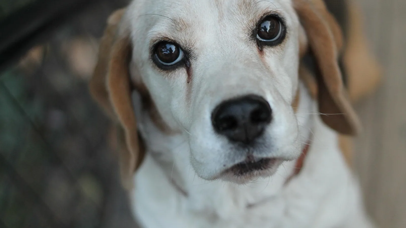
A dog eyelid infection is inflammation or infection of the eyelid skin, the eyelid margin, or the small glands inside the lid. Most cases fall into one of three patterns: blepharitis (diffuse inflammation of the lid, often allergic or bacterial), a stye or hordeolum (a focal infected gland at the lid margin), or an abscess (a painful, fluid-filled pocket under the skin). The pattern matters because the treatment is different for each one. Warm compresses and topical antibiotics suit many styes, abscesses usually need systemic antibiotics and often drainage, and blepharitis is treated by finding and controlling the underlying cause.
This article is educational and is not a substitute for veterinary diagnosis or treatment.
Owner Triage: What to Do First
Look at the eyelid without touching the eyeball. Note four things: whether the swelling is diffuse or a single lump, whether the lid margin has a visible pimple-like bump, whether the surface is red, crusted, or hairless, and whether your dog is squinting, pawing at the face, or holding the eye closed.
Then decide how fast you need help.
- Same-day veterinary care: a swollen lid with a firm, hot, painful lump, yellow or green discharge, fever, lethargy, or a dog that will not let the eye be examined.
- Urgent or emergency care: sudden massive lid swelling with hives or facial swelling elsewhere, difficulty breathing, a bulging eye, a cloudy or blue-tinged cornea, or a wound that goes through the eyelid.
- Routine appointment within a day or two: mild redness and crusting along the lid margin with normal blinking, no corneal cloudiness, and a dog that is eating and behaving normally.
Never squeeze or lance a swollen eyelid at home. Never put human eye drops into a dog's eye without veterinary advice. Both are covered in more detail below.
At a Glance: Blepharitis, Stye, and Abscess
| Feature | Blepharitis | Stye (hordeolum) | Abscess |
|---|---|---|---|
| Onset | Gradual, often weeks | Gradual over days | Rapid, often overnight |
| Distribution | Diffuse, both lids, often both eyes | Single focal bump at the lid margin | Single focal swelling in the lid tissue |
| Appearance | Red, thickened, scaly or crusted lid margin, possible hair loss | Small raised nodule at the margin, may point outward | Firm to fluctuant swelling, hot and painful, may drain pus |
| Discharge | Watery, mucoid, or crusty at the lid line | Minimal, sometimes a bead of pus | Purulent, often thick and yellow-green |
| Pain | Mild to moderate | Mild | Marked, dog resists handling |
| First-line approach | Identify and control the underlying cause, keep the lid clean | Warm compress, topical antibiotic, recheck | Systemic antibiotics, drainage if fluctuant |
| Typical recheck | 2 to 4 weeks | 1 to 2 weeks | 3 to 7 days |
This table is a starting framework, not a diagnosis. A veterinarian confirms the pattern with an examination and, when needed, cytology or culture.
Eyelid Anatomy and Why It Matters
The eyelid is a thin, layered structure. The outer surface is haired skin. Beneath it sits the orbicularis oculi muscle, which closes the lid. Deeper still is the tarsal plate, a firm band of connective tissue that gives the lid its shape. Embedded along the margin are the meibomian glands, which secrete the oily part of the tear film.
That layered design explains the clinical patterns. Inflammation of the skin and margin produces blepharitis. Infection of a single meibomian gland produces a stye, and when that gland infection becomes a walled-off pocket of pus, it behaves like an abscess. Because the meibomian glands sit right at the margin, a small problem there can disrupt the tear film and irritate the cornea. That is why eyelid disease and corneal disease so often travel together.
The meibomian glands are also a documented site of infection. In two dogs with an atypical tumor-like swelling of the upper eyelid, cytology showed neutrophilic inflammation with cocci, and bacterial culture confirmed Staphylococcus pseudintermedius in both [1]. The masses were treated with topical and systemic antibiotics plus anti-inflammatory drugs, and the clinical picture changed when steroids were stopped and antibiotics were chosen based on susceptibility results [1]. The lesson for owners is that a firm lump on the eyelid is not automatically a tumor. Infection of the meibomian glands can mimic one.
Causes of Dog Eyelid Infection
Bacterial Infection
Bacteria are the most common infectious cause of eyelid disease in dogs. Staphylococcus species are the classic offenders, and Staphylococcus pseudintermedius has been confirmed by culture in dogs with meibomian gland infection [1]. Staphylococcal blepharitis can be chronic and recurrent. A study of 44 dogs with chronic staphylococcal blepharitis found that 31 responded to antibiotic and corticosteroid treatment, while 13 needed additional injections of a homologous staph bacterin to control signs after antibiotics and steroids alone failed to prevent recurrence [2]. That study is old, but it established the principle that some staphylococcal lid disease needs more than a single course of medication.
Demodex Mites
Demodex mites live in hair follicles and sebaceous glands. When they overgrow, they cause demodicosis, a severe and common skin disease in dogs [3]. The mites are associated with blepharitis and folliculitis in people, and canine demodicosis is a well-recognized model of mite-driven skin disease [3]. Demodex is a reasonable consideration when blepharitis is chronic, when there is hair loss around the eye, or when the dog has other skin lesions. Diagnosis requires skin scrapings or hair samples examined under a microscope [3].
Allergic and Atopic Disease
Allergy is a leading driver of diffuse blepharitis. Environmental allergens, food allergens, and contact irritants can all inflame the periocular skin. The eyelids are thin and highly vascular, so they react quickly. Allergic blepharitis tends to be bilateral, itchy, and recurrent, and it often comes with redness of the conjunctiva and clear or watery discharge. Because the underlying problem is immune-mediated rather than purely infectious, antibiotics alone do not fix it.
Fungal and Yeast Involvement
Malassezia yeasts are normal inhabitants of canine skin, but they can overgrow and contribute to lid inflammation. A prospective study of 167 eyelids from 84 dogs found Malassezia organisms on the periocular skin of only 3 of 56 clinically normal dogs (5%), while the proportion was significantly higher in eyes with ocular discharge, especially mucoid or mucopurulent discharge [4]. The same study found Malassezia more often on eyes treated with topical ophthalmic medications, particularly aqueous-based products or a combination of oil- and aqueous-based products, than on untreated eyes [4]. This is a useful clinical clue. Chronic topical treatment can change the skin's yeast population, and that change can itself contribute to lid irritation.
Viral Causes
Canine herpesvirus-1 (CHV-1) is an alphaherpesvirus with a host range restricted to canids, and latent infection is endemic in domestic dog populations worldwide [5]. In mature dogs, CHV-1 can produce a range of adnexal and ocular surface lesions, including blepharitis, conjunctivitis, ulcerative keratitis, and nonulcerative keratitis [5]. These can occur during primary infection or when latent virus reactivates [5]. Reactivation has been linked to a variety of triggers, including the administration of immunosuppressive drugs [5]. As more dogs live with chronic immune-mediated disease and receive immunosuppressive therapy, the clinical importance of CHV-1 ocular infection is likely to grow [5].
A 2025 review confirms that CHV-1 infection in adult dogs can manifest as a spectrum of ocular disease from eyelid inflammation (blepharitis) and conjunctival inflammation (conjunctivitis) to more severe corneal conditions, including ulcerative and nonulcerative keratitis [6]. The same review notes that prophylactic topical antimicrobial therapy is recommended to prevent disease progression in dogs with CHV-1 ocular disease [6].
Parasites and Foreign Bodies
Parasites can colonize the conjunctival sac and irritate the lids. Thelazia callipaeda, the eyeworm, has been reported in dogs in Portugal and the Republic of Moldova, where it caused conjunctival hyperemia, serous discharge at the medial canthus, and blepharitis [7][8]. In the Portuguese case, eight worms were collected and identified, and the dog was treated with a subcutaneous injection of ivermectin plus topical ophthalmic fusidic acid drops and oral milbemycin oxime, with resolution of clinical signs one week later [7].
Cutaneous myiasis is a more severe parasitic problem. In two stray dogs in Colombia, Cochliomyia hominivorax larvae were found in a large wound at the palpebral level, and one dog required enucleation because the upper eyelid was destroyed and the cornea was severely damaged [9]. This is an extreme example, but it shows how fast an untreated lid wound can escalate.
Foreign bodies such as plant awns, grass seeds, and grit can lodge under the eyelid and cause focal inflammation. A foreign body should be suspected when blepharitis or conjunctivitis is one-sided and sudden.
Leishmaniasis as a Regional Consideration
Leishmania infantum infection can produce periocular lesions and purulent ocular discharge in dogs. In a study of 77 dogs, 50 (64.9%) had ocular changes, including periocular lesions in 25 dogs (32.5%) and purulent eye discharge in 41 dogs (53.3%) [10]. Conjunctival swabs are a useful non-invasive sample for detecting Leishmania DNA, and PCR positivity was actually higher in dogs without periocular injury than in those with it [10]. Leishmaniasis is not endemic across the United States, but it matters for dogs with travel or import history from endemic regions.
Trauma and Radiation
Direct trauma from a scratch, bite, or rubbing against a rough surface can injure the eyelid and seed infection. Radiation therapy to the head is another documented cause. In dogs receiving fractionated radiation for nasal carcinoma, all dogs developed blepharitis, keratoconjunctivitis, and corneal epithelial atrophy within one month of treatment [11]. Blepharitis has also been reported as a minor transient side effect of soft X-ray therapy used for chronic superficial keratitis [12].
Immune-Mediated Disease
Sterile granuloma and pyogranuloma syndrome (SGPS) is an immune-mediated condition that produces nodular skin lesions. A case report described a dog with a firm 1 cm nodule on the lower palpebral conjunctiva and medial eyelid thickening with ectropion, alongside a prior diagnosis of cutaneous SGPS [13]. Topical and systemic corticosteroids improved the skin lesions but not the conjunctival mass or the eyelid thickening, and the nodule required surgical excision [13]. The eyelid thickening did resolve during a later prednisone taper [13]. This is a reminder that not every eyelid lump is infectious, and some are immune-mediated.
Risk Factors
Several factors raise the odds that a dog will develop eyelid inflammation or infection.
- Breed anatomy. Brachycephalic breeds with a large palpebral fissure and nasal entropion are prone to chronic ocular surface irritation. A review of 601 dogs (1180 eyes) undergoing medial canthoplasty found the Pug was by far the most represented breed, with 403 dogs [14]. Indications included macropalpebral fissure, medial entropion, pigmentary keratitis, trichiasis from a haired caruncle, strabismus after globe prolapse, and facial nerve paralysis [14].
- Skin disease elsewhere. Dogs with atopic dermatitis, demodicosis, or Malassezia overgrowth often have periocular involvement.
- Chronic topical eye medication. As noted above, eyes treated with topical ophthalmic medications had a higher proportion of Malassezia on the periocular skin than untreated eyes [4].
- Immunosuppressive therapy. Reactivation of latent CHV-1 has been linked to immunosuppressive drug administration [5].
- Age. Demodicosis and allergic disease often start in young adulthood, while immune-mediated and neoplastic lid disease becomes more common in middle-aged and older dogs.
- Environment. Dogs that spend time in tall grass, brush, or areas with sand flies face higher exposure to foreign bodies and vector-borne parasites [7][8].
Signs of a Dog Eyelid Infection
Signs can be subtle at first. Owners often notice one eye looks slightly smaller or that the dog is blinking more than usual.
- Swelling. Diffuse puffiness suggests blepharitis. A single raised lump suggests a stye or abscess.
- Redness. The lid margin and conjunctiva look pink to deep red.
- Discharge. Watery discharge points toward allergy or early irritation. Mucoid or mucopurulent discharge is more consistent with infection. In one study, Malassezia was significantly more common on eyes with mucoid or mucopurulent discharge than on eyes without discharge [4].
- Blepharospasm. Squinting and holding the eye closed signals pain. This is a red flag for corneal involvement.
- Alopecia. Hair loss around the eye suggests chronic inflammation, demodicosis, or Malassezia involvement.
- Crusting and scaling. Crusts along the lid margin are typical of staphylococcal blepharitis.
- A visible bump at the lid margin. This is the classic stye appearance.
- Firm, hot, painful swelling. This suggests an abscess, especially if it appeared quickly.
- Third eyelid protrusion. A raised third eyelid often means the eye is painful or the globe is being pushed.
- Ocular discharge with periocular lesions. In dogs with leishmaniasis, periocular lesions and purulent discharge are common findings [10].
How Veterinarians Diagnose Eyelid Infections
Diagnosis starts with a full ophthalmic examination. The veterinarian assesses the eyelids, conjunctiva, cornea, anterior chamber, and tear production. A Schirmer tear test and intraocular pressure measurement are standard when the eye is painful.
Specific tests depend on the pattern.
- Cytology. A swab or impression smear of the lid margin or discharge is examined for bacteria, yeast, and inflammatory cells. In the meibomitis cases, cytology showed neutrophilic inflammation with cocci, which guided culture [1].
- Bacterial culture and susceptibility. Culture is especially useful for recurrent, chronic, or nonresponsive infections. It confirmed Staphylococcus pseudintermedius in both meibomitis cases and guided antibiotic selection [1].
- Skin scrapings and hair samples. These are used to detect Demodex mites when demodicosis is suspected [3].
- Adhesive tape cytology. This non-invasive technique samples the periocular skin surface and can identify Malassezia organisms [4].
- Conjunctival swabs for PCR. Conjunctival swabs can detect Leishmania DNA and are a less invasive, fast, and painless collection method [10]. They have also been used to monitor Leishmania infection in endemic areas [15].
- Biopsy. A persistent or atypical lid mass may need histopathology to rule out neoplasia or confirm immune-mediated disease. In the SGPS case, histopathology revealed a well-demarcated conjunctival stromal nodule with deep extension into the palpebral stroma [13].
- Imaging. Ultrasound or advanced imaging is occasionally used for deep or atypical swellings.
Treatment Decision Points
Treatment follows the pattern. The flowchart below shows the main decision path from owner observation to first-line veterinary action.
flowchart TD
    A[Swollen eyelid] --> B{Diffuse or focal}
    B -->|Diffuse| C[Blepharitis likely]
    B -->|Focal| D{Soft or firm}
    D -->|Soft and fluctuant| E[Abscess likely]
    D -->|Firm margin bump| F[Stye likely]
    C --> G[Find underlying cause]
    G --> H[Allergy or infection control]
    E --> I[Systemic antibiotics]
    I --> J[Drainage if needed]
    F --> K[Warm compress]
    K --> L[Topical antibiotic]
    H --> M[Recheck 2 to 4 weeks]
    J --> N[Recheck 3 to 7 days]
    L --> O[Recheck 1 to 2 weeks]
Blepharitis
Blepharitis is diffuse inflammation, so treatment targets the underlying cause rather than a single spot.
- Allergic blepharitis. The goal is allergen control and reduction of inflammation. This may involve changing the diet, managing the home environment, or using medications prescribed by the veterinarian. Antibiotics alone will not resolve allergic disease.
- Bacterial blepharitis. Topical and systemic antibiotics are used based on cytology and, when indicated, culture and susceptibility results [1]. Chronic staphylococcal blepharitis may recur after antibiotics and corticosteroids are stopped, and some dogs have required additional immunotherapy with a staph bacterin to control signs [2].
- Demodectic blepharitis. Treatment targets the mites. Lotilaner is an isoxazoline drug that was approved in 2017 as an oral veterinary medicine for dogs to treat demodicosis, other mite infections, and tick infections [16]. It works by inhibiting arthropod-selective gamma-aminobutyric acid-gated chloride channels [16].
- Malassezia-associated blepharitis. Reducing yeast overgrowth and reviewing topical medications may help, since Malassezia is more common on eyes treated with certain topical products [4].
- Viral blepharitis. For CHV-1 ocular disease, prophylactic topical antimicrobial therapy is recommended to prevent disease progression [6]. One experimental study of topical cidofovir in dogs with recurrent ocular CHV-1 found that clinical ocular disease scores were significantly higher in the cidofovir group than in the vehicle group, and marked conjunctival pigmentation and blepharitis developed in the cidofovir group but not the vehicle group [17]. That result argues against assuming that every antiviral is helpful for CHV-1 lid disease.
Stye (Hordeolum)
A stye is a focal infection of a lid gland, usually at the margin. First-line care is conservative.
- Apply warm compresses several times daily to encourage drainage and reduce swelling.
- Use a topical antibiotic prescribed by the veterinarian.
- Keep the area clean and prevent the dog from rubbing the eye.
- Recheck in one to two weeks if the stye has not resolved.
If a stye keeps recurring or becomes a firm mass, the veterinarian may reconsider the diagnosis. The meibomitis cases show that a firm lid mass can be infectious rather than neoplastic, and that culture-guided antibiotics plus anti-inflammatory drugs can be effective [1].
Abscess
An abscess is a walled-off collection of pus. It is painful and can rupture on its own.
- Do not squeeze it. Squeezing can push infection deeper into the eyelid and orbit, increase pain, and damage the gland tissue.
- Systemic antibiotics are the foundation of treatment.
- If the abscess is fluctuant, the veterinarian may drain it under sterile conditions, sometimes with sedation or anesthesia.
- Recheck within three to seven days to confirm the infection is resolving.
Surgical and Advanced Options
Surgery is reserved for specific situations. Lip-to-lid subdermal plexus flaps are used to reconstruct lower eyelid defects, most often after tumor removal. A review of 46 cases found that post-oncological reconstruction was the main indication in 87% of dogs and 80% of cats, and optimal flap healing occurred in 10 of 15 dogs (67%) [18]. Complications were common but mostly minor, with flap necrosis in 4 of 15 dogs (27%) and postoperative infection in 1 of 15 dogs (7%) [18]. Blink dysfunction was the most common ophthalmological complication, occurring in five dogs [18].
For deep melting corneal ulcers, freeze-dried amniotic membrane transplantation has been used with good results. In a study of 15 dogs and 5 cats, mean healing time was 17 plus or minus 2 days, and corneal transparency was restored with vision preserved in 90% of cases [19]. This is relevant to eyelid disease only because severe lid problems can lead to corneal damage that requires this level of care.
What Not to Do at Home
Two mistakes cause real harm.
Do not squeeze, pop, or lance a swollen eyelid. An abscess is a contained infection. Squeezing can rupture it into surrounding tissue, spread bacteria, and worsen pain. It can also damage the meibomian glands and leave the lid scarred or deformed.
Do not use human eye drops or ointments without veterinary advice. Human products are formulated for human eyes and may contain preservatives, pH buffers, or active ingredients that are inappropriate for dogs. Some can irritate the cornea or mask a serious problem. Even over-the-counter lubricants can interfere with diagnostic tests if used before an examination.
A third mistake is delaying care for a squinting dog. Squinting usually means corneal pain, and corneal ulcers can worsen quickly.
Prevention
Prevention focuses on the underlying causes and on reducing irritation.
- Control allergy and skin disease. Dogs with atopic dermatitis or recurrent skin infections benefit from consistent management of the primary condition.
- Keep the periocular area clean. Gentle wiping with a veterinarian-approved product removes discharge and crusts.
- Review chronic topical medications. Because Malassezia is more common on eyes treated with certain topical products, ask the veterinarian to reassess any long-term eye medication [4].
- Manage parasites. In regions where eyeworms or sand flies are present, parasite prevention reduces exposure [7][8].
- Protect against trauma. Supervise dogs in tall grass and brush, and address any eyelid wound promptly.
- Address breed anatomy. Dogs with macropalpebral fissure or entropion may benefit from corrective surgery to reduce chronic irritation [14].
- Monitor immunosuppressed dogs. Dogs receiving immunosuppressive therapy have a higher risk of CHV-1 reactivation, so any new eyelid or eye sign deserves prompt evaluation [5].
Prognosis
Prognosis depends on the cause and how quickly treatment starts.
- Simple styes usually resolve with warm compresses and topical antibiotics.
- Abscesses generally respond well to systemic antibiotics and drainage when treated early.
- Bacterial blepharitis can be chronic and recurrent. In one older study, 31 of 44 dogs responded to antibiotic and corticosteroid treatment, while 13 needed additional bacterin injections to control signs [2].
- Allergic blepharitis is manageable but often requires ongoing control of the underlying allergy.
- Viral blepharitis from CHV-1 can recur when latent virus reactivates [5].
- Immune-mediated lid disease may need long-term immunosuppression or surgical excision, and response varies. In the SGPS case, the conjunctival nodule required surgery while the eyelid thickening resolved with a prednisone taper [13].
- Severe trauma or myiasis can require enucleation if the eyelid and cornea are destroyed [9].
Emergency Red Flags
Seek immediate veterinary care if you see any of the following.
- Sudden, severe swelling of the eyelid with hives, facial swelling, vomiting, or difficulty breathing
- A bulging eye or a eye that cannot be pushed back into the socket
- A cloudy, blue, or white patch on the cornea
- A wound that goes through the full thickness of the eyelid
- Yellow or green discharge with fever, lethargy, or loss of appetite
- The dog crying out, pawing at the eye, or refusing to open it
- No improvement after 48 hours of home warm compresses for a suspected stye
Limitations and When to Contact a Veterinarian
This article describes general patterns. Individual dogs vary, and a veterinarian needs to examine the eye to make a diagnosis.
Contact a veterinarian promptly if:
- The swelling is getting worse instead of better
- The dog is squinting or holding the eye closed
- There is any discharge that is thick, yellow, or green
- The eyelid feels hot, firm, or very painful
- The dog has a known history of allergy, demodicosis, or immunosuppressive therapy
- The problem has not improved within 48 hours of warm compresses
- The problem keeps coming back after treatment
Frequently Asked Questions
Is a dog eyelid infection contagious to other pets or to people?
Most bacterial eyelid infections in dogs are not considered contagious to people under normal hygiene. Demodex mites from dogs have been linked to skin lesions in a human owner in one case report, so good hand hygiene is sensible [3]. Leishmaniasis is a zoonotic concern in endemic regions, and dogs are the main domestic reservoir [10]. If your dog has a known infectious cause, ask your veterinarian about household precautions.
Can I use human antibiotic eye drops on my dog?
No, not without veterinary advice. Human eye drops are formulated for human eyes and may contain ingredients or preservatives that irritate a dog's cornea. They can also mask a serious problem or interfere with diagnostic tests. Wait for a veterinarian to prescribe the right product.
How long does a dog eyelid infection take to heal?
It depends on the type. A simple stye often improves within a week with warm compresses and topical antibiotics. An abscess usually needs several days of systemic antibiotics before it clearly improves. Chronic blepharitis can take weeks to months to control, especially when allergy or demodicosis is involved.
Should I squeeze a swollen eyelid to drain it?
No. Squeezing can push infection deeper, spread bacteria, increase pain, and damage the meibomian glands. If an abscess needs drainage, a veterinarian does it under sterile conditions, sometimes with sedation.
What causes a hard lump on a dog's eyelid?
A hard lump can be an infected meibomian gland, a stye, a cyst, or a tumor. In two dogs, firm upper eyelid masses turned out to be antibiotic-responsive Staphylococcus pseudintermedius meibomitis rather than cancer [1]. Any persistent lump should be examined and possibly sampled.
Are some dog breeds more prone to eyelid problems?
Yes. Brachycephalic breeds with a large palpebral fissure and nasal entropion are at higher risk of chronic ocular surface irritation. In a review of medial canthoplasty, the Pug was the most represented breed, with 403 of 601 dogs [14].
Can allergies cause a dog eyelid infection?
Allergies cause blepharitis, which is inflammation of the eyelid. That inflammation can then be complicated by secondary bacterial or yeast infection. Treating the allergy is essential, because antibiotics alone will not resolve allergic lid disease.
When should I go to an emergency vet instead of waiting for an appointment?
Go immediately if the eye is bulging, the cornea looks cloudy or blue, there is a full-thickness eyelid wound, the dog is in severe pain, or there is facial swelling with breathing difficulty. These signs suggest damage that can worsen within hours.
Related Articles
- Chinchilla Respiratory Infections: Causes, Signs, and Treatment
- Guinea Pig Respiratory Infections: Causes, Signs, and Treatment
- Dog Nail Infection: Signs, Causes, and Treatment
- Dog Ear Infection Signs Treatment
- Reptile Respiratory Infections: Causes, Symptoms, and Treatment
- Cat Ear Infection with Odor: Causes and Treatment
- Pannus in Dogs: Causes, Signs, and Treatment
- Staph Infection in Dogs: Signs and Treatment
- Liver Worm in Dogs and Cats: Signs and Treatment
Sources
- Atypical antibiotic-responsive Staphylococcus pseudintermedius-associated tumor-like meibomitis in the upper eyelid: A case report of two dogs.
- Staphylococcal bacterin for treatment of chronic staphylococcal blepharitis in the dog.
- [[A dog related Demodex spp. infestation in a student: a rare Demodex case].](https://pubmed.ncbi.nlm.nih.gov/29933739/)
- Malassezia spp on the periocular skin of dogs and their association with blepharitis, ocular discharge, and the application of ophthalmic medications.
- Canine herpesvirus-1 ocular diseases of mature dogs.
- Prevalence and Clinical Impacts of Canine Herpesvirus-1 (CHV-1) in Dogs: A Review of Reproductive Effects and Ocular Lesions.
- Canine ocular thelaziosis caused by Thelazia callipaeda in Portugal.
- First report of canine ocular thelaziosis in the Republic of Moldova.
- First report of myiasis in dogs caused by Cochliomyia hominivorax (Coquerel 1858) in Colombia.
- Polymerase chain reaction using conjunctival swab samples for detecting Leishmania DNA in dogs.
- Radiation-induced ocular injury in the dog: a histological study.
- Radiotherapy for canine chronic superficial keratitis using soft X-rays (15 kV).
- Nodular conjunctivitis as a novel ocular manifestation of canine sterile granuloma/pyogranuloma syndrome.
- Simplified medial canthoplasty: Technique and early postoperative complications in 601 dogs (1180 eyes).
- Monitoring Leishmania infection and exposure to Phlebotomus perniciosus using minimal and non-invasive canine samples.
- Lotilaner for Demodex Blepharitis: The Journey from Veterinary Use to Human Medicine.
- In Vitro and In Vivo Evaluation of Cidofovir as a Topical Ophthalmic Antiviral for Ocular Canine Herpesvirus-1 Infections in Dogs.
- Outcome of lip-to-lid subdermal plexus flaps to reconstruct defects of the lower eyelid in dogs and cats: 46 cases (2005-2022).
- Surgical Repair of Deep Melting Ulcers With Freeze-Dried Amniotic Membrane Transplantation in Dogs and Cats.