Circumvallate Papillae: Structure and Function
By Dr. Zubair Khalid, DVM, MS, PhD ·
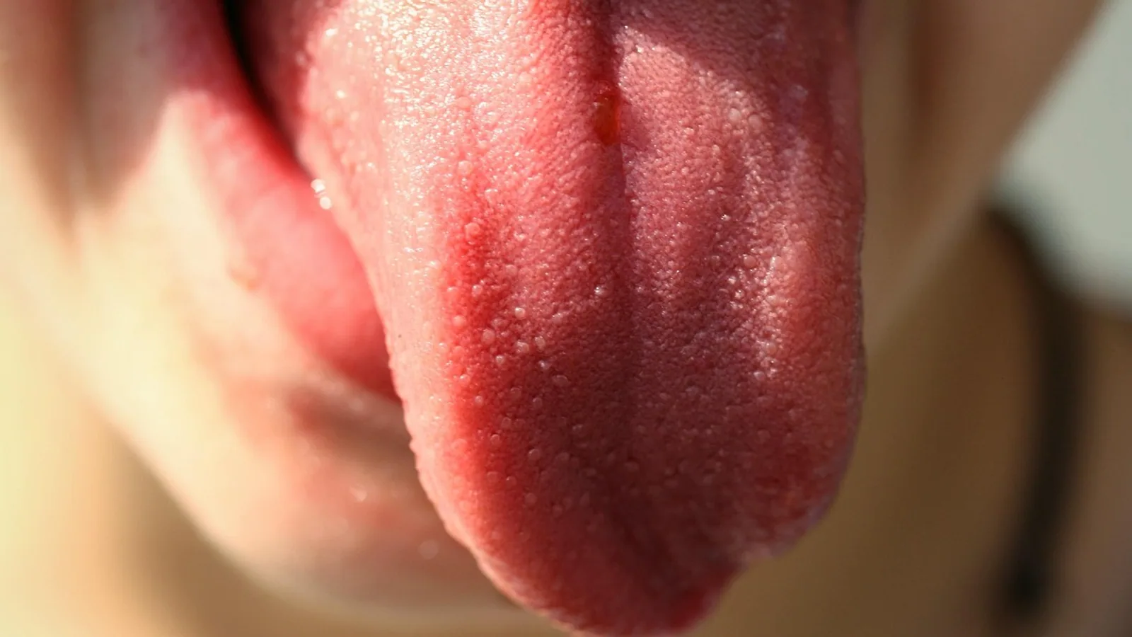
Circumvallate papillae (also written papillae circumvallate, or vallate papillae) are the largest gustatory lingual papillae, arranged in a V- or U-shaped row at the boundary between the body and root of the tongue and encircled by a trench whose base is flushed by serous von Ebner glands. Each papilla carries taste buds on its lateral walls, making it a primary site of bitter and posterior tongue taste detection.
These structures matter because they sit at the crossroads of three systems students often study separately: the gustatory epithelium, the lingual salivary glands, and the immune surveillance network of the tongue. They are also a comparative anatomy landmark. The number, shape, and even presence of circumvallate papillae vary widely across species, from roughly 7 to 12 in humans to several dozen in cattle, and this variation tracks feeding behavior and evolutionary history. For the veterinary student, understanding circumvallate papillae means understanding how a carnivore, a ruminant, and a rodent each sample the chemical world before swallowing.
What Are Circumvallate Papillae?
A lingual papilla is a projection of the lingual mucosa, the stratified squamous epithelium and underlying connective tissue that covers the tongue. Papillae fall into two functional groups. Mechanical papillae, chiefly filiform papillae, provide friction and grip. Gustatory papillae house taste buds and transmit chemical information to the brain via cranial nerves. Circumvallate papillae belong to the gustatory group, along with fungiform and foliate papillae.
The name describes the shape. "Circumvallate" means "surrounded by a wall" or "surrounded by a rampart." Each papilla is a large, dome- or barrel-shaped structure sunk into the tongue surface. A circular trench, or fossa, separates the papilla from a surrounding ridge of tissue called the vallum. Taste buds sit in the epithelium of the papilla's lateral wall, facing into that trench rather than onto the open dorsal surface.
This recessed design has a functional consequence. Tastants dissolved in saliva must flow into the trench to reach the taste pores. The trench is not a dead end. Serous glands open at its base and continuously rinse it, clearing old stimulus and delivering fresh saliva.
Why the Recessed Architecture Matters
Taste is a contact sense. A taste bud only fires when a dissolved molecule reaches its taste pore and interacts with receptors on the apical surface of taste cells. A papilla that sits flush with the tongue surface, like a fungiform papilla, is exposed to whatever passes over it. A circumvallate papilla is different. Its taste buds are tucked into a groove, protected from abrasion by food and from the mechanical shear of chewing and swallowing.
The trench also concentrates stimulus. As saliva and dissolved tastants pool in the groove, the effective concentration at the taste pore can be higher and more sustained than on an exposed surface. The serous glands then reset the system. This combination of protection, concentration, and continuous flushing is the core functional logic of the circumvallate papilla.
Structure of a Circumvallate Papilla
Epithelium and Taste Buds
The papilla is covered by stratified squamous epithelium that is non-keratinized or only lightly keratinized on its lateral walls, in contrast to the heavily keratinized filiform papillae. Taste buds are embedded in this lateral epithelium. A taste bud is an ovoid cluster of 50 to 100 modified epithelial cells that opens to the surface through a narrow taste pore.
Taste buds contain several cell types. Type I cells have a supporting or glial-like role. Type II cells detect sweet, bitter, and umami stimuli through G protein-coupled receptors. Type III cells form conventional synapses with gustatory nerve fibers and mediate sour taste. In mice, type III cells depend on the vesicular protein SNAP25 for both synaptic transmission and long-term maintenance, and loss of SNAP25 reduces type III cell numbers and abolishes sour and ammonium responses in the chorda tympani nerve [1]. Recent work in mice shows that the salty taste channel TMC4 is broadly expressed across all three taste cell types in the circumvallate papillae, with over 98% of TMC4-EGFP positive cells colocalizing with markers for type I, II, and III cells [2].
Connective Tissue Core
Beneath the epithelium lies a connective tissue core, sometimes called the lamina propria core. This core is a vascular and neural scaffold. It carries capillaries that nourish the epithelium and nerve fibers that will synapse with taste cells. In cats, three-dimensional reconstruction of gustatory papillae shows that the connective tissue core of circumvallate papillae has a characteristic mushroom-like shape, distinct from the four core types seen in fungiform papillae [3]. The core shape is not decorative. It determines the surface geometry of the papilla and therefore the volume and flow pattern of the trench.
The Trench and von Ebner Glands
The trench, or fossa, is the defining feature. At its base, ducts from von Ebner glands open into the groove. Von Ebner glands are minor salivary glands, serous in secretion, located in the connective tissue deep to the circumvallate papillae. Their secretion is watery and enzyme-rich, and it serves two roles. It flushes the trench to clear lingering tastants, and it dissolves hydrophobic molecules so they can reach taste receptors.
The glands are not passive plumbing. In mice, Lgr5-expressing cells are concentrated in the excretory ducts of von Ebner glands during the first postnatal week, coincident with the appearance of differentiated taste buds. These Lgr5+ ductal cells behave as candidate stem cells for the turnover of posterior tongue taste buds, which are replaced continuously throughout life [4]. The duct is therefore both a conduit and a stem cell niche.
The ducts also participate in immune surveillance. Single-cell RNA sequencing of mouse circumvallate papillae shows that von Ebner gland duct cells strongly express markers characteristic of microfold (M) cells, the specialized epithelial cells of gut-associated lymphoid tissue. These ducts can transcytose fluorescently labeled E. coli and nanobeads, which are then taken up by underlying immune cells [5]. The taste pore, which must remain open to the lingual surface to sample tastants, is a potential portal for pathogens, and the duct system appears to patrol it.
Innervation
Circumvallate papillae are innervated by the glossopharyngeal nerve (cranial nerve IX). This is a key contrast with the anterior tongue. Fungiform papillae on the anterior two-thirds of the tongue are innervated by the chorda tympani branch of the facial nerve (cranial nerve VII). Taste buds on the soft palate and epiglottis receive fibers from cranial nerves VII and X respectively. Lectin histochemistry in the rat shows that taste buds in circumvallate and foliate papillae, both innervated by cranial nerve IX, stain strongly with Bandeiraea simplicifolia lectin-I, while fungiform and soft palate taste buds, innervated by cranial nerve VII, stain strongly with Ulex europaeus agglutinin-I [6]. The glycoconjugate profile of a taste bud correlates with its nerve supply, which suggests that innervation helps define the molecular identity of the taste epithelium.
The Four Lingual Papilla Types Compared
| Feature | Filiform | Fungiform | Foliate | Circumvallate |
|---|---|---|---|---|
| Primary role | Mechanical, friction and grip | Gustatory | Gustatory | Gustatory |
| Location | Apex, body, and often root | Scattered on dorsal surface, concentrated at apex | Lateral folds at caudal tongue margin | V- or U-shaped row at body-root boundary |
| Keratinization | Heavy, keratinized | Light, non-keratinized on taste surface | Light | Light on lateral walls |
| Taste buds | Absent | Few per papilla | Many, in folds | Many, on lateral walls |
| Gland association | None specific | None specific | Serous lingual glands | Serous von Ebner glands |
| Species notes | Dense in ruminants and cats, with multiple subtypes | Present in most mammals, variable density | Prominent in rabbits, absent in some felids | Large and few to numerous depending on species |
Filiform papillae are the workhorses of the tongue surface. They are keratinized, conical or hair-like, and contain no taste buds. Their function is mechanical: they rasp, grip, and move food. In cattle, six filiform subtypes have been described, varying from long rod-like forms at the tip to leaf-like and conical forms on the body, with accessory processes numbering from one pair to four pairs depending on subtype [7]. In cats, five filiform subtypes plus conical papillae on the root create a tongue surface adapted for grooming and for scraping meat from bone [8]. The dense, caudally directed filiform papillae of cats are the reason a cat's tongue feels rough.
Fungiform papillae are mushroom-shaped and scattered among the filiform papillae, most densely at the tongue tip. They carry a small number of taste buds, typically on their dorsal surface. They are the papillae most often counted in human taste research because they are visible in vivo, and automated software has been developed to count them from digital images to reduce researcher bias [9]. In veterinary species, fungiform papillae vary in number and shape. Arab Zebu cattle have two fungiform subtypes, oval and round, with three to five taste pores on the oval form and five to nine on the round form [7].
Foliate papillae are parallel folds or ridges on the caudolateral margin of the tongue. They contain many taste buds in the epithelium of the folds and are associated with serous glands at the base of the grooves. They are prominent in rabbits. Their presence is variable across species. In a comparative study of four wild felids, foliate papillae were absent in the lion subspecies Panthera leo bleyenberghi and in the Eurasian lynx, while the clouded leopard had delicate smooth folds separated by parallel grooves but without taste buds [8]. This is a useful reminder that the "four papilla types" framework describes a general mammalian plan, not a fixed checklist for every species.
Comparative Anatomy Across Species
Humans
Humans have roughly 7 to 12 circumvallate papillae arranged in a V near the sulcus terminalis. Each papilla contains hundreds of taste buds, and the total taste bud population of the circumvallate row is substantial, though exact counts vary with age and method.
Ruminants
Cattle have many more circumvallate papillae than humans, with counts in the range of roughly 34 to 40 reported in standard anatomy texts. Arab Zebu cattle show ovoid circumvallate papillae surrounded by a U-shaped annular pad and round forms surrounded by a circular pad, and the papillae are concentrated on the lateral surfaces of the torus linguae [7]. The high count fits the ruminant feeding strategy. Ruminants sample large volumes of plant material and benefit from extensive bitter detection to avoid toxic plants.
Carnivores
Cats have relatively few circumvallate papillae, but each is large and well supplied with taste buds. Three-dimensional reconstruction in short-hair cats shows diversity in size and internal connective tissue microstructure of fungiform and vallate papillae, with taste buds either evenly distributed or grouped. The estimated total number of taste buds on the cat tongue is approximately 8,265, distributed across fungiform and vallate papillae, with the caudal tongue region specialized for final tasting before swallowing [3]. In the American mink, four to six circumvallate papillae sit at the border between body and root, and a pair of lateral organs replaces foliate papillae at the caudal edge of the corpus [10].
Rodents
Rodents typically have a single midline circumvallate papilla or a small number, and they lack the lateral asymmetry seen in some other mammals. The rat circumvallate papilla is a standard laboratory model for taste research. Histological and immunohistochemical studies in Sprague-Dawley rats from 6 to 72 weeks of age show that the size of the circumvallate papilla increases with age while the density of taste bud area decreases, even though the number of taste bud sections in a coronal plane stays constant. The number of cells co-expressing the senescence marker SMP30 with the type II cell marker PLCβ2 and the type III marker SNAP-25 declines with age [11]. This is one of the clearest demonstrations that taste decline in aging is cellular, not simply a loss of taste buds.
Primates and Other Mammals
The red ruffed lemur has six to eight vallate papillae arranged in a Y-shape at the border between body and root, alongside foliate papillae at the posterior lateral border [12]. The Abyssinian black-and-white colobus has just three vallate papillae at the body-root boundary [13]. Pallas's squirrel has two or three rounded or elongated vallate papillae, and its tongue shows transitional features between rodents and other mammals, lacking a lingual torus and having connective tissue cores somewhat similar to those of other orders [14]. The desert hedgehog has both mechanical (conical and filiform) and gustatory (fungiform and circumvallate) papillae, with the highest keratinization at the apex and no lingual glands at the apex [15].
The takeaway is that circumvallate papilla number is species-specific and functionally meaningful. It should never be quoted as a single universal figure.
How Circumvallate Papillae Are Studied
Histology
Standard hematoxylin and eosin staining of a longitudinal tongue section shows the circumvallate papilla as a large epithelial dome with a deep trench on each side and a connective tissue core beneath. Taste buds appear as pale ovoid structures within the lateral epithelium, with a narrow pore opening into the trench. Von Ebner glands appear as clusters of serous acini deep to the papilla, with ducts running toward the trench base. The Histology Guide tongue slide is a standard teaching resource for this architecture [16].
Scanning Electron Microscopy and Connective Tissue Cores
Scanning electron microscopy reveals surface detail that light microscopy cannot. After removing the epithelium with a chemical treatment, the underlying connective tissue core can be imaged directly. This technique has been used across many species to compare core morphology. In the red ruffed lemur, the core of the foliate papilla has numerous tubular projections along the groove with a salivary gland conduit at the base, while the fungiform core is a longitudinally ridged cylindrical structure [12]. In the colobus monkey, filiform core morphology differs between juvenile and senescent individuals, with juvenile cores immature and senescent cores forming a concavity surrounded by auxiliary cores [13].
Three-Dimensional Reconstruction
Serial section reconstruction allows researchers to build a spatial model of a papilla and count taste buds accurately. In short-hair cats, this method produced the first spatial visualization of fungiform and vallate papilla connective tissue cores and taste bud arrangement in a carnivore [3]. The method is labor-intensive but gives the most complete picture of taste bud number and distribution.
Molecular and Transcriptomic Methods
Transcriptomic analysis of circumvallate papillae has expanded the picture of what these structures do. In pigs, RNA sequencing of circumvallate papillae from 12 animals identified significant expression of 10 of the 12 known bitter taste receptor genes (T2R3, T2R4, T2R7, T2R9, T2R10, T2R16, T2R20, T2R39, T2R41, and T2R60) and led to the discovery of two novel T2R transcripts, T2R61 and T2R62 [17]. This work shows that the circumvallate papilla is not just a bitter detector in general but a site where a large fraction of the species' bitter receptor repertoire is expressed.
Lectin Histochemistry
Lectin histochemistry uses plant or animal lectins that bind specific sugar residues to map glycoconjugate distribution. A comparative study across five mammalian orders (cow, horse, monkey, dog, and mouse) found that lectin binding patterns differ among circumvallate, foliate, and fungiform papillae, among taste bud cell types, and among species [18]. This suggests that the glycocalyx of taste cells contributes to species differences in taste sensitivity.
Clinical Relevance, Limitations and Common Mistakes
Circumvallate papillae are clinically relevant in several ways. Because they sit at the back of the tongue, they are sometimes mistaken for pathological lesions by owners who catch a glimpse of them during oral examination. The normal circumvallate row can look like a series of raised bumps or even like a row of small ulcers to an untrained eye. A veterinarian who recognizes the normal V-shaped arrangement avoids unnecessary diagnostics.
Taste dysfunction in animals is difficult to assess directly, but the cellular basis is becoming clearer. Age-related decline in taste sensitivity in rats is associated with decreased density of taste bud area in the circumvallate papilla and reduced numbers of cells co-expressing type II and type III markers with the senescence marker SMP30, even though total taste bud number in a coronal section is preserved [11]. This means that a normal-looking taste bud count does not guarantee normal function.
Dietary state affects the gustatory system. In mice, calorie restriction increases fungiform papilla density by up to 35% compared with controls, alongside increased expression of the sweet receptor genes Tas1r2 and Tas1r3 and higher sweet preference [19]. The study measured fungiform papillae at the tongue tip and gene expression in circumvallate papillae, so it demonstrates that systemic metabolic state can alter both anterior and posterior taste structures.
Immune surveillance at the circumvallate papilla is an active area of research. Von Ebner gland ducts express M cell markers and can transcytose bacteria and beads, which are then taken up by underlying immune cells [5]. This suggests that the circumvallate papilla is not only a sensory organ but also an immune checkpoint at the back of the tongue.
Common mistakes students make include the following. First, confusing filiform papillae with gustatory papillae. Filiform papillae are mechanical and keratinized and contain no taste buds. Second, assuming that all mammals have the same number of circumvallate papillae. The count ranges from one or a few in rodents to several dozen in cattle. Third, forgetting that circumvallate papillae are innervated by cranial nerve IX while fungiform papillae are innervated by cranial nerve VII. Fourth, treating foliate papillae as universally present. They are absent in some felids and replaced by lateral organs in the mink [8][10]. Fifth, overlooking the role of von Ebner glands. These glands are not incidental. They flush the trench, dissolve tastants, house stem cells for taste bud turnover, and participate in immune surveillance [5][4].
This article is educational and is not a substitute for veterinary diagnosis or treatment.
Quick Review
- Circumvallate papillae are the largest gustatory lingual papillae, arranged in a V or U at the body-root boundary.
- Taste buds sit on the lateral walls facing a trench, not on the exposed dorsal surface.
- Serous von Ebner glands open into the trench base, flushing it and dissolving tastants.
- Innervation is by cranial nerve IX, unlike fungiform papillae (cranial nerve VII).
- Filiform papillae are mechanical, keratinized, and have no taste buds.
- Circumvallate papilla number is species-specific, roughly 7 to 12 in humans and roughly 34 to 40 in cattle.
- Von Ebner gland ducts house Lgr5+ stem cells for taste bud turnover and express M cell markers for immune surveillance.
Frequently Asked Questions
What are circumvallate papillae?
Circumvallate papillae are large gustatory structures on the back of the tongue, arranged in a V- or U-shaped row and surrounded by a trench that contains taste buds on its inner wall.
How many circumvallate papillae do animals have?
The number varies by species. Humans have roughly 7 to 12, cattle roughly 34 to 40, cats have relatively few large ones, and rodents typically have one or a small number.
Do circumvallate papillae have taste buds?
Yes. They carry many taste buds on their lateral walls, facing into the trench, and are a major site of bitter and posterior tongue taste detection.
What do von Ebner glands do?
Von Ebner glands are serous salivary glands that open into the trench of each circumvallate papilla. They flush the trench, dissolve tastants, and house stem cells and immune surveillance cells.
What is the difference between filiform and circumvallate papillae?
Filiform papillae are mechanical, heavily keratinized, and contain no taste buds. Circumvallate papillae are gustatory, lightly keratinized on their lateral walls, and contain many taste buds.
Which nerve supplies circumvallate papillae?
Circumvallate papillae are innervated by the glossopharyngeal nerve, cranial nerve IX.
Related Articles
- G-Quadruplex Telomere Structure and Function
- Equine Hoof Anatomy: Structures and Function
- Plasmid in Bacteria: Structure, Types, and Functions
- Histone Variants: Structure, Function, and Implications
- Chromatin in a Cell: Structure, Function, and Dynamics
- Nucleosome Protein: Structure, Function, and Assembly
- Papillary Dermis: Structure and Function Explained
- Condyloid Joint: Structure and Movement
- Anatomical Heart: Structure and Chambers
Sources
- Dual functions of SNAP25 in mouse taste buds.
- TMC4 localizes to multiple taste cell types in the mouse taste papillae.
- Three-dimensional reconstruction of gustatory papillae and its taste buds in short-hair cats (Felis Catus domestica, felidae, Carnivora).
- Lgr5+ ductal cells of von Ebner's glands: Candidate stem cells for turnover of posterior tongue taste buds.
- Taste Papillae-associated Salivary Gland Ducts Contribute to Immune Surveillance.
- Glycoconjugate in rat taste buds.
- Biological aspects of the lingual papillae of the Arab Zebu cattle: a new perspicuity of its chad ecological adaptations.
- Comparative Study of Lingual Papillae, Lingual Glands and Lyssa of the Tongue of Selected Wild Felids (Carnivora, Felidae) in Biological Aspects.
- TongueSim: Development of an Automated Method for Rapid Assessment of Fungiform Papillae Density for Taste Research.
- Comparative morphology of the lingual papillae and their connective tissue cores in the tongue of the American mink, Neovison vison.
- Histological and immunohistochemical studies of the fungiform and the circumvallate papillae through the life stages from 6- to 72-week-old Sprague-Dawley male rats.
- The red ruffed lemur, Varecia rubra (É. Geoffroy Saint-Hilaire, 1812): a comparative morphology investigation of lingual papillae and connective tissue cores.
- Comparative morphology of the lingual papillae and their connective tissue cores in the tongue of the Abyssinian black-and-white colobus (Colobus guereza).
- Comparative Morphology of the Lingual Papillae and Their Connective Tissue Cores in the Tongue of Pallas's Squirrel (Callosciurus erythraeus thai, Kloss, 1917).
- Tongue microarchitecture and functional characterization of the lingual papillae in the desert hedgehog (Paraechinus aethiopicus).
- Tongue - Gastrointestinal Tract
- The Bitter Taste Receptor (T2R) Gene Repertoire in the Porcine Circumvallate Papillae Consists of Fourteen Genes, Including Two Newly Validated T2R61 and T2R62.
- Comparative lectin histochemical studies on taste buds in five orders of mammals.
- Weight loss induced by calorie restriction in mice is associated with a higher sweet taste response and increased fungiform papillae density.