Hippocampus Function: Memory and Spatial Navigation
By Dr. Zubair Khalid, DVM, MS, PhD ·
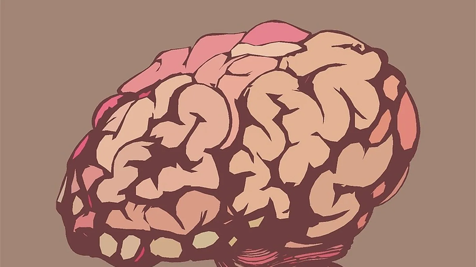
The hippocampus is a paired archicortical structure in the medial temporal lobe whose principal function is to build flexible, relational representations of events and places, allowing an animal to encode where something happened, what happened, and in what order. Its purpose is best summarized as the conversion of fragmented cortical experience into a coherent, retrievable, and predictive map that can guide future behavior.
That single sentence hides an enormous amount of anatomy. The hippocampus is one of the few brain regions where the wiring diagram is known well enough that students can trace a signal from the neocortex to a single principal neuron and back out again. It is also the region where the link between a synapse and a behavior is clearest, largely because of long-term potentiation, the leading cellular model of learning. For a veterinary student, the hippocampus matters for three practical reasons. It explains how animals solve navigation problems, it explains why anesthesia and certain toxins disturb memory, and it provides the anatomical vocabulary you need to read the comparative cognition literature on dogs, birds, bats, and fish.
This article is educational and is not a substitute for veterinary diagnosis or treatment.
Why the Hippocampus Matters in Veterinary Science
Spatial cognition is not an abstract laboratory curiosity. A dog that learns the route to a familiar park, a horse that anticipates a specific gate on a training track, and a mouse that avoids a corridor where it once received an air puff are all expressing hippocampal function. When that function is disrupted, the deficit is often behavioral rather than obviously neurologic. Animals may still walk, eat, and respond to commands while failing to update routes, discriminate similar locations, or retrieve a recently learned avoidance.
The hippocampus also sits at the center of a translational bridge. Rodent spatial tasks such as the Morris water maze are the standard instruments for detecting hippocampal impairment, and those same paradigms have been adapted into desktop virtual mazes for humans to compare mnemonic discrimination across species [1]. Understanding the circuit lets you interpret those results instead of memorizing them.
Core Anatomy of the Hippocampal Formation
The hippocampal formation includes the dentate gyrus, the Ammon's horn (fields CA3, CA2, and CA1), the subiculum, and the adjacent entorhinal cortex. The principal neurons differ by subfield. The dentate gyrus is built from granule cells. CA3 and CA1 are built from pyramidal cells. Both regions contain a vast network of local interneurons that shape when and how those principal cells fire [2].
The dentate gyrus and CA1 are not interchangeable. They differ in cell morphology, synaptic plasticity, signaling molecules, capacity for neurogenesis, vulnerability to insult, and response to drugs [2]. This matters clinically. Anesthetic agents do not silence the whole hippocampus uniformly. Under isoflurane, the local field potential shifts toward delta frequency and single-unit firing rates fall in both CA1 and dentate gyrus, but the dentate gyrus is affected more strongly and recovers its theta power more slowly when the animal wakes [3]. If you are reasoning about post-anesthetic cognitive effects, the gateway region is the one to watch.
The Trisynaptic Circuit Step by Step
The defining feature of hippocampal wiring is a unidirectional trisynaptic pathway that begins in the entorhinal cortex and runs through the dentate gyrus, then CA3, then CA1 [2]. Trace it in order.
- Entorhinal cortex to dentate gyrus (perforant path). Axons from the entorhinal cortex perforate the subicular cortex and synapse on granule cell dendrites in the molecular layer of the dentate gyrus. This is the first synapse of the circuit and the main cortical entry point.
- Dentate gyrus to CA3 (mossy fibers). Granule cell axons, the mossy fibers, project to CA3 pyramidal cells. This is a small, powerful, highly convergent connection.
- CA3 to CA1 (Schaffer collaterals). CA3 pyramidal axons branch as Schaffer collaterals and synapse on CA1 pyramidal dendrites in the stratum radiatum.
- CA1 to subiculum and back to cortex. CA1 is the primary efferent of the trisynaptic loop, projecting onward to the subiculum and returning output toward the entorhinal cortex and neocortex.
There is also a monosynaptic shortcut. Entorhinal axons project directly to CA1, bypassing the dentate gyrus and CA3 entirely. This matters functionally. The trisynaptic pathway and the monosynaptic pathway appear to support different computations, with the trisynaptic route implicated in pattern separation and the direct route implicated in extracting regularities across many experiences [4].
The circuit is not strictly feed-forward. CA3 sends back-projections to the dentate gyrus, and causal analysis in rodent epilepsy models shows these back-projections participate in the network dynamics that generate pathological fast ripples in CA1 [5]. That is a reminder that the tidy textbook arrow diagram is a scaffold, not the whole truth.
flowchart TD
    A[Entorhinal cortex] --> B[Dentate gyrus granule cells]
    A --> C[CA1 direct path]
    B --> D[CA3 pyramidal cells]
    D --> E[CA1 pyramidal cells]
    C --> E
    E --> F[Subiculum]
    F --> G[Entorhinal cortex]
    G --> H[Neocortex]
    D --> B
The diagram shows the main forward route from cortex through dentate gyrus, CA3, and CA1, the direct entorhinal shortcut to CA1, and the CA3 back-projection to the dentate gyrus.
Table: Hippocampal Subfields at a Glance
| Subfield | Main input | Main output | Principal cell type | Functional emphasis |
|---|---|---|---|---|
| Entorhinal cortex | Neocortex, hippocampal return | Dentate gyrus, CA3, CA1 | Stellate and pyramidal cells | Gateway, grid and predictive spatial signals |
| Dentate gyrus | Entorhinal cortex via perforant path | CA3 via mossy fibers | Granule cells | Pattern separation, sparse coding |
| CA3 | Dentate gyrus, entorhinal cortex | CA1 via Schaffer collaterals, back to dentate gyrus | Pyramidal cells | Associative completion, recurrent network |
| CA1 | CA3 via Schaffer collaterals, entorhinal cortex directly | Subiculum, deep entorhinal layers | Pyramidal cells | Comparison of pathways, output generation |
| Subiculum | CA1 | Entorhinal cortex, subcortical targets | Pyramidal cells | Relay of hippocampal output |
The table condenses the anatomy. The functional column is where the interesting disagreements live, and those disagreements are the reason the circuit is studied so heavily.
Spatial Representation: Place Cells, Grid Cells, and Head-Direction Cells
Three cell classes carry most of the spatial signal, and students frequently blur them together.
Place cells are hippocampal pyramidal neurons that fire when the animal occupies a specific location in an environment, the place field. Place cell density is not fixed. It is dynamically modulated by rewards and objects, and computational models reproduce object-centered overrepresentation of place fields in two-dimensional environments and reward-centered overrepresentation in one-dimensional environments [6]. In other words, place cells encode a map that is weighted by what matters to the animal, not a neutral survey.
Grid cells are entorhinal neurons whose firing fields form a repeating hexagonal lattice across space. They provide a metric, a coordinate system that lets the animal estimate distance and direction independent of any single landmark. A recent refinement is the predictive grid cell, an entorhinal neuron whose spatial firing field shifts systematically relative to the animal's direction of travel, so that its activity precedes the animal's future location [7]. These cells are candidates for transmitting prospective spatial information from entorhinal cortex to hippocampus and for organizing theta sequences [7].
Head-direction cells fire as a function of which way the head is pointing, independent of location. They act as an internal compass. They are typically recorded in regions upstream of or parallel to the hippocampus rather than in CA1 itself.
The division of labor is roughly this. Grid cells and head-direction cells supply a metric and a heading, the scaffolding of a coordinate system. Place cells bind that scaffolding to a specific environment and to the salient objects and rewards within it. The hippocampus proper is where self-location and object location converge into a population code that supports memory-based, goal-directed navigation [8].
Allothetic and Idiothetic Cues
Navigation depends on two cue classes. Allothetic cues are external, such as landmarks and distal visual features. Idiothetic cues are internal, generated by self-motion, including vestibular and proprioceptive signals. The medial entorhinal cortex is mainly associated with distal visual cues, while the lateral entorhinal cortex handles proximal cues, and their convergence in hippocampal place cells builds the coordinate system that guides decisions [8].
Theta oscillation organizes the timing of this activity. Late phases of theta support prospective representation through phase precession, in which a place cell fires progressively earlier in the theta cycle as the animal crosses its field. Early phases have been proposed to support retrospective representation through phase procession and the encoding of new associations. Experimental evidence shows this phase code is multiplexed. When rats learned new associations between landmarks and self-motion cues, phase precession stayed intact and continued to predict future positions, while phase procession diminished, matching its proposed role in encoding [9].
Memory Function: Episodic Memory and Pattern Separation
Spatial navigation and episodic memory are often treated as one function because they share medial temporal lobe machinery. They are related but not identical. Natural spatial navigation requires moving the body through space, while episodic memory does not, and functional imaging in children and adults shows that entorhinal and perirhinal activity differs between tour-based and room-based encoding conditions [10]. The medial temporal lobe processes both, but the regions interact somewhat differently in navigational versus episodic contexts and at different developmental stages [10].
One computation that links the two is pattern separation, the creation of distinct representations from similar inputs. It is what lets an animal treat two nearly identical corridors as different places. The dentate gyrus is central to this. In an individual with highly selective bilateral dentate gyrus lesions that disrupt the trisynaptic pathway, pattern separation was significantly impaired on mnemonic similarity tasks, while statistical learning of regularities across many inputs remained intact on implicit measures [4]. That dissociation supports the division of labor between the trisynaptic and monosynaptic pathways.
Dentate gyrus granule cells are well suited to this job because they are sparse and heterogeneous. Most granule cells receive spatially tuned synaptic input, but only a minority convert that input into spatially tuned output, and mature granule cells vary widely in dendritic morphology and intrinsic excitability [11]. This heterogeneity expands the computational capacity of the network and supports pattern separation [11]. The dentate gyrus also receives both "where" information from the medial entorhinal cortex and "what" information from the lateral entorhinal cortex, which places it strategically to gate cortical information into the hippocampal network [11].
Predictive Maps and Trajectory Scanning
The hippocampus does more than record the present. Predictive map theory formalizes its function as a successor representation, a learned estimate of which states are likely to follow the current one [6]. Trajectory scanning extends this idea. Rodent recordings show alternating left-right scans of space in medial entorhinal cortex and place cell firing that scans toward goal locations, providing a mechanism for planning routes to remembered goals and for encoding sequences of vectors that correspond to episodic memories of events at a specific time and place [12]. The same scanning process could generate egocentric boundary and bearing responses consistent with human imaging findings [12].
Long-Term Potentiation: The Leading Plasticity Model
Long-term potentiation (LTP) is a persistent increase in synaptic strength following brief, high-frequency stimulation of a pathway. It is the leading cellular model for how learning is stored, and the hippocampus is where it was characterized most thoroughly. The Schaffer collateral synapse onto CA1 pyramidal cells is the classic preparation because it is laminar, accessible, and robust.
LTP matters for this article because it supplies the mechanism that connects the anatomy to the behavior. A circuit that can strengthen specific synapses can bind a particular combination of place, cue, and outcome into a representation that is retrievable later. The subfields differ in how readily they express plasticity. CA1 and the dentate gyrus differ in synaptic plasticity, signaling molecules, and vulnerability to insult [2], so a manipulation that alters plasticity in one subfield will not produce identical behavioral effects in the other.
How Hippocampal Function Is Tested
Behavioral testing is where hippocampal function becomes measurable, and the paradigms are worth knowing because they appear throughout the comparative literature.
The Morris water maze requires an animal to find a hidden platform using distal spatial cues. Escape latency is the standard readout, and prolonged latency indicates impaired spatial learning or navigation [13].
The spatial similarity task measures mnemonic discrimination by parametrically varying the distance between locations, producing a spatial distance memory function. The same paradigm has been adapted into an open-source desktop virtual maze for humans, deliberately modeled on rodent tasks to bridge the behavioral translation gap [1].
Inhibitory avoidance with route choice tests whether an animal updates a learned route after an aversive experience. In an air puff-based task, water-restricted mice first learned to prefer a short path for water reward, then received an air puff at the center of that path. Mice given only three air puffs showed weak avoidance at a 6-hour memory test, while mice that continued receiving air puffs until they rarely chose the short path showed strong avoidance at both 6 and 24 hours. Chemogenetic suppression of hippocampal activity 30 minutes before the 6-hour test impaired retrieval of the aversive memory [14]. This design is valuable because it separates route learning from route updating and shows the hippocampus is required for retrieval, not only acquisition.
Targeted stimulation is the interventional counterpart. Mice receiving 72 mV at 90 Hz through bidirectional microelectrode arrays aimed at place cell ensembles reached control-level performance after one week of training that took control mice two weeks. The stimulated group showed a 19.7 percent increase in learning trajectory success rate, a 50.2 percent increase in spatial information, and a 25.1 percent reduction in place field area compared with non-stimulated controls [15].
Comparative Notes Across Species
Most of what is taught about hippocampal function comes from rodents, and that provenance should shape how confidently you generalize.
Rodents. Rats and mice provide the core dataset for place cells, grid cells, theta phase coding, and the behavioral tasks above. The virtual reality and electrophysiology work on multiplexed theta phase coding, for example, was performed in rats [9].
Primates, including humans. The same medial temporal lobe structures support episodic and spatial processing, but the interaction between entorhinal and perirhinal cortex changes with age. Connectivity between those regions for episodic memory decreased with age in a developmental study, and exploratory findings suggested behavior-driven connectivity among hippocampus, entorhinal cortex, and lateral occipital cortex during reconstruction of spatial context [10]. Route efficiency in a virtual navigation task has been used as an early indicator of cognitive decline, reflecting the medial temporal lobe's vulnerability in neurodegeneration [16].
Bats. Bats are a natural comparative model because they navigate in three dimensions over large distances, which stresses the two-dimensional assumptions built into most rodent mazes.
Birds. Birds that cache food and retrieve it months later provide some of the strongest evidence that hippocampal specialization tracks navigational demand rather than body plan.
Fish. The teleost forebrain has no structure called a hippocampus, but it has proposed homologs in the pallium. This is where comparative claims need care. Lesions to the lateral or medial telencephalic pallium have been reported to impair allocentric spatial navigation in goldfish, but a critical reexamination argues the behavioral tasks lacked essential controls such as systematic maze rotations and probe trials needed to rule out non-allocentric strategies, and that the lesions were poorly localized, in some cases extending into subpallial regions and the mesencephalic optic tectum [17]. The same commentary notes that the ventral subdivision of the lateral pallium has been linked to map-like spatial memory, while the medial pallium is associated primarily with emotional and motivational processes rather than spatial cognition [17]. The lesson for students is methodological. A lesion that impairs navigation does not prove the lesioned structure is a spatial map unless the task rules out simpler strategies and the lesion is confined.
Clinical Relevance, Limitations and Common Mistakes
The hippocampus is highly plastic, and that plasticity has a cost. Its high degree of neuroplasticity and its control over limbic dynamics make it intrinsically prone to seizure generation, which is why it is the structure most affected in temporal lobe epilepsy models [5]. Hippocampal involvement also appears in developmental conditions. In a mouse model of Fragile X syndrome, astrocyte-specific deletion of Fmr1 altered inhibitory circuit development in CA1, reduced synaptic GABA-A receptor subunits and perisomatic GABAergic synapse density, yet increased the amplitude of spontaneous inhibitory postsynaptic currents in pyramidal cells, with corresponding effects on social and spatial navigation behavior [18]. The finding that astrocytes, not just neurons, shape hippocampal inhibition is a useful corrective to purely neuron-centric thinking.
Environmental insults reach the hippocampus too. Mice exposed to 4.3 GHz microwave radiation at 10 or 30 mW/cm2 for 30 minutes showed prolonged escape latency in the Morris water maze, indicating impaired spatial learning or navigation, alongside hippocampal molecular changes identified by integrated RNA sequencing and proteomic analysis [13].
The limitations are real. Almost all circuit-level claims rest on rodent recordings, and the mapping from rodent to dog, horse, or cat is inferred rather than demonstrated. Hippocampal lesions rarely abolish memory in general. They impair the ability to form and retrieve certain kinds of flexible, relational, and spatially embedded memories while leaving many others intact, which is exactly why the dentate gyrus lesion case showed impaired pattern separation with preserved implicit statistical learning [4].
Common mistakes students make:
- Treating the hippocampus as the memory center. It is a critical node in a distributed system, not a storage vault.
- Confusing place cells with grid cells. Place cells are hippocampal and environment-specific. Grid cells are entorhinal and provide a repeating metric.
- Assuming the trisynaptic circuit is the only route. The direct entorhinal to CA1 projection is functionally important [4].
- Forgetting that dentate gyrus and CA1 respond differently to the same insult or drug [5, 8].
- Reading a lesion study as proof of function without checking whether the task controlled for non-spatial strategies [17].
- Assuming the fish pallium and the mammalian hippocampus are equivalent rather than homologous candidates under active debate [17].
Quick Review
- The trisynaptic circuit runs entorhinal cortex to dentate gyrus via the perforant path, to CA3 via mossy fibers, to CA1 via Schaffer collaterals [2].
- A direct entorhinal to CA1 path bypasses the dentate gyrus and CA3 and is linked to statistical learning rather than pattern separation [4].
- Place cells are hippocampal and environment-specific. Grid cells are entorhinal and metric. Head-direction cells signal heading.
- Predictive grid cells fire ahead of the animal's future location and may carry prospective spatial information into the hippocampus [7].
- The dentate gyrus supports pattern separation through sparse, heterogeneous granule cell firing [11].
- Long-term potentiation at the Schaffer collateral synapse is the leading cellular model of learning.
- Most circuit evidence comes from rodents, so cross-species generalization needs explicit justification [1, 18].
Frequently Asked Questions
What is the main function of the hippocampus?
The hippocampus builds flexible relational representations of places and events, supporting spatial navigation and episodic memory. It converts cortical input into a predictive map that guides future behavior.
What is the difference between place cells and grid cells?
Place cells are hippocampal neurons that fire at one specific location in one environment. Grid cells are entorhinal neurons whose firing fields form a repeating hexagonal lattice that provides a distance and direction metric.
What is the trisynaptic circuit?
It is the unidirectional pathway from entorhinal cortex to dentate gyrus to CA3 to CA1. Each leg uses a named fiber tract: perforant path, mossy fibers, and Schaffer collaterals.
Does the hippocampus store memories permanently?
No. It is required for forming and retrieving certain flexible, relational memories, but long-term storage is distributed across cortical networks. Hippocampal lesions impair specific memory operations rather than erasing memory globally.
Why do rodents dominate hippocampal research?
Their spatial behavior is easy to quantify in mazes, and their laminar hippocampal anatomy is favorable for electrode recording. Rats and mice supply most place cell, grid cell, and theta phase data.
Can hippocampal function be improved?
Targeted electrical stimulation of place cell ensembles accelerated spatial learning in mice, with a 19.7 percent increase in learning trajectory success rate and a 25.1 percent reduction in place field area compared with controls [15]. This is an experimental result, not a clinical protocol.
Related Articles
- Purpose of scoping review
- Orthopedic Dog Bed Purpose
- Newborn Screening: Purpose, Process, and What It Detects
- Buffer Solutions: Types, Purpose, and Applications in Biology
- The Purpose of Meta-Analysis: Why Synthesize Studies Quantitatively?
- Computational Resource Optimization for Variant Calling Pipelines: CPU, Memory, and Storage Considerations
- Why Horses Wear Horseshoes: Purpose and Care
- Dual Purpose Chicken Breeds: Comparison Table for Homesteads
Sources
- The Spatial Similarity Task: A cross-species approach to spatial memory.
- Cellular and Molecular Differences Between Area CA1 and the Dentate Gyrus of the Hippocampus.
- Electrophysiological activity pattern of mouse hippocampal CA1 and dentate gyrus under isoflurane anesthesia.
- Dentate Gyrus Integrity Is Necessary for Behavioral Pattern Separation But Not Statistical Learning.
- Causal relationship of CA3 back-projection to the dentate gyrus and its role in CA1 fast ripple generation.
- A predictive map learned from diverse entorhinal inputs explains the role of context-dependent reorganization of hippocampal place cells.
- Predictive grid cells: Future spatial representations in the hippocampal-entorhinal circuit.
- Mechanistic Integration of Distal and Proximal Cues in the Rodent Entorhinal-Hippocampal Circuit: Insights From a Biorobotics Model.
- Allothetic and idiothetic spatial cues control the multiplexed theta phase coding of place cells.
- Neural Organization of Episodic Memory and Navigation in Children and Adults.
- Integration of spatial and non-spatial information by heterogeneous dentate gyrus granule cells.
- Trajectory scanning as a predictive coding mechanism for goal-directed navigation, obstacle avoidance and episodic memory.
- Sensitive Molecules Involved in Spatial Learning and Memory Impairment of Mice Induced by 4.3 GHz Microwave Radiation.
- An alternative inhibitory avoidance task for studying hippocampus-dependent spatial aversive memory in mice.
- Modulation of place cells using targeted stimulation with bidirectional microelectrode arrays enhances spatial learning speed in mice.
- Route efficiency in spatial navigation as an early indicator of cognitive decline in amnestic mild cognitive impairment.
- Investigating map-like navigation and hippocampus homologs in teleost fish: Comment on Givon et al., 2023 "Lateral and medial telencephalic pallium lesions impair spatial memory in goldfish".
- Hippocampal Astrocytes Impact Postnatal Development of Inhibitory Connections, Parvalbumin Levels, Social, and Spatial Navigation Behaviors in a Mouse Model of Fragile X Syndrome.