Eyesight in Dogs: How Dogs See the World
By Dr. Zubair Khalid, DVM, MS, PhD ·
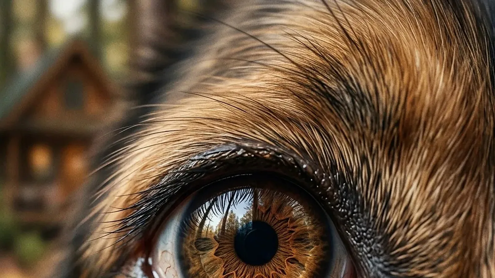
Dogs see the world in two colors, not one, and they see it about four to eight times less sharply than a person with normal vision. Their eyes are built for motion and dim light rather than fine detail, so a dog notices a squirrel's movement long before it can resolve the squirrel's outline.
This is not a defect. Canine eyesight is a different set of trade-offs, tuned by evolution for a crepuscular hunter that tracks moving prey across open ground. Understanding those trade-offs explains a great deal of everyday dog behavior, from the way a dog loses a thrown ball against grass to the way it tracks a hand signal at distance.
This article is educational and is not a substitute for veterinary diagnosis or treatment.
The Short Answer: What Dogs Actually See
Three facts anchor everything else in this article.
First, dogs are dichromats. Their retinas contain two classes of cone photopigment, with spectral peaks near 429 nm and 555 nm [1]. Those peaks correspond roughly to blue-violet and yellow-green. A dog distinguishes blue from yellow, but red and green look similar to each other, closer to shades of yellow-gray. The common claim that dogs see in black and white is wrong. It has been wrong in the scientific literature since at least 1989, when behavioral discrimination experiments in three dogs confirmed two cone pigments and dichromatic color vision [1].
Second, canine visual acuity is modest by human standards. Behavioral testing of whippets, pugs, and a Shetland sheepdog in bright light produced spatial frequency thresholds between 5.5 and 19.5 cycles per degree, while humans tested in the same setup reached 32.1 to 44.2 cycles per degree [2]. In practical terms, a human resolves detail from roughly three times the distance a dog can in both bright and dim light [2]. On a Snellen-style scale, that places typical canine acuity near 20/75, meaning a dog must be about 20 feet from an object to see what a person with 20/20 vision sees at 75 feet.
Third, dogs have a reflective layer behind the retina called the tapetum lucidum. It bounces light back through the photoreceptors for a second chance at absorption, which dramatically improves dim-light sensitivity. The cost is optical scatter. Light reflected off the tapetum is not perfectly aligned, so it smears the image slightly and reduces the sharpness of fine detail.
The Canine Retina: Rods, Cones, and the Area Centralis
Two photoreceptor types with different jobs
The retina is a thin sheet of neural tissue lining the back of the eye. It contains photoreceptors, the light-sensing cells, of two kinds. Rods are numerous, highly sensitive, and operate in dim light. They do not carry color information. Cones are less numerous, need brighter light, and provide color and fine spatial resolution.
Dogs have a rod-dominant retina. Rods far outnumber cones, which is the anatomical basis for their strong scotopic (night) vision and their relatively poor photopic (daylight) detail vision. Cones are not evenly spread across the canine retina. Using peanut agglutinin lectin staining and antibodies against long/medium-wavelength and short-wavelength cone opsins, researchers mapped cone distribution in beagle retinas and identified a visual streak of high density running superior to the optic disc, with a temporal area of peak density called the area centralis [3].
The area centralis is the canine equivalent of the human fovea, the region of sharpest vision. It matters clinically because, unlike the human macula, the canine area centralis is not visible on routine funduscopic examination [3]. A veterinarian looking at a dog's retina through an ophthalmoscope sees the optic disc and the tapetum, but the point of best vision is not marked by an obvious landmark.
Ganglion cells and the wiring behind the eye
Photoreceptors pass their signals to bipolar cells, then to retinal ganglion cells, whose axons form the optic nerve. Ganglion cell types in the dog retina closely correspond to those in the cat. Intracellular staining with Lucifer Yellow identified alpha cells with large cell bodies and large, densely branched dendritic trees, beta cells with medium cell bodies and small, densely branched trees, and several other types with smaller cell bodies [4].
Alpha cells are thought to carry brisk-transient responses, the kind that signal motion and change. Beta cells are thought to carry sustained responses, the kind that carry detail. Dendritic field sizes increase from the central retina to the periphery. Alpha cell dendritic fields grow from 160 to 200 microns centrally to about 1,100 microns peripherally, and beta cell fields grow from 25 microns to about 360 microns [4]. That gradient is the neural signature of a retina that resolves detail in the center and detects motion at the edges.
What the cone pigments measure
The two canine cone pigments peak near 429 nm and 555 nm [1]. A separate electroretinography study using silent substitution stimuli confirmed short-wavelength cones peaking at 429 to 435 nm and long/medium-wavelength cones peaking at 555 nm, and showed that long/medium cone responses peak at high temporal frequencies around 32 Hz under bright light, while rod responses peak at low frequencies around 4 Hz under dim light [5].
That frequency separation is not a laboratory curiosity. It reflects a retina that can hand off from rods to cones as light levels change, and it explains why dogs detect fast motion so well in daylight.
Dog Vision Versus Human Vision: A Direct Comparison
| Feature | Dog | Human |
|---|---|---|
| Cone photopigments | Two (dichromat), peaks near 429 nm and 555 nm [1] | Three (trichromat), short, medium, and long wavelengths |
| Color perception | Blue and yellow-green distinguishable, red and green similar [1] | Full red, green, and blue discrimination |
| Visual acuity (behavioral) | 5.5 to 19.5 cycles per degree in bright light [2] | 32.1 to 44.2 cycles per degree in the same setup [2] |
| Approximate Snellen equivalent | About 20/75 | About 20/20 |
| Acuity in dim light | 1.8 to 3.5 cycles per degree [2] | 5.9 to 9.9 cycles per degree [2] |
| Tapetum lucidum | Present, improves dim-light sensitivity, scatters light | Absent |
| Binocular visual field | About 60 degrees | About 120 degrees |
| Total visual field | About 240 to 250 degrees | About 180 to 200 degrees |
| Flicker detection | Higher than human, roughly 70 to 80 Hz | Roughly 50 to 60 Hz |
| Area of sharpest vision | Area centralis, not visible on funduscopy [3] | Fovea, visible as the macula |
The flicker fusion figures reflect the temporal resolution of the canine retina. A dog integrates visual information in shorter time slices than a person does, so a flickering light that looks steady to a human can still look like a flicker to a dog. This is one reason older fluorescent tubes and some LED fixtures can appear to flicker to a dog when they look constant to the owner.
Field of View, Binocularity, and Eye Position
Eye position on the skull determines how much of the world a dog sees and how much of it is seen with both eyes at once.
Dogs have eyes set more laterally than humans. The total visual field is wider, roughly 240 to 250 degrees compared with about 180 to 200 degrees in humans. The binocular overlap, the region seen by both eyes at once, is narrower, about 60 degrees compared with about 120 degrees in humans.
Binocular overlap is what supports stereopsis, the brain's ability to compute depth from the slight difference between the two eyes' images. A narrower binocular field means a narrower zone of good depth perception, concentrated directly in front of the face. Outside that zone, a dog relies on monocular cues such as motion parallax, relative size, and occlusion to judge distance.
This arrangement suits a predator that needs to detect movement across a wide arc and then fixate on a target. It also explains why a dog may misjudge a jump it approaches from the side, and why a dog tracks a moving object more reliably than it judges a stationary one at distance.
Breed Variation: Brachycephalic Dogs and Sighthounds
The canine skull is remarkably plastic, and eye position varies with it.
Brachycephalic breeds, the flat-faced dogs such as pugs and French bulldogs, have shortened skulls and more forward-facing eyes. That geometry increases binocular overlap and can improve depth perception in the frontal field, but it also compresses the orbit and the nasal cavity. The behavioral acuity study that tested whippets, pugs, and a Shetland sheepdog found large individual variation, with dogs discriminating spatial frequencies between 5.5 and 19.5 cycles per degree [2]. The whippets in that study were among the higher performers, consistent with the sighthound body plan: a long muzzle, laterally placed eyes, and a visual streak adapted for scanning open ground.
Sighthounds such as greyhounds and whippets have a pronounced visual streak, the horizontal band of high cone and ganglion cell density that supports wide-field motion detection. Brachycephalic dogs have a more compressed retina and a shorter visual axis. Neither arrangement is superior in absolute terms. Each matches the hunting and social behavior the breed was developed for.
Breed also matters for inherited retinal disease. Mutations affecting photoreceptors are widespread across breeds, and the clinical course differs by mutation. In dogs with a PRCD variant, outer nuclear layer thinning was detectable in all quadrants by 0.8 years of age, with the fovea-like region spared until about 2.3 years, and the cone-rich visual streak showing only about 22 percent loss by 4.8 years while the inferior retina lost 92 percent [6]. That pattern of regional sparing is why some dogs with inherited retinal degeneration retain useful vision longer than the degree of retinal thinning alone would suggest.
The Tapetum Lucidum: Bright Eyes, Blurred Detail
The tapetum lucidum is a layer of reflective tissue between the retina and the choroid. Light that passes through the photoreceptors without being absorbed hits the tapetum and reflects back through the retina, giving the photoreceptors a second chance to capture it. This roughly doubles the effective light path and is the main reason dogs see better than humans in dim conditions.
The trade-off is resolution. Reflected light is not perfectly collimated, so it scatters as it passes back through the retina. The result is a brighter but slightly softer image. The tapetum also produces the eyeshine seen in photographs and in car headlights, which is a normal reflection and not a sign of disease.
The behavioral data confirm the dim-light advantage is real but modest. In a dim-light condition of 0.0087 cd m⁻², dogs reached acuities of 1.8 to 3.5 cycles per degree while humans reached 5.9 to 9.9 cycles per degree [2]. Dogs gain sensitivity, but humans still resolve more detail in the same dim light. The tapetum buys photons, not sharpness.
Why Motion Matters More Than Detail
A dog's visual system is weighted toward detecting change. The alpha ganglion cells with their large dendritic fields and brisk-transient responses are built to signal movement [4]. The rod-dominant retina extends useful vision into twilight. The wide total field catches motion across a broad arc.
The practical consequence is that a stationary object at distance may be invisible to a dog while the same object moving is obvious. A dog that fails to notice a ball lying in grass may find it immediately once it rolls. A dog that seems to ignore a person standing still at the far end of a field may react the moment that person waves.
This also explains why visual cues in dog-directed communication tend to be large and moving. A pointing gesture is a moving arm. A hand signal is a sweeping motion. Research that altered photographs to approximate canine vision, reducing color information and acuity, found that human participants had difficulty identifying the direction of a glance and followed gaze cues less accurately and more slowly than when viewing the original images [7]. The study authors argued that experimenters should design visual stimuli that fit canine perceptual limits rather than assuming dogs see what people see [7].
Clinical Relevance, Limitations and Common Mistakes
What owners commonly get wrong
The most common error is assuming dogs are colorblind in the sense of seeing only grayscale. They are not. They have dichromatic color vision with two cone pigments [1], and they distinguish blue from yellow-green reliably. A blue toy on green grass is more visible to a dog than a red toy on green grass, because red and green fall close together in canine color space.
The second common error is overestimating how much detail a dog can resolve at distance. A dog that fails to come when called from across a field may not have recognized the owner's face. It may have recognized the owner's motion, gait, or silhouette. Dogs rely heavily on non-visual cues, particularly scent and sound, and a dog with reduced vision may compensate so well that the owner does not notice the loss for months.
The third error is assuming that a dog with cloudy eyes has lost vision. Corneal or lenticular opacity reduces the clarity of the optical path, but the retina and visual pathways may still function. Conversely, a dog with clear eyes can have severe retinal disease. The only way to distinguish is a veterinary ophthalmic examination, often with electroretinography to assess retinal function directly.
Recognizing vision loss at home
Veterinary guidance on recognizing vision loss in pets emphasizes changes in behavior rather than the appearance of the eye. Signs include bumping into objects, reluctance to move in unfamiliar environments, hesitation on stairs, increased startle responses, and changes in how a dog navigates at night [8]. A dog that navigates the family home perfectly well may still be losing vision, because familiar environments can be memorized.
Any sudden change in vision is an emergency. Sudden acquired retinal degeneration, glaucoma, and optic neuritis can all cause rapid vision loss, and several of these conditions are painful or have systemic implications. A dog that becomes suddenly blind, especially with dilated pupils that do not respond to light, needs same-day veterinary assessment.
Inherited retinal disease
Canine inherited retinal degenerations are a major area of both clinical and research interest because dogs serve as models for human retinal genetic disorders [3]. Progressive retinal atrophy describes a group of conditions in which photoreceptors degenerate over time. The clinical course varies by mutation and breed. In one well-characterized form caused by an RPGRIP1 insertion, cone function was lost as early as 2 months of age while rod function was preserved, and by 9 months of age neither cone nor rod function could be detected by electroretinography, yet functional visual assessment showed that affected dogs could still avoid obstacles until about 11 months of age [9]. That gap between electrophysiological loss and observable behavioral loss is a recurring theme in canine retinal disease and a reason that owners often underestimate the severity of a diagnosis.
What remains uncertain
Canine visual acuity has been measured by several methods, and the numbers do not always agree. Pattern visual evoked potential studies in beagles have produced estimates ranging from roughly 0.54 to 2.14 cycles per degree in one study [10] to 14.29 cycles per degree or better in another that corrected for refractive error [11]. Cortical and retinal evoked potential thresholds in dogs under neuromuscular block averaged 12.59 and 11.61 cycles per degree respectively [12]. Behavioral testing has produced values from 5.5 to 19.5 cycles per degree [2]. The spread reflects differences in method, refractive correction, stimulus luminance, and individual variation. The most defensible statement is that canine acuity is substantially lower than human acuity and that the exact figure depends on how it is measured.
The relationship between retinal anatomy and behavior also varies by breed in ways that are not fully mapped. The area centralis is not visible on funduscopy [3], so clinical assessment of the region of best vision relies on indirect methods. For an individual dog, a veterinarian can assess vision and retinal health but cannot give a precise Snellen-equivalent number the way an optometrist can for a person.
Frequently Asked Questions
Are dogs colorblind?
No. Dogs are dichromats with two cone pigments peaking near 429 nm and 555 nm, so they see blue and yellow-green but confuse red with green [1].
Do dogs see in black and white?
No. That claim is a persistent myth. Behavioral color discrimination experiments confirmed two cone pigments and dichromatic color vision in dogs [1].
How sharp is a dog's vision compared with a human's?
Behavioral testing found dogs resolve 5.5 to 19.5 cycles per degree in bright light while humans in the same setup reached 32.1 to 44.2 cycles per degree [2]. That corresponds to roughly 20/75 vision, about three times less detailed than typical human vision.
Can dogs see in the dark?
Dogs see better than humans in dim light because of the tapetum lucidum and a rod-dominant retina, but they still resolve less detail than humans at the same low light level [2].
Why do dogs' eyes glow in photographs?
The glow is light reflecting off the tapetum lucidum, the reflective layer behind the retina that improves dim-light sensitivity. It is normal.
Do dogs see better than humans at detecting motion?
Yes. Dogs have a higher flicker fusion threshold than humans and a retina weighted toward motion detection, with alpha ganglion cells specialized for brisk-transient responses [4].
Do flat-faced dogs see differently from sighthounds?
Yes. Brachycephalic dogs have more forward-facing eyes and greater binocular overlap, while sighthounds have a pronounced visual streak suited to scanning wide open ground [2].
How can I tell if my dog is losing vision?
Watch for bumping into objects, hesitation on stairs, reluctance to move in unfamiliar places, and increased startle responses [8]. A dog can memorize a familiar home well enough to hide gradual vision loss, so changes in new environments are often the first clue.
Related Articles
- Snake Senses: How Snakes Hear, See, and Smell the World
- Rna World Hypothesis
- 50 Cutest Dog Breeds in the World (With Photos & Vet Care Guide)
- RNA World Theory: Origin of Life and Early Evolution
- Real World Example of RNA: From mRNA Vaccines to CRISPR
- Ethical Dilemmas in Research: Real-World Case Studies and Solutions
- Do Dogs See in the Dark? Canine Night Vision
- Dog Eye Shut: Causes and When to See a Vet
- What Colors Can Cats See? Feline Vision Explained
Sources
- Color vision in the dog.
- High visual acuity revealed in dogs.
- Topographical characterization of cone photoreceptors and the area centralis of the canine retina.
- Morphological types of ganglion cells in the dog and wolf retina.
- In vivo electroretinographic differentiation of rod, short-wavelength and long/medium-wavelength cone responses in dogs using silent substitution stimuli.
- PRCD-associated retinitis pigmentosa in dogs and humans.
- Do you see what I see? The difference between dog and human visual perception may affect the outcome of experiments.
- signs of vision loss in pets
- The RPGRIP1-deficient dog, a promising canine model for gene therapy.
- Pattern-stimulated visual evoked potential in dog: changes in elicited response with pattern size and calculation of visual acuity.
- Measurement of visual acuity in Beagle dog by visual evoked potential with pattern stimulation.
- Canine visual acuity: retinal and cortical field potentials evoked by pattern stimulation.