Compact Bone: Structure and Function Explained
By Dr. Zubair Khalid, DVM, MS, PhD ·
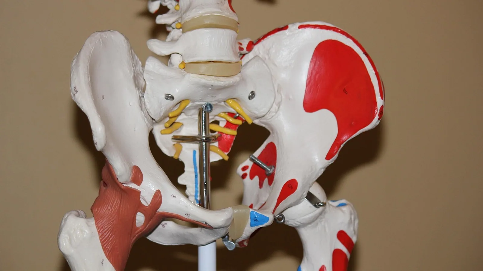
Compact bone is the dense, solid outer layer of a bone that forms a hard shell around the softer spongy bone inside, and it is built from repeating cylindrical units called osteons, each made of concentric lamellae wrapped around a central Haversian canal that carries blood vessels and nerves. Its main function is to resist bending, torsion, and compression while housing osteocytes that maintain the bone matrix and remodel it throughout life.
Understanding compact bone matters because it is where most of a skeleton's mechanical strength lives. It is the tissue a surgeon drills through to place an implant, the tissue a radiologist measures to judge bone quality, and the tissue that fractures when a dog jumps from a height or a horse gallops on hard ground. For veterinary students, compact bone is also the clearest place to learn how bone is organized, because its repeating structure can be seen, counted, and measured under a microscope.
This article is educational and is not a substitute for veterinary diagnosis or treatment.
What Is Compact Bone? Definition and Key Terms
The compact bone definition is straightforward: it is the dense outer layer of bone tissue, also called cortical bone, that forms a solid shell around the marrow cavity and the spongy bone within. The definition of compact bone usually includes two ideas, that it is dense and that it is the outer, load-bearing layer.
The definition for compact bone in comparative anatomy adds a third idea. Compact bone is not uniform. It is a living tissue with a blood supply, a nerve supply, and a resident cell population, and it is continuously rebuilt.
A few terms need to be clear before the structure makes sense.
- Cortical bone. Another name for compact bone. The word cortical comes from cortex, meaning outer layer.
- Cancellous bone. Also called spongy or trabecular bone. It is the honeycomb-like tissue inside the bone, made of a network of struts and plates [1].
- Osteon. The structural and functional unit of compact bone, sometimes called a Haversian system [1].
- Lamella. A thin layer of bone matrix. Lamellae are laid down in concentric rings in an osteon.
- Haversian canal. The central channel of an osteon that carries blood vessels and nerves [1].
- Lacuna. A small cavity in the bone matrix that holds one osteocyte.
- Canaliculus. A microscopic channel that connects one lacuna to its neighbors, allowing osteocytes to communicate.
- Volkmann's canal. A channel that runs across osteons and links the Haversian canals to each other and to the outer surface of the bone.
- Osteoblast. A bone-forming cell.
- Osteoclast. A bone-resorbing cell.
Compact bone makes up roughly 80 percent of skeletal mass in the body, with spongy bone making up the remainder. That figure is a standard textbook value and reflects the fact that the dense outer shell is the dominant tissue by weight even though spongy bone has a much larger surface area.
Why Compact Bone Matters in Veterinary Practice
Every long bone in a dog, cat, horse, or cow has a similar basic plan. A tube of compact bone forms the shaft, called the diaphysis. The ends, called the epiphyses, are capped with a thin shell of compact bone over a core of spongy bone. The compact shell is thickest in the middle of the shaft, where bending forces are greatest, and thinnest near the ends.
That arrangement is not accidental. Compact bone is strongest when it resists bending and twisting, and the shaft of a long bone is exactly where those forces concentrate during locomotion. A greyhound's radius and a horse's third metacarpal are both essentially tubes of compact bone optimized for stiffness.
The clinical importance follows directly. Fractures of the diaphysis are fractures of compact bone. Surgical implants are anchored in compact bone. Bone strength measurements, whether by imaging or by mechanical testing, usually target compact bone. When a veterinarian evaluates bone health in a patient with metabolic disease, kidney disease, or a fracture, cortical bone is often the tissue being assessed.
The Osteon: The Structural Unit of Compact Bone


The osteon is the defining feature of compact bone in mammals. Each osteon is a cylinder, typically a few hundred micrometers across, made of concentric lamellae around a central Haversian canal [1]. The canal carries one or more blood vessels and accompanying nerves, which is why compact bone is not dead tissue despite its mineralized appearance.
The osteon has four elements that work together: osteocytes, lamellae, the lacuno-canalicular network, and the Haversian canal [1]. Osteocytes sit in lacunae between lamellae. Their long cell processes run through canaliculi, forming the lacuno-canalicular network that connects every osteocyte to its neighbors and to the Haversian canal. This network is the route by which nutrients reach osteocytes and by which osteocytes sense mechanical load.
The lamellae themselves are not simple sheets. At the nanoscale, collagen fibrils within a lamella are arranged in a twisted plywood pattern, and mineral clusters follow that pattern [2]. This means the mineral phase is not randomly scattered. It is organized along the collagen, which is one reason compact bone can resist cracking in multiple directions.
Osteons are not the only structure in compact bone. Between osteons are interstitial lamellae, which are remnants of older osteons that have been partially resorbed and replaced. Around the outer and inner surfaces of the bone are circumferential lamellae, which run around the whole circumference rather than around a central canal.
Lamellae, Lacunae, and Canaliculi: How the Parts Fit
The lamellae are the layers of mineralized matrix. In an osteon they form rings, and each ring contains osteocytes in lacunae. The number of lamellae per osteon varies, but the pattern is consistent: a central canal, then concentric rings of matrix with cells embedded between them.
Osteocytes are the most numerous cells in mature bone. Each osteocyte occupies its own lacuna, and its processes extend through canaliculi to contact the processes of neighboring osteocytes. The result is a dense, interconnected network. Fluid moving through this network is thought to be the main way osteocytes detect mechanical load, because fluid flow generates shear stress on the osteocyte membrane [3]. The lacuno-canalicular network is therefore both a transport system and a sensor.
The Haversian canal is the supply line. It contains capillaries, sometimes larger vessels, and nerve fibers. Because compact bone is thick, diffusion alone cannot keep osteocytes alive. The canalicular network brings the blood supply close to every cell, and the Haversian canal brings the blood supply into the bone.
Volkmann's canals run roughly perpendicular to the Haversian canals. They connect adjacent osteons and link the deep Haversian system to the periosteal and endosteal surfaces. Without Volkmann's canals, each osteon would be an isolated island with its own blood supply and no connection to the rest of the bone. In scaffold engineering, researchers compare circumferential and radial arrangements of Volkmann-like canals because the arrangement affects both mechanical strength and fluid permeability [4].
How Compact Bone Is Observed and Measured
Compact bone is studied at several scales.
At the macroscopic scale, cortical bone thickness is measured on radiographs, computed tomography, or cone beam computed tomography. In a study of the mandibular buccal shelf, cortical bone thickness was measured at 5 mm and 7 mm heights from the alveolar crest and at 70 and 80 degree angulations from the occlusal plane, and thickness varied significantly among skeletal growth patterns [5]. That kind of measurement is used clinically to decide where a bone screw can be placed safely.
At the microscopic scale, osteons are counted and measured on undecalcified histological slides. Researchers measure Haversian canal area, perimeter, and circularity, and they use techniques such as lacunarity and fractal dimension to describe how the canal network is distributed through the tissue [6]. In one study of the human mandible, canal area and perimeter were larger in older individuals than in younger ones, and circularity was lower in the older group [6].
At the nanoscale, techniques such as focused ion beam scanning electron microscopy reveal mineral clusters within lamellae. In mineralized osteonal bone, these clusters averaged 600 to 700 nanometers in diameter, with about eight clusters per lamella, and they followed the twisted plywood arrangement of collagen fibrils [2].
At the mechanical scale, cortical bone is tested for stiffness, strength, and fracture behavior. Osteon orientation affects elasticity and fracture behavior, much as fiber orientation affects a composite material [7]. In one numerical study, stress intensity factor values increased as applied load increased from 15 to 35 megapascals and as crack length increased from 1.8 to 2.2 millimeters, and osteon orientation significantly influenced fracture characteristics [7].
Compact Bone vs. Spongy Bone: A Direct Comparison
The two bone tissues are often confused, so the differences are worth setting out clearly.
| Feature | Compact (cortical) bone | Spongy (cancellous) bone |
|---|---|---|
| Structure | Dense, solid matrix organized into osteons with concentric lamellae around Haversian canals [1] | Honeycomb-like network of trabeculae with no Haversian systems [1] |
| Location | Outer shell of all bones, thickest in the diaphysis of long bones | Interior of epiphyses, metaphyses, and flat bones, and lining the medullary cavity |
| Porosity | Low porosity, roughly 5 to 10 percent | High porosity, often 50 to 90 percent |
| Cells | Osteocytes in lacunae connected by canaliculi, supplied by Haversian and Volkmann's canals | Osteocytes on the surface of trabeculae, supplied by diffusion from marrow |
| Function | Resists bending, torsion, and compression, provides mechanical strength [1] | Distributes load, absorbs energy, provides a large surface for mineral exchange |
| Remodeling | Remodels continuously through osteons and basic multicellular units | Remodels on trabecular surfaces |
| Proportion of skeletal mass | About 80 percent | About 20 percent |
The two tissues are not independent. They are continuous, and load is transferred from one to the other at the junction between the cortical shell and the trabecular core.
How Compact Bone Remodels
Compact bone is not static. It is rebuilt throughout life by two cell types working in sequence. Osteoclasts resorb a tunnel or a pit in the bone, and osteoblasts fill it in with new lamellae. In cortical bone, this process creates and replaces osteons. The unit of cells that carries out one cycle of resorption and formation is called a basic multicellular unit, or BMU [8].
The Haversian canal-to-osteon area ratio is a common way to describe remodeling activity. It reflects the balance between resorption and formation during a BMU cycle [8]. When the ratio is measured, the method matters: pooling measurements from many canals and osteons usually returns a lower ratio than pairing each canal with its own osteon, and the two methods can lead to different interpretations of how much bone was formed [8].
Remodeling is influenced by many factors. Physical activity and diet are key determinants [9]. In a study of human femur midshafts, cortical-to-total area and the Haversian canal-to-osteon ratio showed negative correlations, meaning that as the cortical area increased, the canal-to-osteon ratio tended to decrease [9]. The authors also found that femur size and biomechanical properties had a dimensional effect on the size and density of secondary osteons produced during remodeling [9].
In dogs, cortical bone remodeling has been studied directly because dogs have a well-developed Haversian system, similar to humans [10]. Using micro-computed tomography and spatial mapping of Haversian canals, researchers detected morphometric differences between different dosing regimens of a bone-forming drug, and the daily higher regimen specifically increased the number of eroded pores creating spaces between existing canals [10].
Comparative Notes Across Domestic Species
The basic plan of compact bone is conserved across mammals, but there are species differences worth knowing.
Dogs have a well-developed Haversian system, which makes them a useful model for studying cortical bone remodeling [10]. Their osteonal bone is organized similarly to human bone, with secondary osteons replacing older tissue over time.
Horses have dense cortical bone in the third metacarpal and metatarsal bones, and the thickness of that cortex is a major determinant of fracture risk during high-speed exercise. The same principles of osteon orientation and lamellar organization apply.
Cattle and sheep have cortical bone that is often used in mechanical testing because the tissue is relatively uniform and easy to prepare. Porcine cortical bone is frequently used in drilling studies. In one such study, 268 porcine cortical bone specimens were drilled at a constant feed rate of 0.5 mm per second to study how force and torque signals change as a drill approaches the far cortex [11].
Birds have a different cortical bone organization, with a higher proportion of woven and parallel-fibered bone and fewer classic osteons. That difference is relevant when comparing avian fracture healing to mammalian fracture healing.
Clinical Relevance, Limitations and Common Mistakes
Compact bone is central to several clinical problems.
Fracture risk depends on the balance between load and bone capacity. When loads exceed bone capacity, fracture occurs [12]. A framework for estimating forearm flexural rigidity from cortical bone mechanics technology was developed to place measured stiffness on a fall-demand scale, and the required rigidity varied widely with body size, surface compliance, and whether the fall was caught with one hand or two [12]. For median-body-size older-adult women, the required rigidity ranged from 14.1 to 22.9 newton-meters squared for two-hand loading and 28.2 to 45.7 newton-meters squared for single-hand loading [12]. These numbers illustrate how much the demand changes with the mechanics of the fall.
Surgical drilling is another area where cortical bone structure matters. Excessive drill advancement after cortical breakthrough is a safety concern in orthopedic procedures, and researchers have used force and torque signals to estimate drill-tip position before breakthrough [11]. Thermal necrosis and microcrack formation are also risks during drilling, and drill geometry affects the forces and temperatures generated [13].
Inflammatory disease can erode cortical bone. In an experimental arthritis model, synovial inflammation preceded histologically detectable cartilage and bone erosion, and transcriptional activation of inflammatory and osteoclastogenic genes defined a pre-erosive molecular phase before structural damage was measurable [14]. That sequence matters because it suggests a window in which bone loss might be prevented.
Common mistakes students make with compact bone include the following.
- Confusing compact bone with the whole bone. Compact bone is a tissue, not an organ.
- Thinking osteons are present in all bone. Osteons are characteristic of compact bone in mammals, and spongy bone does not have them [1].
- Assuming compact bone is inert. It is vascular, innervated, and continuously remodeled.
- Forgetting Volkmann's canals. They connect osteons and link the Haversian system to the bone surfaces.
- Treating cortical thickness as a fixed property. It varies by site, by age, by sex, and by growth pattern [5].
- Assuming all species have the same cortical organization. Dogs and humans have well-developed Haversian systems, while birds do not [10].
Individual patients vary, and any clinical decision about bone health, fracture risk, or surgery needs a veterinarian who can examine the animal and interpret imaging in context.
Quick Review
- Compact bone, or cortical bone, is the dense outer layer of bone that surrounds spongy bone.
- The osteon is its structural and functional unit, made of concentric lamellae around a central Haversian canal [1].
- Osteocytes sit in lacunae and connect through canaliculi, forming the lacuno-canalicular network [1].
- Volkmann's canals link osteons to each other and to the bone surfaces.
- Compact bone makes up roughly 80 percent of skeletal mass.
- It remodels continuously through the coordinated action of osteoclasts and osteoblasts in basic multicellular units [8].
- Its main function is mechanical support, resisting bending, torsion, and compression [1].
Frequently Asked Questions
What is compact bone in simple terms?
Compact bone is the hard, dense outer layer of a bone. It forms a solid shell around the softer spongy bone inside and is built from repeating cylindrical units called osteons.
What is the main function of compact bone?
The main function of compact bone is to provide mechanical strength. It resists bending, torsion, and compression, and it supports the weight of the body during movement [1].
How is compact bone different from spongy bone?
Compact bone is dense and organized into osteons, while spongy bone is a honeycomb-like network of trabeculae without Haversian systems [1]. Compact bone forms about 80 percent of skeletal mass and is found in the outer shell of bones.
What is an osteon?
An osteon is the structural and functional unit of compact bone. It consists of concentric lamellae around a central Haversian canal that carries blood vessels and nerves [1].
Do all animals have the same compact bone structure?
No. Dogs and humans have well-developed Haversian systems, which makes dogs useful models for cortical bone research [10]. Birds have a different cortical organization with fewer classic osteons.
Does compact bone change over time?
Yes. Compact bone remodels continuously throughout life. Osteoclasts resorb old bone and osteoblasts replace it, and the balance between these processes changes with age, activity, diet, and disease [9][8].
Related Articles
- G-Quadruplex Telomere Structure and Function
- Equine Hoof Anatomy: Structures and Function
- Plasmid in Bacteria: Structure, Types, and Functions
- Histone Variants: Structure, Function, and Implications
- Chromatin in a Cell: Structure, Function, and Dynamics
- Nucleosome Protein: Structure, Function, and Assembly
- Jaw Bone Anatomy: Mandible Structure and Landmarks
- Spongy Bone: Structure, Function, and Histology
- Condyloid Joint: Structure and Movement
Sources
- A 3-Dimensional Scaffolding System Recapitulates the Hierarchical Osteon Structure.
- Mesoscale mineral clusters in osteonal bone follow the twisted plywood structure of collagen.
- Multiscale interstitial fluid computation modeling of cortical bone to characterize the hydromechanical stimulation of lacunar-canalicular network.
- A scaffold design method for femoral defects incorporating Haversian system-inspired architecture: Performance comparison of circumferential and radial canal arrangements.
- Estimation of Mandibular Buccal Shelf Cortical Bone Thickness at Variable Sites, Heights and Angulations in Three Different Growth Patterns-A Cone Beam Computed Tomography Study.
- Morphology and spatial distribution of cortical bone canals: Evaluation of shape parameters, lacunarity, and fractal dimension in the human irradiated mandible.
- Cortical bone fracture analysis including the combined influence of osteon orientations, applied load and crack lengths: A numerical investigation.
- Evaluating Haversian canal-to-osteon ratios for reconstructing bone remodelling activity: balancing precision and efficiency in data collection.
- Scaling relationships between Haversian canal-to-secondary osteon and midshaft femur cortical-to-total area in a human autopsy sample.
- Evaluation of cortical bone remodeling in canines treated with daily and weekly administrations of teriparatide by establishing AI-driven morphometric analyses and GIS-based spatial mapping.
- Feasibility of Drill-Tip Position Estimation During Cortical Bone Drilling Using Force and Torque Signals.
- Interpreting forearm flexural rigidity from cortical bone mechanics technology under forward-fall loading: A scenario-based load-to-capacity framework.
- Investigation of drilling and necrosis zone performance of split point drills in surgical cortical bone drilling.
- Inflammation-driven bone erosion and reactive remodeling in experimental arthritis revealed by cortical bone surface analysis.