Condyloid Joint: Structure and Movement
By Dr. Zubair Khalid, DVM, MS, PhD ·
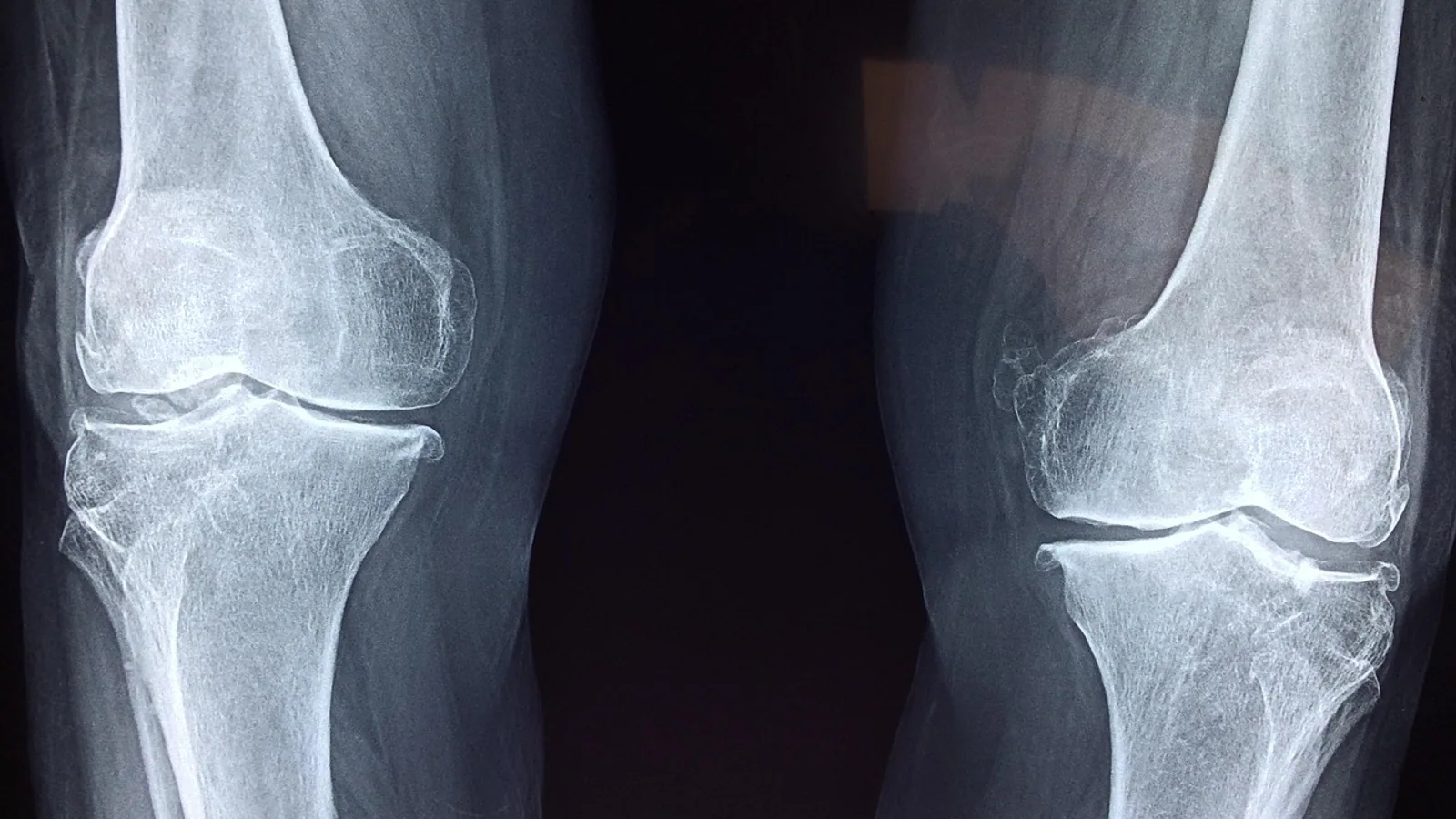
A condyloid joint is a biaxial synovial joint in which an oval, convex articular surface fits into an elliptical, concave surface, permitting flexion and extension in one plane and abduction and adduction in a perpendicular plane. The shape is often described as an ellipsoid, so the terms condyloid joint, ellipsoid joint, and ellipsoidal joint refer to the same structural class.
This joint type matters in veterinary practice because it sits at the center of normal locomotion and weight bearing in dogs, cats, horses, and food animals. The metacarpophalangeal and metatarsophalangeal joints are condyloid joints, and they carry the full load of the limb during every stride. When a condyloid joint is injured, infected, or destabilized, the animal loses more than range of motion. It loses the ability to bear weight comfortably and to place the paw or hoof correctly.
What Defines a Condyloid Joint
Synovial joints are classified by the geometry of their articular surfaces and by the number of axes around which motion occurs. A condyloid joint has two axes, which makes it biaxial. The convex member is oval or egg-shaped rather than spherical. The matching concave member is elliptical. Because the two surfaces are curved in two directions but not in three, the joint can move freely in two planes and only grudgingly in the third.
The classic description of the human thumb metacarpophalangeal joint captures this geometry well. The head of the first metacarpal is spindle-shaped, with two flexures that determine its mobility. The ratio of the semidiameter of the radio-ulnar flexure to the semidiameter of the dorso-palmar flexure describes how the head is shaped, and only flexion and extension maintain perfect contact between the articular surfaces [1]. Transverse motions and axial motions are possible because the articular cartilage deforms and because the joint cavity is relatively small compared with the head, but those motions are accessory rather than primary [1].
That last point is the one students most often miss. A condyloid joint does not have a true rotational axis. Any rotation that occurs is passive, limited, and produced by the slack in the capsule and the deformability of cartilage, not by a dedicated rotational surface.
The Two Axes
The first axis runs transversely through the joint and produces flexion and extension. In the metacarpophalangeal joint, flexion curls the digit toward the palm or sole, and extension straightens it.
The second axis runs dorsoventrally and produces abduction and adduction. Abduction moves the digit away from the midline of the limb, and adduction brings it back.
Because these two axes are perpendicular, the joint can combine the motions into circumduction, a circular sweep of the distal segment. Circumduction is not a third axis. It is the product of two axes working in sequence.
Structure, Step by Step


Articular Surfaces
The proximal bone contributes the convex condyle. The distal bone contributes the concave glenoid or fossa. Both surfaces are covered by hyaline cartilage, which is smooth, avascular, and slightly compressible. The cartilage thickness is greatest where load is highest, which is why the central contact area of a weight-bearing condyloid joint is the thickest part of the cartilage layer.
Joint Capsule
A fibrous capsule surrounds the joint and attaches near the margins of both articular surfaces. The capsule is lined internally by synovial membrane, which produces synovial fluid for lubrication and nutrition of the cartilage. The capsule of a condyloid joint is relatively loose to permit motion in two planes, but it is not so loose that the joint dislocates under normal load.
Collateral Ligaments
Collateral ligaments run along the sides of the joint and resist abduction and adduction beyond the physiologic range. They also limit rotation. In the metacarpophalangeal joint, the collateral ligaments are taut when the joint is flexed and slack when it is extended, a pattern that stabilizes the digit during weight bearing. The ligaments are the primary restraint against varus and valgus stress.
Accessory Structures
Some condyloid joints contain a meniscus or intra-articular disc that improves congruence between the two surfaces. The temporomandibular joint of dogs is formed between the condyloid process of the mandible and the mandibular fossa of the temporal bone, and its anatomy varies with skull type [2]. The meniscus in that joint acts as a barrier that prevents fusion of the condylar fragment with the glenoid fossa after trauma, which is why disruption of the meniscus position is linked to ankylosis [3].
Movement at a Condyloid Joint
Flexion and Extension
Flexion decreases the angle between the two bones, and extension increases it. This is the primary and most extensive motion at every condyloid joint. In the metacarpophalangeal joint of a dog, flexion and extension dominate the gait cycle. In the radiocarpal joint, flexion and extension position the paw during the stance phase.
Abduction and Adduction
Abduction and adduction occur around the second axis. These motions are smaller in amplitude than flexion and extension and are limited by the collateral ligaments. In the human thumb metacarpophalangeal joint, the amplitude of transverse motion does not correlate with the intensity of the radio-ulnar flexure of the head, which means the range of abduction and adduction is not simply predicted by the shape of the condyle [1].
Rotation
Rotation at a condyloid joint is passive and limited. It occurs because the articular cartilage deforms under load and because the capsule permits a small amount of twist. There is no true rotational axis. When a clinician tests rotation at a condyloid joint, the motion felt is the sum of cartilage deformation, capsular slack, and ligamentous give.
Circumduction
Circumduction is the conical sweep produced by combining flexion, extension, abduction, and adduction in sequence. It is a useful clinical descriptor because it demonstrates that both axes are functional.
Comparative Examples Across Species
Metacarpophalangeal and Metatarsophalangeal Joints
These are the archetypal condyloid joints in companion animals. In dogs and cats, the metacarpophalangeal joints are the joints between the metacarpal bones and the proximal phalanges, and the metatarsophalangeal joints are the equivalent joints in the hind limb. Both are biaxial and both bear weight during standing and locomotion.
Radiocarpal Joint
The radiocarpal joint is the articulation between the distal radius and the proximal row of carpal bones. It functions as a condyloid joint in many species, allowing flexion and extension of the carpus along with a limited amount of abduction and adduction. The midcarpal joint, which is separate, shows a complex relationship between the medial and lateral parts during the dart thrower's motion, and the lateral part of that joint is ellipsoid and accommodates the arc of movement [4].
Femorotibial Joint
The femorotibial joint is the articulation between the femoral condyles and the tibial plateau. In some species and in some descriptions it is classified as a condylar or condyloid joint because the femoral condyles are oval and the tibial surface is relatively flat with menisci improving congruence. In dogs, the femorotibial joint is a complex joint with a wide range of motion, and its classification varies with how strictly the ellipsoid criterion is applied. The key structural feature is that the femoral condyles are convex in two directions and the tibial surface is concave or flat, which produces biaxial motion with limited rotation.
Temporomandibular Joint
The temporomandibular joint is formed between the condyloid process of the mandible and the mandibular fossa of the temporal bone [2]. It is a condylar joint with a fibrocartilaginous disc. The condylar process is the mandibular condyle, and its shape and position vary with skull type. In a study of dog skulls, the facial index was useful for classifying skull types, while the rotational angle was of limited use for assessing the temporomandibular joint until normal breed values are established [2]. Radiographic positioning requires about 10 degrees of rotation in either axis to project the joints independently of each other, and lateral rotational angles of 10 to 30 degrees are most useful in mesaticephalic and dolichocephalic breeds, while 20 to 30 degrees are most useful in brachycephalic breeds [2].
First Tarsometatarsal Joint
In humans, about 90% of first tarsometatarsal joints are screw-shaped, with an axis directed upward and forward that couples plantar flexion with adduction and pronation. About 10% are ellipsoid-shaped, with a distal articular surface on the medial cuneiform shaped like an ovoid head, and a strong ligament near the lateral edge produces the same coupled motion [5]. The medial cuneonavicular joint is always ellipsoid-shaped, with the head formed by the medial facet of the distal articular surface of the navicular bone [5]. Both joints have considerable mobility [5]. This is a useful reminder that joint shape varies between individuals and that the same functional motion can be produced by different geometries.
Comparison With Other Synovial Joint Types
| Joint type | Number of axes | Motion permitted | Classic examples |
|---|---|---|---|
| Condyloid (ellipsoid) | Two | Flexion-extension, abduction-adduction, limited passive rotation | Metacarpophalangeal, metatarsophalangeal, radiocarpal, temporomandibular |
| Hinge (ginglymus) | One | Flexion-extension only | Elbow, interphalangeal joints |
| Ball-and-socket (spheroidal) | Three | Flexion-extension, abduction-adduction, axial rotation | Hip, shoulder |
| Saddle (sellaris) | Two | Flexion-extension, abduction-adduction, opposition | First carpometacarpal joint of the thumb |
The hinge joint is the one most often confused with the condyloid joint. A hinge joint has a single axis and permits only flexion and extension. A condyloid joint has two axes and permits abduction and adduction as well. If a joint can move side to side under its own power, it is not a pure hinge.
The ball-and-socket joint has three axes and a spherical head. It permits true axial rotation. A condyloid joint does not.
The saddle joint has two axes and reciprocally concave-convex surfaces. It permits motion in two planes like a condyloid joint, but the surface geometry is different. In a saddle joint, each surface is concave in one direction and convex in the other.
How Joint Type Is Tested and Observed
Physical Examination
A veterinarian assesses a condyloid joint by moving it through flexion and extension, then through abduction and adduction, and then attempting rotation. The range of motion in each plane is recorded. Pain, crepitus, swelling, and instability are noted. The collateral ligaments are tested by applying varus and valgus stress with the joint in slight flexion, which is the position in which the ligaments are taut.
Imaging
Radiography is the first-line imaging modality. For the temporomandibular joint, positioning is critical. A study of dog skulls found that 10 degrees of rotation in either axis was required to project the joints independently of each other, and that lateral rotational angles of 10 to 30 degrees in mesaticephalic and dolichocephalic breeds and 20 to 30 degrees in brachycephalics were most useful, with long axis rotational views of 10 to 30 degrees depending on the region of interest [2]. This level of detail matters because the condyloid process of the mandible is superimposed on the skull in standard lateral views.
Advanced imaging includes computed tomography and cone beam computed tomography, which allow three-dimensional reconstruction of the joint surfaces and measurement of joint space. In human dentistry, cone beam CT has been used to measure condylar volume, condylar superficial area, and joint space in patients with malocclusion, and these measurements show that condylar position and morphology differ between skeletal classes [6] [7]. Similar techniques are used in veterinary dentistry and oral surgery.
Biomechanical Modeling
Researchers use ellipsoid models to represent joint surfaces in kinematic simulations. A study of the scapulothoracic sliding plane assessed whether an ellipsoid surface could model that plane and found that ellipsoid radii varied by up to 10.2 cm in the antero-posterior direction, 3.9 cm in the medio-lateral direction, and 18.4 cm in the cranio-caudal direction across movements and participants [8]. When all frames of a movement were used for calibration, the median scapula-to-ellipsoid distance was about 0.5 mm [8]. This shows that ellipsoid modeling is a practical tool for representing curved joint surfaces, though the parameters must be calibrated carefully.
Comparative Species Differences
Digitigrade Versus Plantigrade Stance
Dogs and cats are digitigrade, meaning they walk on their digits with the metacarpophalangeal and metatarsophalangeal joints held in a partially flexed position. Humans are plantigrade, meaning the entire foot contacts the ground and the metacarpophalangeal joints are not weight bearing. This difference changes the mechanical demands on the condyloid joints of the paw. In digitigrade animals, the metacarpophalangeal joints are load bearing and their collateral ligaments must resist repeated cyclic stress. In plantigrade humans, the metacarpophalangeal joints are primarily manipulative.
Unguligrade Stance
Horses and cattle are unguligrade, meaning they walk on the tips of their digits enclosed in hooves. The metacarpophalangeal joint (fetlock) is a condyloid joint that is heavily loaded and has an extensive range of flexion and extension. The joint capsule and collateral ligaments are strong, and the sesamoid bones at the palmar aspect of the joint act as pulleys that increase the mechanical advantage of the flexor tendons.
Skull Type and Temporomandibular Joint
In dogs, skull type affects the anatomy of the temporomandibular joint. The facial index provides a useful method of classifying skull types, and the rotational angle is of limited use in assessment of the temporomandibular joint until normal breed values are established [2]. This means that a radiographic view that works well for a mesaticephalic dog may not be optimal for a brachycephalic dog.
Clinical Relevance, Limitations and Common Mistakes
Condyloid joints are clinically important because they are common sites of injury and disease. Fractures of the condylar process of the mandible can lead to ankylosis if the meniscus is displaced and no longer separates the condylar fragment from the glenoid fossa [3]. Condylar resorption is an aggressive degenerative disease of the temporomandibular joint seen mostly in adolescent and young women, and it can result in a shorter mandibular condyloid process, ramus, and body, with compensatory growth at the gonial angle and coronoid process [9]. Management options include oral appliances, orthodontics, medical management, orthognathic surgery with or without disc repositioning, and alloplastic temporomandibular joint replacement [9].
In veterinary orthopedics, condylar fractures of the humerus are repaired through a craniolateral approach to the elbow that provides direct visualization of the distal humeral articular surface while preserving the primary supporting ligamentous and tendinous structures, which allows a stable postoperative joint with normal range of motion [10]. This principle applies to condyloid joints generally: preserving the capsule and collateral ligaments improves outcomes.
The most common mistakes students make are these. First, confusing condyloid with hinge joints. A hinge joint has one axis. A condyloid joint has two. Second, assuming that rotation at a condyloid joint is active. It is passive and limited. Third, forgetting that the collateral ligaments are the primary restraint against abduction and adduction. Fourth, treating all condyloid joints as identical. The metacarpophalangeal joint, the radiocarpal joint, and the temporomandibular joint have different accessory structures and different mechanical demands. Fifth, ignoring species differences in stance. A digitigrade animal loads its metacarpophalangeal joints in a way that a plantigrade animal does not.
This article is educational and is not a substitute for veterinary diagnosis or treatment.
Quick Review
- A condyloid joint is a biaxial synovial joint with an oval convex surface fitting an elliptical concave surface.
- It permits flexion-extension and abduction-adduction, but not true axial rotation.
- Rotation is passive and limited, produced by cartilage deformation and capsular slack.
- The joint capsule and collateral ligaments restrict movement and stabilize the joint.
- Classic examples include the metacarpophalangeal and metatarsophalangeal joints, the radiocarpal joint, and the temporomandibular joint.
- A hinge joint has one axis. A ball-and-socket joint has three. A saddle joint has two but different surface geometry.
- Digitigrade animals load their metacarpophalangeal joints during standing and locomotion, while plantigrade animals do not.
Frequently Asked Questions
What is a condyloid joint?
A condyloid joint is a biaxial synovial joint in which an oval convex surface fits into an elliptical concave surface. It allows flexion and extension in one plane and abduction and adduction in a perpendicular plane.
What is the difference between a condyloid joint and a hinge joint?
A hinge joint has one axis of motion and permits only flexion and extension. A condyloid joint has two axes and also permits abduction and adduction.
Can a condyloid joint rotate?
A condyloid joint does not have a true rotational axis. Any rotation is passive and limited, produced by deformation of the articular cartilage and slack in the joint capsule.
Which joints in dogs are condyloid?
The metacarpophalangeal and metatarsophalangeal joints are condyloid, as is the radiocarpal joint. The temporomandibular joint is also a condylar joint with a fibrocartilaginous disc.
Why does stance matter for condyloid joints?
Digitigrade animals such as dogs and cats bear weight on their metacarpophalangeal and metatarsophalangeal joints, so those joints are load bearing. Plantigrade animals such as humans do not bear weight on those joints, so they are primarily manipulative.
What ligaments limit movement at a condyloid joint?
Collateral ligaments run along the sides of the joint and resist abduction and adduction beyond the physiologic range. They also limit rotation. The joint capsule provides additional restraint.
Related Articles
- Cattle Alleyway Design for Efficient Movement
- Sheep Handling Facilities and Low-Stress Movement
- Bison Handling Facility Design for Low-Stress Movement
- Cervid Handling Facility Design for Low-Stress Movement
- Swine Handling Facility Design for Safe Pig Movement
- Sheep Handling Equipment: Design and Use for Low-Stress Movement
- Myoneural Junction: Structure and Function
- Synovial Joints and Synovium: Structure and Function
- Ball and Socket Joints: Examples, Movement, and Anatomy
Sources
- [[The basic human thumb joint--an egg-shaped joint].](https://pubmed.ncbi.nlm.nih.gov/7445945/)
- The effect of obliquity on the radiographic appearance of the temporomandibular joint in dogs.
- Role of the meniscus in the etiology of posttraumatic temporomandibular joint ankylosis.
- A composite 3D printed model of the midcarpal joint.
- [[Biomechanics of the joints of the large toe. Shape and movement of the first tarsometatarsal joint and of the medial cuneonavicular joint].](https://pubmed.ncbi.nlm.nih.gov/1514356/)
- A pilot investigation of condylar position and asymmetry in patients with unilateral posterior scissors-bite malocclusion based on three-dimensional reconstructive imaging technique.
- [[Comparison of condylar position between Angle Class I and Class II malocclusion in teenagers].](https://pubmed.ncbi.nlm.nih.gov/28275792/)
- Is an ellipsoid surface suitable to model the scapulothoracic sliding plane?
- Idiopathic Condylar Resorption: What Should We Do?
- Craniolateral approach to the canine elbow for repair of condylar fractures or joint exploration.