Jaw Bone Anatomy: Mandible Structure and Landmarks
By Dr. Zubair Khalid, DVM, MS, PhD ·
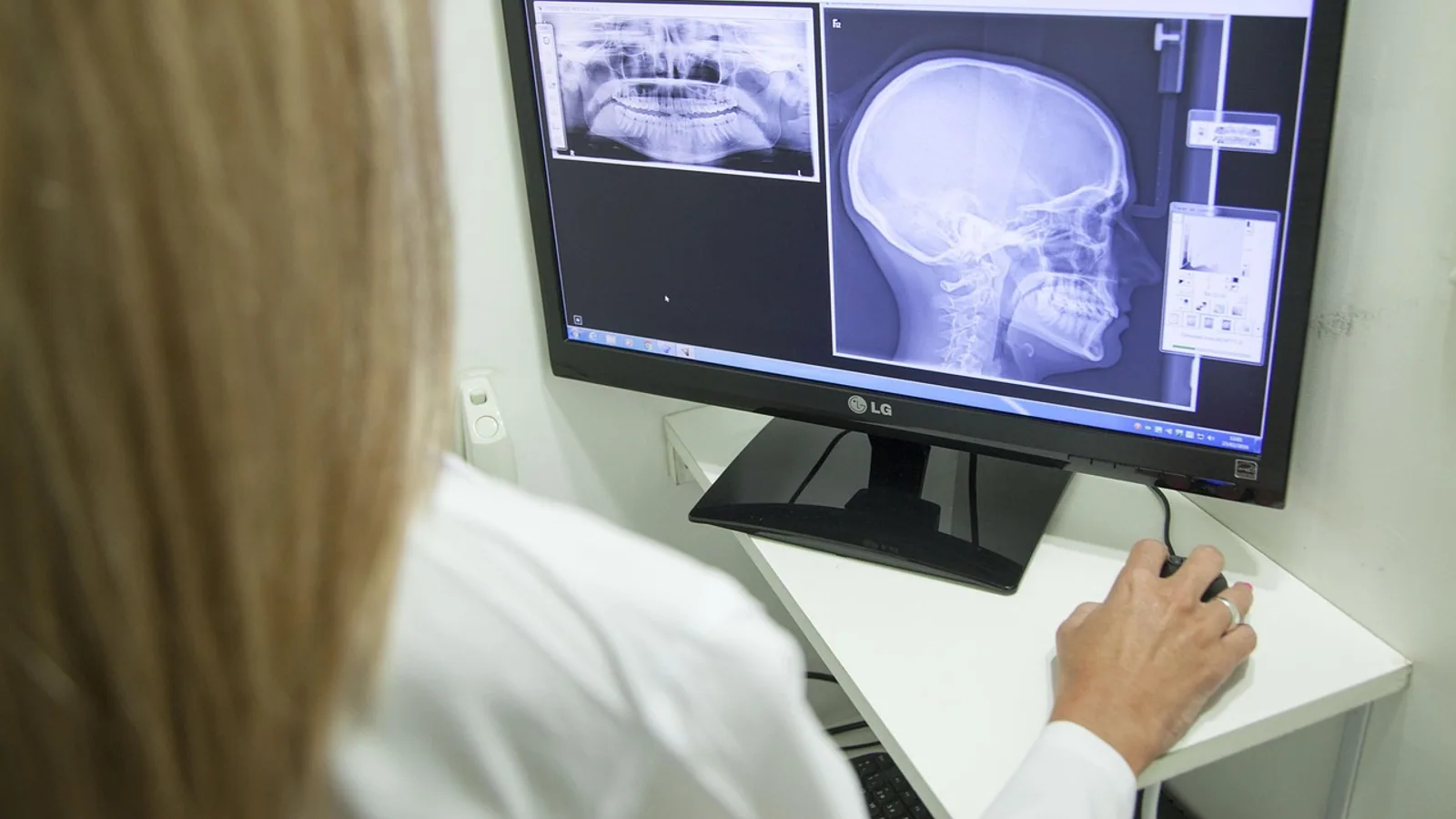
The mandible is the single mobile bone of the skull, forming the lower jaw and articulating with the temporal bone at the temporomandibular joint. It carries the lower dental arcade, transmits the inferior alveolar nerve and vessels through the mandibular canal, and serves as the insertion site for the muscles of mastication.
Why the Mandible Matters in Veterinary Practice
Every oral examination, dental extraction, intubation, and maxillofacial procedure depends on a working knowledge of the mandible. The mandibular jaw bone is the thickest bone of the face in most domestic species, yet it is also one of the most commonly fractured. Its canal houses a neurovascular bundle that supplies all lower teeth and the lower lip, so a misplaced needle or drill can cause permanent sensory loss. In dogs and cats, the mandible symphysis is a fibrocartilaginous joint that allows slight independent movement of the two halves, a feature that is absent in horses and ruminants. Recognizing these differences prevents procedural errors and improves diagnostic accuracy on radiographs.
This article is educational and is not a substitute for veterinary diagnosis or treatment.
Core Terminology: What the Mandible Is and Is Not
The mandible bone (also called the mandibular bone) is a paired, dermal bone that develops partly by intramembranous ossification and partly by endochondral ossification at the condylar process. In domestic mammals, the two halves (hemimandibles) fuse at the mandibular symphysis, a midline joint that is cartilaginous in young animals and becomes fibrous or partially ossified with age.
The term "mental bone" is not standard veterinary or human anatomy. It appears occasionally in lay writing and translated texts, but it has no defined structure in Nomina Anatomica Veterinaria or standard human anatomy references. The correct terms are mandible, mandibular body, and mandibular ramus. The related adjective "mental" is legitimate only in specific structures: the mental foramen, mental nerve, and mental protuberance (in humans). Students should avoid "mental bone" entirely in written work and clinical communication.
Summary Table: Mandible Terms and What They Refer To
| Term | Definition | Common Confusion |
|---|---|---|
| Mandible | The entire lower jaw bone, paired in domestic mammals | Sometimes called "jaw bone," which also includes the maxilla |
| Mandibular body | Horizontal portion bearing the lower teeth | Confused with the ramus |
| Mandibular ramus | Vertical portion extending toward the temporomandibular joint | Confused with the angle |
| Mandibular angle (gonion) | Caudalventral junction of body and ramus | Confused with the angular process in rodents |
| Condylar process | Articular process forming the temporomandibular joint | Confused with the coronoid process |
| Coronoid process | Dorsal process for temporalis muscle insertion | Confused with the condylar process |
| Mandibular foramen | Opening on the medial ramus leading into the mandibular canal | Confused with the mental foramen |
| Mental foramen | Opening on the lateral body for the mental nerve | Confused with the mandibular foramen |
| Mandibular canal | Internal canal carrying the inferior alveolar nerve and vessels | Sometimes called the inferior alveolar canal |
| "Mental bone" | Not a recognized anatomical term | Should not be used |
Structure of the Mandible: Step by Step
The Mandibular Body
The body (corpus mandibulae) is the horizontal part of the mandible that holds the lower teeth. In the dog, the body is relatively slender and curves gently from the symphysis to the angle. In the horse, the body is deep and robust, accommodating the long roots of the lower cheek teeth. The body has two surfaces: a lateral surface facing the cheek and a medial surface facing the tongue. The alveolar process sits on the dorsal aspect and contains the tooth sockets (alveoli). The ventral border is the thickest part and is the preferred site for bone plates in fracture repair.
The mandibular symphysis joins the two hemimandibles rostrally. In dogs and cats, this is a fibrocartilaginous joint with limited mobility. In horses and cattle, the symphysis ossifies early, creating a rigid lower jaw. The incisive portion of the body (the incisive part) holds the lower incisors and, in ruminants, the dental pad opposes the upper incisors.
The Mandibular Ramus
The ramus is the vertical or caudally directed portion of the mandible. It extends from the angle to the temporomandibular joint. The lateral surface of the ramus provides insertion for the masseter muscle, while the medial surface provides insertion for the medial pterygoid muscle. The mandibular foramen sits on the medial surface of the ramus, roughly midway between the condylar and coronoid processes in the dog. This foramen is the entry point for the inferior alveolar nerve and vessels into the mandibular canal.
The ramus thickness varies by species and individual. In humans, cone-beam computed tomography studies show a mean ramus thickness of approximately 6.5 mm at the level of the mandibular lingula [1]. The mandibular lingula (spine of Spix) is a small bony projection on the medial ramus that serves as the primary landmark for inferior alveolar nerve blocks [2]. A comprehensive review of over 11,900 mandibular sides found five distinct lingula shapes: triangular, truncated, nodular, assimilated, and bridge or M-shaped [2].
The Mandibular Angle
The angle of the mandible (angulus mandibulae) is the region where the ventral border of the body meets the caudal border of the ramus. In dogs, the angle is rounded and lies near the level of the last lower molar. In horses, the angle is more angular and provides attachment for the digastricus muscle. The angle is a palpable landmark on the lateral aspect of the head in most species. In humans, the great auricular nerve passes near the angle of the mandible, and a cadaver study found that the ratio of the angle-to-mastoid distance divided by the angle-to-nerve distance averaged 0.457, providing a reliable surgical guide [3].
The Condylar Process
The condylar process (processus condylaris) is the caudalmost projection of the ramus. It ends in the mandibular condyle, a transversely elongated articular surface that articulates with the mandibular fossa of the temporal bone. This forms the temporomandibular joint, the only synovial joint of the skull. The condyle in dogs is roughly cylindrical and allows hinge-like opening and closing plus some lateral grinding. In horses, the condyle is flatter and permits more extensive lateral excursion during chewing. Fractures of the mandibular condyle are common maxillofacial injuries, and research in rat models shows that fracture repair involves osteogenic and chondrogenic signaling pathways, including TGF-beta/Smad signaling [4].
The Coronoid Process
The coronoid process (processus coronoideus) is the dorsal projection of the ramus, rostral to the condylar process. It serves as the insertion point for the temporalis muscle, one of the major jaw-closing muscles. In carnivores, the coronoid process is tall and blade-like. In herbivores, it is broader and lower. The coronoid process is separated from the condylar process by the mandibular notch, a concave area that allows passage of the masseteric nerve and vessels.
The Mandibular Canal and Foramina
The mandibular canal is a bony tunnel that runs through the body of the mandible, beginning at the mandibular foramen on the medial ramus and ending at the mental foramen on the lateral body. It carries the inferior alveolar nerve (a branch of the mandibular division of the trigeminal nerve) and the inferior alveolar artery and vein. These structures supply the lower teeth, the bone of the mandible, and the lower lip and chin.
The mandibular foramen is the entrance to the canal. Its position varies by species and age. In dogs, it lies on the medial surface of the ramus, approximately midway between the condylar and coronoid processes. In horses, it is located further caudally and ventrally. The mental foramen is the exit point on the lateral surface of the body. Its position varies significantly across species, which is why species-specific knowledge is essential for nerve blocks and surgical planning.
The mandibular incisive canal is an anterior extension of the mandibular canal beyond the mental foramen. A cone-beam computed tomography study of 345 patients found that the incisive canal was identifiable in 74.5% of patients and 60.6% of images, with a mean length of 13.7 mm and a mean diameter of 1.65 mm [5]. The canal's superior border lay 6.46 mm from premolar apices, 7.10 mm from canine apices, and 8.99 mm from incisor apices [5]. These measurements matter in veterinary dentistry when placing implants or performing apical surgery.
How the Mandible Is Examined and Imaged
Physical Examination
In dogs and cats, the mandible is assessed by palpation along the ventral border from the symphysis to the angle. The examiner checks for symmetry, swelling, crepitus, and pain. The temporomandibular joint is palpated just ventral to the zygomatic arch. In horses, the mandible is examined externally and through the oral cavity, with attention to the incisive region and the cheek teeth arcades.
Radiography
Lateral and dorsoventral or ventrodorsal views are standard. Intraoral radiography provides detail of the tooth roots and alveolar bone. The mandibular canal appears as a radiolucent band running through the body. The mental foramen is visible as a small radiolucency on the lateral aspect of the body near the premolars.
Advanced Imaging
Cone-beam computed tomography (CBCT) is increasingly used for detailed mandibular assessment. CBCT can measure ramus thickness, locate the mandibular lingula, and trace the mandibular canal in three dimensions. A study of 412 hemimandibles found that the mandible was slightly thicker 4 mm above the lingula (6.54 mm) than at the level of the lingula (6.53 mm), and that the buccal and lingual cortical plates fused approximately 12 mm posterior to and 8 mm above the lingula [1]. These morphometric data help surgeons plan sagittal split osteotomies and avoid unfavorable splits.
Histology and Bone Quality Assessment
Mandibular bone quality can be assessed non-invasively using fractal dimension analysis on panoramic radiographs. Fractal dimension quantifies trabecular complexity. A study of pediatric chronic kidney disease patients found significantly lower fractal dimension values across three regions of interest compared with controls, with the largest effect in the gonial region (Cohen d = -1.39) [6]. Mandibular cortical width, panoramic mandibular index, and superior foraminal distance did not differ between groups [6]. This suggests that trabecular changes may be detectable earlier than cortical changes on panoramic imaging.
In another study, type 2 diabetes was associated with higher fractal dimension values (1.409) than controls (1.386), indicating increased microarchitectural complexity in mandibular trabecular bone [7]. Hypertension showed a similar but non-significant trend [7]. These findings are preliminary and require validation with established reference methods.
Comparative Mandible Anatomy: Dog, Cat, Horse, Cow, and Human
The mandible varies considerably across species in shape, size, and dental formula. The table below summarizes key features.
| Feature | Dog | Cat | Horse | Cow | Human |
|---|---|---|---|---|---|
| Mandible shape | Elongated, slender body; tall coronoid | Short, robust body; prominent coronoid | Deep, massive body; long diastema | Deep body; wide diastema; angular process | U-shaped body; prominent chin |
| Mandibular symphysis | Fibrocartilaginous, mobile | Fibrocartilaginous, mobile | Ossified, rigid | Ossified, rigid | Fused in adulthood |
| Condylar process | Transverse cylinder | Transverse cylinder | Flattened, wide | Flattened, wide | Ellipsoid |
| Coronoid process | Tall, blade-like | Tall, blade-like | Broad, low | Broad, low | Triangular, pointed |
| Mandibular foramen position | Mid-ramus, medial | Mid-ramus, medial | Caudalventral ramus | Caudalventral ramus | Mid-ramus, medial |
| Mental foramen position | Lateral body, near premolars | Lateral body, near premolars | Lateral body, rostral | Lateral body, rostral | Lateral body, below premolars |
| Dental formula (permanent) | I 3/3, C 1/1, P 4/4, M 2/3 = 42 | I 3/3, C 1/1, P 3/2, M 1/1 = 30 | I 3/3, C 1/1 (canine), P 3/3, M 3/3 = 40-42 | I 0/3, C 0/1, P 3/3, M 3/3 = 32 | I 2/2, C 1/1, P 2/2, M 3/3 = 32 |
| Jaw movement | Hinge plus lateral grinding | Hinge plus lateral grinding | Extensive lateral excursion | Extensive lateral excursion | Hinge plus lateral grinding |
Note: Dental formulas are presented as upper/lower for each tooth type. The horse has a long diastema between the incisors and cheek teeth. The cow lacks upper incisors and has a dental pad instead.
Clinical Relevance, Limitations and Common Mistakes
The mandible is clinically important in dentistry, oral surgery, anesthesia, and oncology. Inferior alveolar nerve blocks are among the most common procedures in veterinary dentistry. The mandibular lingula is the primary landmark for these blocks, but its shape varies. A comprehensive review found that dry bone studies predominantly report triangular lingula shapes (39%), while CBCT studies reveal nodular predominance (50.6%) in many populations [2]. The internal oblique or mylohyoid ridge has emerged as a reliable alternative landmark with 95.7% visibility and superior block success rates (97.3%) compared with traditional methods (approximately 85%) [2].
Mandibular fractures are common in dogs and cats, often resulting from falls, vehicle trauma, or animal bites. The ventral border of the body is the thickest area and the preferred site for bone plates. The mandibular canal must be avoided during implant placement or fracture repair to prevent nerve damage.
Mandibular bone can be affected by systemic disease. Chronic kidney disease in children reduces mandibular trabecular complexity [6]. Selective serotonin reuptake inhibitors have been studied in rats for their effects on mandibular alveolar bone and salivary glands, with histological and biochemical changes observed [8]. Mandibular metastasis from hepatocellular carcinoma is rare, occurring in less than 1% of cases, and can be misdiagnosed as a benign dental condition [9].
Bone regeneration research uses mandibular defect models. A rat study showed that secretome from human exfoliated deciduous teeth stromal cell sheets facilitated mandibular bone defect repair by shifting macrophage inflammatory states [10]. Bovine bone graft inlay versus onlay techniques have been compared for mandibular ridge augmentation, with onlay showing greater horizontal bone gain [11].
Common mistakes in mandible anatomy include confusing the mandibular foramen with the mental foramen, misidentifying the coronoid and condylar processes, and using the non-standard term "mental bone." Another frequent error is assuming that the mandibular symphysis is fused in all species. In dogs and cats, it is mobile, which affects how forces are distributed during chewing and how fractures are stabilized.
This article is educational and is not a substitute for veterinary diagnosis or treatment.
Quick Review: 7 Points to Memorize
- The mandible is the only mobile skull bone, articulating at the temporomandibular joint.
- The mandibular body holds the lower teeth. The ramus extends toward the joint.
- The condylar process articulates with the temporal bone. The coronoid process is for temporalis insertion.
- The mandibular canal carries the inferior alveolar nerve and vessels from the mandibular foramen to the mental foramen.
- The mental foramen position varies by species and is a key landmark for nerve blocks.
- The term "mental bone" is not standard anatomy and should be avoided.
- Dental formula and mandible shape differ markedly across dogs, cats, horses, cows, and humans.
Frequently Asked Questions
What is the mandible bone?
The mandible is the lower jaw bone. It is the only mobile bone of the skull and holds the lower teeth.
Is the mental bone a real anatomical structure?
No. "Mental bone" is not a recognized term in veterinary or human anatomy. The correct terms are mandible, mandibular body, and mandibular ramus.
What runs through the mandibular canal?
The mandibular canal carries the inferior alveolar nerve and the inferior alveolar artery and vein.
Where is the mental foramen located?
The mental foramen is on the lateral surface of the mandibular body. Its exact position varies by species.
How does the mandible differ between dogs and horses?
Dogs have a slender, mobile mandible with a fibrocartilaginous symphysis. Horses have a deep, rigid mandible with an ossified symphysis and a long diastema.
What is the mandibular lingula?
The mandibular lingula is a bony projection on the medial ramus. It is a key landmark for inferior alveolar nerve blocks.
Related Articles
- Bovine Heart Anatomy and Auscultation Landmarks
- Equine Hindlimb Anatomy: Bones, Joints, and Ligaments
- Do Frogs Have Bones? Skeletal Anatomy and Pet Care
- Equine Hoof Anatomy: Structures and Function
- Canine Liver Anatomy and Lobular Structure
- Snake Anatomy: Do Snakes Have Teeth, Spines, and Other Structures?
- Compact Bone: Structure and Function Explained
- Spongy Bone: Structure, Function, and Histology
Sources
- In search of a safe zone for sagittal split ramus osteotomy: a cone-beam computed tomography study of the mandibular ramus anatomy.
- The mandibular lingula (spine of Spix) in contemporary research: A comprehensive critical review of global morphological variations and clinical implications.
- Identifying Great Auricular Nerve via Bony Landmarks: A Cadaver Study.
- METTL3-mediated m6A methylation to promote mandibular condyle fracture repair via TGF-β/Smad signaling pathway.
- Morphometric analysis of the mandibular incisive canal using cone beam computed tomography images.
- Evaluation of Mandibular Bone Structure Using Fractal Analysis and Radiomorphometric Indices in Pediatric Chronic Kidney Disease.
- Mandibular trabecular fractal dimension in individuals with type 2 diabetes and hypertension: a retrospective cross-sectional panoramic radiography study.
- Impact of selective serotonin reuptake inhibitors on major salivary glands and mandibular alveolar bone; a histological, histochemical and biochemical study.
- A Rare Presentation of Mandibular Bone Metastasis from Primarily Diagnosed Hepatocellular Carcinoma: A Case Report.
- Secretome of SHED Sheets Facilitates Mandibular Bone Defect Repair by Shifting Macrophages Inflammatory State.
- Bovine bone graft inlay versus onlay for mandibular ridge augmentation (randomized clinical trial).