Third Eyelid Showing in Dogs: Causes and When to Worry
By Dr. Zubair Khalid, DVM, MS, PhD ·
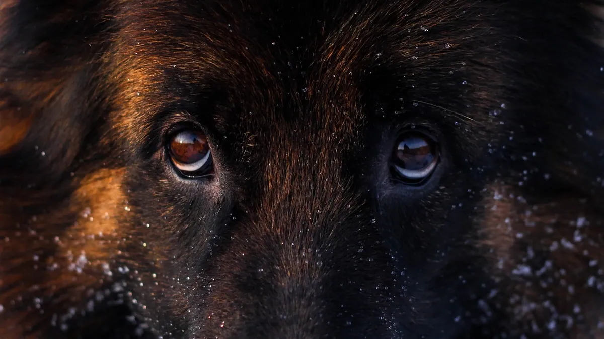
A dog's third eyelid showing means the pink or white membrane in the inner corner of the eye has slid across part of the eyeball. It can happen for simple reasons like eye pain, dehydration, or tiredness, and it often goes back to normal on its own. But when the third eyelid stays out, covers a large part of the eye, or appears with other symptoms, it can point to a condition that needs veterinary care.
This article is educational and is not a substitute for veterinary diagnosis or treatment.
Quick Answer
- A brief third eyelid peek in a tired or squinting dog is common and usually harmless.
- One eye affected often points to a local problem, such as pain, a scratch, or a foreign object.
- Both eyes affected at once more often suggests a whole-body issue, such as dehydration, illness, or a nerve problem.
- A third eyelid that stays out for more than a few hours, or that comes with a sunken eye, a droopy lid, or a small pupil, needs same-day veterinary care.
- Never try to push the third eyelid back or apply human eye drops without veterinary advice.
What the Third Eyelid Looks Like and What Is Normal
The third eyelid (also called the nictitating membrane) is a thin, pale pink to white sheet of tissue tucked into the inner corner of each eye. It normally stays hidden, but it can slide partway across the eye for short periods. You might notice it when your dog is drifting off to sleep, when the eye is dry, or when your dog is squinting.
The membrane also holds a tear-producing gland. When that gland pops out and stays visible as a pink, round bump, owners often call it cherry eye. That is a different problem from a flat third eyelid that simply slides across the eye, and it has its own causes and treatments. You can read more about that in Cherry Eye in Dogs: Causes, Surgery Options, and Recovery.
A normal third eyelid is smooth and lies flat against the eye. It should not look swollen, red, or lumpy. If the tissue looks puffy or has a growth on it, that is a reason to call your vet.
Third Eyelid Showing in Dogs: The Main Causes
The list below covers the most common reasons owners see this sign. Some are mild and pass quickly. Others are serious and need prompt care.
| Cause | Typical clues | How urgent |
|---|---|---|
| Pain or squinting (corneal ulcer, scratch, foreign body) | One eye, tearing, pawing at the face, light sensitivity | Same-day vet visit |
| Dehydration or fatigue | Both eyes, mild, improves after rest or fluids | Monitor, call vet if it persists |
| Entropion (eyelid rolls inward) | Eyelashes rub the eye, squinting, redness, common in certain breeds | Routine to same-day, depending on comfort |
| Conjunctivitis (pink eye) | Red eye, swelling, discharge, mild discomfort | Routine visit |
| Horner's syndrome (nerve problem) | One eye sunken, droopy upper lid, small pupil, third eyelid out | Same-day vet visit |
| Tetanus | Stiff jaw, rigid muscles, third eyelid out in both eyes | Emergency |
| Retrobulbar mass (growth behind the eye) | Eye pushed forward, bulge, slow onset, one side | Same-day vet visit |
| Zygomatic sialadenitis (salivary gland inflammation) | Pain when opening the mouth, eye bulge, third eyelid out | Same-day vet visit |
| Tumor of the third eyelid itself | Firm nodule or lump on the membrane | Same-day vet visit |
Pain and Eye Irritation
Eye pain is one of the most common reasons a third eyelid slides out. When the surface of the eye hurts, the muscles that hold the third eyelid back can relax, and the membrane drifts forward. Causes include scratches on the cornea, ulcers, foreign objects like grass awns, and dry eye.
Entropion is a related cause. In entropion, the eyelid edge rolls inward so the lashes and fur rub against the eye surface. It is the most frequent inherited eyelid defect in many dog breeds, and it can also follow scarring or severe winking from eye pain [1]. The rubbing causes discomfort and irritation of the conjunctiva and cornea, and long-term entropion can lead to scarring, abnormal coloring, and slow-healing sores on the cornea [1]. Early spasms of entropion may reverse if the cause is removed or the pain is eased, but established entropion usually needs surgery [1].
Conjunctivitis and Other Surface Problems
The conjunctiva is the thin membrane that lines the inside of the eyelids and covers the white of the eye. It helps make tears, protects the eye, and supports healing after injury [2]. When it becomes inflamed, the result is conjunctivitis, or pink eye. Signs include redness, swelling around the cornea, discharge, and mild discomfort [2]. Because many different problems can look alike, your vet often needs a full history, an exam, and tests such as conjunctival scrapings or a Schirmer tear test to pin down the cause [2]. If your dog's eye is red or weepy along with the third eyelid, see Why Are My Dog's Eyes Red? and Dog Eye Discharge: When Should You Worry?.
Dehydration and Fatigue
When a dog is low on fluids or very tired, the fat pad behind the eye can shrink slightly. That lets the eye sit a little deeper and the third eyelid drift forward. This often shows in both eyes at once and improves once your dog rests or drinks. It is the mildest reason on this list, but if it does not clear within a few hours, have your vet take a look.
Horner's Syndrome
Horner's syndrome is a nerve problem that affects one side of the face. The classic pattern is a sunken eye, a droopy upper eyelid, a small pupil, and a third eyelid that slides out. It usually affects just one eye. The underlying cause can be anything from an ear infection to a mass in the chest, so a vet exam is needed to look for the source.
Tetanus
Tetanus is rare but serious. It causes muscle rigidity, and the third eyelid can protrude in both eyes. You may also see a stiff jaw, an odd stance, or trouble eating. This is an emergency.
Retrobulbar Mass and Other Growths Behind the Eye
A mass behind the eyeball pushes the eye forward, and the third eyelid can slide out as a result. In a review of four dogs with tumors around the eye, the most common signs were slowly progressive eye bulge (all four dogs), excessive tearing (three dogs), and prolapse of the third eyelid gland (two dogs) [3]. These growths tend to develop slowly and often affect one side.
Zygomatic Sialadenitis
This is inflammation of the salivary gland near the eye. In a study of 20 dogs, common signs included pain when opening the mouth (18 of 20), redness of the conjunctiva (16 of 20), eye bulge (15 of 20), pain around the eye (15 of 20), and third eyelid protrusion (11 of 20) [4]. Labradors were overrepresented in that group [4]. If your dog yelps when it opens its mouth to eat or yawn, mention that to your vet.
Tumors of the Third Eyelid Itself
Growths can form directly on the third eyelid. Lymphoma is the most common blood cell cancer in dogs, but cases that start mainly in the third eyelid are rarely reported [5]. In one report, a 10-year-old Poodle had a nodule on the left third eyelid for two months, and the mass came back 19 days after surgery and spread into the surrounding eye tissues [5]. Any firm lump on the membrane deserves a prompt exam.
Red Flags That Mean Emergency Care Now
Call an emergency clinic right away if you see any of these:
- The third eyelid covers most of the eye and does not move back within a few hours.
- The eye looks sunken, the upper lid droops, or the pupil is much smaller than the other side.
- The eyeball is bulging out of the socket.
- Your dog has a stiff jaw, rigid legs, or trouble swallowing.
- There is heavy bleeding, a deep wound, or a visible object stuck in the eye.
- Your dog is crying, pawing hard at the face, or cannot open the eye at all.
- The eye looks cloudy, has a blue or white spot, or the surface seems dented.
These signs can mean nerve damage, a growth behind the eye, or a serious eye injury. Fast care protects vision.
Safe Things You Can Do at Home While You Wait
You cannot fix a protruding third eyelid at home, but you can keep your dog comfortable and avoid making things worse.
- Do not try to push the membrane back with your fingers or a cotton swab.
- Do not use human eye drops, saline, or leftover pet medications unless your vet tells you to.
- Keep your dog from rubbing the eye. A soft cone (Elizabethan collar) can help.
- Offer water. Mild dehydration can make the third eyelid show, and drinking may help.
- Keep the area clean with a warm, damp cloth if there is discharge, and use a fresh cloth for each eye.
- Note the timing. Write down when you first saw it, whether one or both eyes are involved, and any other symptoms.
- Limit bright light and let your dog rest in a quiet space.
If the membrane is still out after a few hours, or if anything on the red flag list appears, stop waiting and call your vet.
What Your Veterinarian Will Check and Test
Your vet will start with a full eye exam. They will look at the third eyelid, the conjunctiva, the cornea, and the pupil size on both sides. They will also check how the eye moves and whether it can be pushed gently back into the socket.
Common tests include:
- Schirmer tear test to measure tear production [2]
- Fluorescein stain to look for scratches or ulcers on the cornea
- Conjunctival scrapings or swabs to check for infection [2]
- A full physical exam, including the mouth, ears, and nerves of the face
- Blood work if a whole-body cause is suspected
If a mass behind the eye is possible, your vet may recommend imaging such as CT or MRI. In the tumor review, CT was used to map the location and extent of the lesion and to plan surgery [3]. For salivary gland inflammation, MRI or CT of the head helped confirm the diagnosis [4].
Treatment depends on the cause. Eye pain from entropion may be managed first with stitches, lid injections, or nerve blocks to ease discomfort, and very young puppies with entropion may sometimes be treated with temporary stitches removed in 2 to 3 weeks [1]. Established entropion usually needs surgery [1]. Some eye surgeries do not require routine antibiotics afterward, based on a study of 96 animals that found low complication rates when antibiotics were given only when specific criteria were met [6]. Your vet will decide what is right for your dog.
Prevention and Monitoring
You cannot prevent every cause of a third eyelid showing, but you can lower the odds and catch problems early.
- Keep up with routine eye exams, especially if your breed is prone to eyelid or tear problems.
- Watch for squinting, redness, or discharge, and treat these early before they worsen.
- Keep your dog hydrated, particularly in hot weather or after hard exercise.
- Check the eyes during grooming. Look for lumps, redness, or a membrane that stays out.
- If your dog has a known condition like entropion or dry eye, follow your vet's monitoring schedule.
- After any eye surgery, follow the discharge instructions closely and report swelling, discharge, or a returning third eyelid.
Some eyelid and third eyelid problems have a genetic component. A 2024 genome-wide study of over 1,400 dogs found genetic regions linked to prolapse of the third eyelid gland, including a region containing a gene tied to eye development [7]. That does not mean every dog with a family history will be affected, but it is useful information if you are choosing a breed or a breeder.
Limitations and When to Contact a Veterinarian
This guide covers common patterns, but every dog is different. A third eyelid showing can look the same whether the cause is mild or serious, and only a veterinarian can tell them apart with an exam and tests.
Contact your vet the same day if the third eyelid stays out for more than a few hours, if only one eye is affected, or if you see squinting, redness, or discharge.
Go to an emergency clinic now if the eye is bulging, the pupil sizes do not match, the jaw is stiff, or your dog is in obvious pain.
Schedule a routine visit if the sign comes and goes over days, if both eyes are mildly affected, or if your dog has a known eye condition that seems to be changing.
Frequently Asked Questions
Is a third eyelid showing in a puppy always serious?
No, it is not always serious. Puppies can show a brief third eyelid when they are tired, dehydrated, or have mild eye irritation. However, puppies can also have inherited eyelid defects, and very young puppies with entropion may need temporary stitches to protect the eye. If the membrane stays out or the puppy squints, see your vet.
Why did my dog's third eyelid suddenly show up?
Sudden appearance usually points to eye pain, a scratch, a foreign object, or a nerve problem. A single eye that suddenly shows the membrane along with a droopy lid and small pupil suggests Horner's syndrome. Both eyes appearing suddenly can follow dehydration, fatigue, or a whole-body illness. Sudden changes deserve a prompt exam.
What does it mean if the third eyelid is showing in only one eye?
One eye affected usually means the problem is local to that eye or the nerves on that side. Common causes include a corneal ulcer, a foreign body, entropion, or a growth behind the eye. Because these can threaten vision, a same-day vet visit is the safest choice.
Can dehydration really make both third eyelids show?
Yes, dehydration can cause both third eyelids to slide forward. When fluid levels drop, the fat pad behind the eye shrinks and the eye sits deeper, which lets the membrane drift across. Offer water and rest, but if the sign does not clear within a few hours, call your vet.
How is a protruding third eyelid treated?
Treatment depends on the cause. Eye pain may be managed with stitches, lid injections, or nerve blocks, and established entropion usually needs surgery. Infections may need targeted medication, and masses may need imaging and surgical removal. Your vet will build a plan based on the exam and test results.
Related Articles
- Dog Eye Discharge: When Should You Worry?
- Eye Goop In Dogs
- Why Are My Dog's Eyes Red?
- Cherry Eye in Dogs: Causes, Surgery Options, and Recovery
- Dog Skin Rash on Face: Causes and When to Worry
- Dog Hair Loss on Nose: Causes and When to Worry
- Why Is My Dog Not Opening One Eye? Causes and When to Worry
- Dog Eye Infection: Symptoms, Treatment, and When to Worry
- Growth on a Dog's Eyelid: Types and When to Remove
References
- Disorders of the Eyelids in Dogs - Dog Owners. Merck Veterinary Manual.
- Disorders of the Conjunctiva in Dogs - Dog Owners. Merck Veterinary Manual.
- Surgical and Conservative Management of Periorbital Tumors in Four Dogs. Veterinary sciences, 2026.
- Canine bilateral zygomatic sialadenitis: 20 cases (2000-2019). The Journal of small animal practice, 2025.
- Primary third eyelid lymphoma with aggressive local behavior in a dog. Topics in companion animal medicine, 2024.
- Reassessing Antibiotic Use in Canine and Feline Eyelid and Third Eyelid Surgeries: An Observational Study Supporting Selective Prophylaxis. Veterinary ophthalmology, 2026.
- Genome-wide association studies with prolapsed gland of the third eyelid in dogs. Frontiers in veterinary science, 2024.