Growth on a Dog's Eyelid: Types and When to Remove
By Dr. Zubair Khalid, DVM, MS, PhD ·
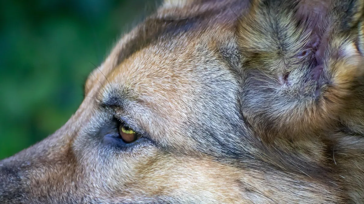
Most growths on a dog's eyelid turn out to be benign tumors of the eyelid glands, and surgical removal is often curative [1][2]. Some are malignant, however, and can invade the eyelid or irritate the eye surface, so any new lump on the lid deserves a veterinary exam. This article is educational and is not a substitute for veterinary diagnosis or treatment.
Quick Answer
- Eyelid tumors are the most common eye tumors in dogs, and meibomian gland tumors (adenoma and adenocarcinoma) are the most frequent type [1].
- In one study of 119 excised eyelid masses, about 67% were benign, about 25% were non-tumors, and about 8% were malignant [2].
- Most eyelid tumors are removed surgically because they irritate the eye and become disfiguring, and surgery is usually successful [1].
- A biopsy is needed to identify the tumor type and guide treatment, since benign and malignant masses can look similar [1].
- See a veterinarian promptly if the mass touches the eyeball, the eye is red or cloudy, or your dog is squinting or pawing at the face.
What Is a Growth on a Dog's Eyelid?
A growth on a dog's eyelid is any lump, bump, or raised area on the upper or lower lid, including the lid margin where the eyelashes sit. The eyelids are thin, layered structures, and the tissues within them can develop primary tumors or become a site where tumor cells settle [1].
Eyelid masses are the most common ocular disease in dogs [2]. They range from harmless overgrowths of gland tissue to cancers that destroy surrounding tissue. Because the lid sits directly against the cornea, even a small benign mass can cause real discomfort if it rubs the eye.
Some eyelid problems are not growths at all. Entropion, the turning in of the eyelid edge so lashes and fur rub the eye, is the most frequent inherited eyelid defect in many breeds [3]. It can look like a swelling or thickening of the lid, but it is a structural problem, not a tumor. Long-term entropion can cause scarring, abnormal coloring, and slow-healing sores on the cornea [3]. If your dog's eye issue involves a red, prolapsed gland at the inner corner instead of the lid itself, see Cherry Eye in Dogs: Causes, Surgery Options, and Recovery.
Types of Eyelid Growths in Dogs
Meibomian Gland Tumors
The meibomian glands line the eyelid margin and produce the oily part of the tear film. Tumors of these glands are the most common lid tumors in dogs [1]. They include:
- Meibomian adenoma, a benign glandular tumor
- Meibomian adenocarcinoma, a malignant glandular tumor
- Meibomian epithelioma and meibomian hyperplasia, which are also common findings [2]
In one study, meibomian epithelioma made up about 35% of excised lid masses, meibomian adenoma about 29%, and meibomian hyperplasia about 19% [2]. Adenocarcinomas are locally invasive and harmful, but they are not known to spread elsewhere in the body [1].
Melanomas
Lid melanomas appear as spreading colored (often dark) masses on the eyelid and are usually removed surgically [1]. A case report described a malignant dermal and conjunctival melanoma on a dog's lower eyelid that was removed completely with clean margins and reconstructed with a flap, with no recurrence over 8 months of follow-up [4].
Papillomas and Viral Warts
Papillomas are among the frequent eyelid tumors in dogs [1]. Some are linked to viral papillomatosis. In one reported case, a papilloma on a dog's lower eyelid transformed into a squamous cell carcinoma over about 5 months, changing from an ovoid brown mass to an irregular flesh-colored one and eventually causing a corneal ulcer beneath it [5]. That case shows why a changing lid mass should be rechecked.
Histiocytomas, Mast Cell Tumors, and Others
Histiocytoma, mastocytoma, and papilloma are other common eyelid tumors [1]. Rare tumors occur too. One report described an adenosquamous carcinoma of the upper eyelid in a 12-year-old dog, a tumor containing both glandular and squamous malignant cells [6].
Non-Cancerous Masses
About a quarter of excised eyelid masses in one study were not tumors at all [2]. These include cysts, inflammatory swellings, and glandular hyperplasia. They can look identical to tumors on exam, which is why tissue testing matters.
| Growth Type | Benign or Malignant | Typical Behavior |
|---|---|---|
| Meibomian adenoma | Benign | Slow growing, often on the lid margin |
| Meibomian epithelioma | Usually benign | Common, can irritate the eye |
| Meibomian adenocarcinoma | Malignant | Locally invasive, does not usually spread [1] |
| Melanoma | Can be malignant | Pigmented, spreading mass on the lid [1] |
| Papilloma | Usually benign | May regress, but can transform [5] |
| Histiocytoma | Usually benign | Often young dogs, may shrink over time |
| Mast cell tumor | Often malignant | Requires staging and wide removal |
| Adenosquamous carcinoma | Malignant | Rare, invasive, ulcerated [6] |
Signs Owners Notice
Eyelid masses are visible, so most owners spot them before anything else goes wrong. Watch for:
- A bump, wart, or raised area on the upper or lower lid, including the lid edge
- A mass that touches or presses on the eyeball
- Redness, tearing, or thick discharge from the eye
- Squinting, blinking, or holding the eye closed
- Pawing at the face or rubbing the eye on furniture
- A corneal ulcer or cloudy spot on the eye surface, which can form under an irregular mass [5]
- A change in the mass itself, such as new darkness, ulceration, or faster growth [5]
Discharge and crusting around the eye have many causes. For help telling ordinary eye goop from something more serious, see Eye Goop In Dogs and Dog Eye Goop Clear.
Causes and Risk Factors
The exact cause of most eyelid tumors in dogs is not well established in veterinary research. What is known:
- Age. In one study, the mean age of dogs with eyelid masses was 9.4 years [2].
- Sex and neuter status. Spayed females had a significantly higher prevalence of eyelid masses than intact females in that same study [2].
- Breed. Some eyelid problems, such as entropion, are inherited and more common in certain breeds [3].
- Viral infection. Viral papillomatosis has been linked to eyelid papillomas, and at least one such mass transformed into squamous cell carcinoma [5].
- Immune and skin disease. One dog that developed an eyelid carcinoma had a long history of atopic dermatitis and long-term topical tacrolimus and steroid treatment, though a direct cause-and-effect link was not established [6].
- Irritation and injury. Eyelid problems can follow injury, infection, skin disease, body-wide disease, or exposure to irritants [3].
If your dog has growths elsewhere on the body along with the eyelid mass, see Skin Growths On Dogs: Comprehensive Veterinary Reference Guide.
How Veterinarians Diagnose an Eyelid Growth
Your veterinarian starts with a full eye exam, checking the mass size, location, and whether it touches the cornea. They also look for corneal ulcers, conjunctival redness, and third eyelid changes [6].
A biopsy is the key step. Your veterinarian will take a sample of the tumor to determine the best treatment and likely outcome [1]. This can be a fine-needle aspirate, an incisional biopsy, or removal of the whole mass with testing afterward. In one melanoma case, the owner declined aspirate and incisional biopsy, and the mass was removed whole and sent for histopathology, which revealed the diagnosis [4].
Imaging and lab work may be recommended before surgery, especially in older dogs. One reported case included a complete blood count, serum biochemistry, urinalysis, chest X-rays, and abdominal ultrasound as part of the workup [6]. Your veterinarian decides which tests your dog needs.
Size matters for planning. In one study, malignant eyelid tumors were significantly larger than benign lesions [2].
Treatment Options
Surgical Removal
Most eyelid tumors require surgery [1]. Removal serves two purposes: it treats the mass and it stops the mass from rubbing the eye. Surgery is usually successful for meibomian tumors [1].
Small masses may be removed with a simple excision. Larger ones need reconstruction. Techniques described in the veterinary literature include:
- Free labial mucocutaneous grafts, in which tissue from the lip is transplanted to rebuild the eyelid margin after a large mass is removed [7]
- Mucocutaneous subdermal plexus flaps, used to close a defect after complete excision of a malignant melanoma [4]
- Modified house-inverted-triangle blepharoplasty, with or without a sliding flap, for defects from tumor removal or eyelid agenesis [8]
In the graft study, all four dogs healed with an esthetic and functional reconstruction, though all had superficial graft necrosis and depigmentation, with total healing taking up to 8 weeks [7].
Cryosurgery Under Local Anesthesia
For dogs that are poor candidates for general anesthesia, cryosurgery with local anesthesia is an option. In one study of 30 dogs, masses were debulked and then frozen with a cryosurgical device under local anesthesia and restraint [9]. About 26% of masses recurred, with an average time to recurrence of 6 months [9]. This approach may suit dogs with high anesthesia risk, but recurrence is a real possibility.
When Removal Is Recommended
Removal is generally advised when:
- The mass touches or irritates the eye
- The mass is growing, changing color, or ulcerating
- The mass is large, since malignant tumors tend to be larger [2]
- The mass is on the lid margin and interferes with blinking or tear spread
- A biopsy shows a malignant tumor
Your veterinarian will weigh your dog's age, anesthesia risk, and the tumor type before recommending a plan.
Recovery, Outlook, and Long-Term Management
Recovery depends on the size of the mass and the reconstruction used. After surgery, expect to use topical medication, antibiotics, and anti-inflammatory ointment as directed [9]. Your dog may need an Elizabethan collar to prevent rubbing.
Healing timelines vary. In the graft study, total healing took up to 8 weeks, with some superficial tissue loss and color change at the graft site [7]. In the melanoma flap case, short-term complications included corneal ulceration and eschar formation, which resolved by the 1-month recheck, and long-term mild trichiasis with tearing and staining [4].
The outlook for most eyelid masses is good. The majority are benign, and surgery prevents further eye irritation with a good prognosis [2]. Malignant meibomian adenocarcinomas are locally invasive but are not known to spread through the body [1].
Follow-up matters. After surgery for conjunctival tumors, dogs should be rechecked regularly because these tumors may regrow [1]. Cryosurgery cases also need monitoring for recurrence, which averaged about 6 months in one study [9]. Your veterinarian will set a recheck schedule based on the tumor type.
Prevention
There is no proven way to prevent most eyelid tumors, since their cause is not well established. What you can do:
- Check your dog's eyelids weekly as part of a home exam, especially in dogs over 8 years old [2]
- Book a veterinary visit for any new lid lump instead of waiting to see if it grows
- Keep up with treatment for skin allergies and other skin disease, since chronic lid irritation has many sources [3]
- Ask your veterinarian about breed-related eyelid defects if you have a breed prone to entropion [3]
- Follow recheck schedules after any mass removal, since regrowth is possible [1]
Limitations and When to Contact a Veterinarian
This article cannot tell you whether your dog's specific growth is benign or malignant. Only a veterinarian can examine the eye, test the tissue, and set a treatment plan for your pet.
Contact a veterinarian the same day if:
- The eye is suddenly swollen shut
- There is thick yellow or green discharge with pain
- Your dog is squinting hard or holding the eye closed
- The mass is bleeding or has suddenly gotten bigger
Seek emergency care now if:
- The eyeball itself looks displaced, sunken, or damaged
- Your dog is in obvious severe pain or cannot open the eye at all
- There is a deep wound or puncture near the eye
Schedule a routine visit soon if:
- You find any new growth on the eyelid, even a small one
- An existing mass changes color, shape, or size [5]
- Your dog has ongoing tearing, staining, or discharge from one eye
Frequently Asked Questions
Are most eyelid growths on dogs cancerous?
No. In one study of excised eyelid masses, about two thirds were benign tumors and about a quarter were not tumors at all [2]. Only a small minority were malignant. Even so, benign masses can irritate the eye and often still need removal [1].
Can a benign eyelid tumor turn into cancer?
It can happen, though it is uncommon. One reported case involved a papilloma on a dog's lower eyelid that progressed to squamous cell carcinoma over about 5 months [5]. Because of this possibility, any changing lid mass should be rechecked by a veterinarian.
Does my dog need surgery for a small eyelid bump?
Not always right away, but most eyelid tumors are eventually removed because they irritate the eye and become disfiguring [1]. A small, stable mass may be monitored with regular exams. Your veterinarian will decide based on the exam and, if needed, a biopsy.
Will my dog's eyelid grow back normally after tumor removal?
In most cases, yes. Veterinarians use reconstruction techniques such as lip grafts and skin flaps to rebuild the lid margin, and reported outcomes include esthetic and functional results [7][4]. Healing can take several weeks, and mild scarring or tear staining is possible [4].
Is anesthesia safe for an older dog with an eyelid mass?
It depends on your dog's overall health, and your veterinarian will assess that before surgery. For dogs who cannot safely have general anesthesia, cryosurgery under local anesthesia is an option, though recurrence rates are higher with that approach [9]. Discuss the risks and benefits with your veterinarian.
Related Articles
- Cherry Eye in Dogs: Causes, Surgery Options, and Recovery
- Dog Eye Goop Removal
- Eye Goop In Dogs
- Skin Growths On Dogs: Comprehensive Veterinary Reference Guide
- Dog Eye Goop Clear
- Dog Scratching Ears And Eyes
- Lump on Your Dog's Eyelid or Face: Causes and Next Steps
- Skin Tags on Dogs: Eyes, Ears, Mouth and More
- Dog Eye or Skin Zit: What Those Bumps Are and What to Do
References
- Cancers and Tumors of the Eye in Dogs - Dog Owners. Merck Veterinary Manual.
- The investigation of histopathology and locations of excised eyelid masses in dogs. Veterinary record open, 2019.
- Disorders of the Eyelids in Dogs - Dog Owners. Merck Veterinary Manual.
- Mucocutaneous subdermal plexus flap for complete excision of a malignant dermal and conjunctival melanoma in a dog. Veterinary ophthalmology, 2023.
- Malignant transformation of a putative eyelid papilloma to squamous cell carcinoma in a dog. Veterinary ophthalmology, 2013.
- Adenosquamous Carcinoma of the Upper Eyelid in a Dog. Topics in companion animal medicine, 2020.
- Free labial mucocutaneous graft for eyelid reconstruction in four dogs. Veterinary ophthalmology, 2025.
- Modified house-inverted-triangle blepharoplasty for eyelid reconstruction in dogs and cats: a retrospective study of 18 cases (2018-2025). The Journal of small animal practice, 2026.
- Application of local anaesthesia and cryosurgery for eyelid masses in dogs. Veterinary medicine and science, 2022.