Horner's Syndrome in Dogs: Causes, Signs, and Recovery
By Dr. Zubair Khalid, DVM, MS, PhD ·
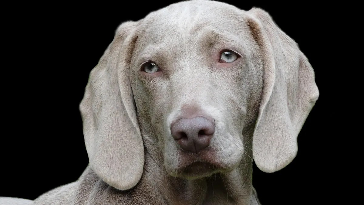
Horner's syndrome in dogs is a nerve problem, not an eye disease. It happens when the sympathetic nerve pathway that runs from the brain to the eye is interrupted, which makes one eye look droopy, small, and sunken. Most dogs improve, but the timeline and the outlook depend on what caused the nerve damage in the first place, so horner's syndrome in dogs treatment always starts with finding that cause.
This article is educational and is not a substitute for veterinary diagnosis or treatment.
Quick Answer
- The classic sign is a triad in one eye: a small pupil, a drooping upper eyelid, and a sunken appearance with the third eyelid raised.
- Causes range from harmless and temporary to serious, including inner ear disease, neck or spinal disease, tumors, and blood vessel problems.
- A sudden droopy eye is worth a same-day veterinary visit, especially if your dog also has trouble walking, head tilt, or trouble eating.
- Treatment targets the underlying cause. The eye signs themselves often need no direct treatment.
- Many dogs recover over weeks to months, but some keep the eye changes permanently if the nerve damage does not heal.
What Horner's Syndrome Looks Like and What Is Normal
Horner's syndrome is a collection of eye and facial changes caused by loss of sympathetic nerve supply to the eye. The medical term for this is oculosympathoparesis, and the core signs are miosis (small pupil), ptosis (drooping upper eyelid), and facial anhydrosis (reduced sweating) [1]. In dogs, sweating matters less than in people, so you will notice the eye changes most.
The three-part sign owners see
Look at your dog's face in good light. In the affected eye you may see:
- A pupil that is much smaller than the other one (this is called anisocoria).
- The upper eyelid drooping, so the eye looks half closed.
- The eyeball sitting back a little, with the pink third eyelid pushing up from the inner corner.
Some dogs also have a slight droop of the lip or ear on the same side. The white of the eye may look redder than usual.
What is normal and what is not
It is normal for a dog's pupils to change size with light and excitement. Both pupils should still match each other. A pupil that stays small while the other one stays wide is not normal. A droopy eyelid from tiredness should pass in minutes, not days. If one eye looks different from the other for more than a few hours, that is a reason to call your veterinarian.
One harmless look-alike is idiopathic unilateral transient miosis. This causes brief episodes of a small pupil, usually after sleep or exercise, and it clears on its own. In one study of 78 dogs, Australian Shepherds, Australian Shepherd mixes, and Miniature Australian Shepherds were heavily overrepresented, the merle coat pattern appeared in 70 percent of cases, and the median episode lasted only 30 minutes [2]. If your dog's eye returns to normal within an hour and stays normal, that pattern is different from true Horner's syndrome.
Horner's Syndrome in Dogs Causes
The sympathetic nerve path is long. It starts in the brain, travels down the spinal cord through the neck, exits into the chest, and runs back up the neck to the eye. Damage anywhere along that path can cause the same eye signs. This is why your veterinarian has to look well beyond the eye.
Common and important causes
| Cause | Typical clues | How urgent |
|---|---|---|
| Inner or middle ear disease | Head tilt, ear odor, scratching at the ear, trouble balancing | Same day |
| Neck or spinal cord disease | Neck pain, stiffness, wobbling, weakness in the legs | Same day to emergency |
| Non-compressive spinal cord disease | Neck signs with Horner's syndrome and no obvious disk compression on imaging | Same day |
| Tumor along the nerve path | Slow onset, other nerve deficits, weight loss | Same day to urgent |
| Blood vessel problem in the neck | Sudden onset with no other neurologic signs | Urgent |
| Trauma or surgery near the neck or ear | Recent injury or procedure | Urgent |
| Unknown (idiopathic) | Only the eye signs, everything else normal | Routine to same day |
In dogs with neck disease, Horner's syndrome is a meaningful clue. In one study of 93 dogs with cervical myelopathy and Horner's syndrome, non-compressive disease inside the spinal cord tissue was found much more often than in dogs with neck disease but no Horner's syndrome (58 percent versus 9 percent) [3]. That means the eye sign can point your veterinarian toward a specific type of spinal problem.
Blood vessel problems are a less common but real cause. A 5-year-old male golden retriever developed sudden left-sided Horner's syndrome with no other neurologic abnormalities, and imaging found a dissection (a tear) in the left internal carotid artery [4]. This is a reminder that a normal-looking dog with only eye signs can still have a serious underlying problem.
In people, Horner's syndrome has been reported after a minimally invasive procedure called radiofrequency ablation of a benign thyroid nodule, appearing the day after the procedure [5]. This shows how delicate the nerve supply near the neck is, and why any recent procedure or injury near the neck or ear belongs in the history you give your veterinarian.
Middle cranial fossa syndrome is a broader problem that can include Horner's syndrome along with weakness of several eye muscles and loss of pupil light response. In dogs and cats, cancer is the most common cause of that syndrome, though infections have also been reported [6].
Red Flags That Make It an Emergency
Call an emergency clinic right away if your dog has any of these along with the eye changes:
- Sudden weakness or inability to stand.
- Wobbling, circling, or a strong head tilt.
- A pupil that does not respond to light in either eye, or pupils that are both very wide.
- Trouble swallowing, drooling, or a change in bark or voice.
- Pain when you touch the neck, or a stiff, guarded neck.
- Breathing that looks labored or noisy.
- A known or suspected head or neck injury.
These signs suggest the problem is not limited to the eye. They need same-day imaging and a full neurologic exam. If your dog also has balance problems, your veterinarian may consider other conditions that affect balance, such as Canine Vestibular Syndrome: Differential Diagnosis and Management.
Safe Things You Can Do at Home While You Wait
You cannot treat Horner's syndrome at home, but you can keep your dog comfortable and give your veterinarian better information.
Protect the eye
A droopy eye may not blink fully, which can dry the surface. Keep the eye moist with a plain artificial tear product made for pets or people, and ask your veterinarian before using anything medicated. Watch for redness, discharge, cloudiness, or squinting, which can mean the cornea is irritated.
Keep a simple log
Write down the date and time you first noticed the change, which eye is affected, and whether the pupil size changes with light. Note any recent ear infection, neck injury, dental work, surgery, or travel. Note any change in appetite, balance, or energy. This history often shortens the diagnostic path.
Avoid neck strain
Skip collar walks and use a harness instead. Avoid rough play, tug games, and jumping on and off furniture until your veterinarian has examined your dog. If neck disease is the cause, this matters.
Do not give human medications
Never give human eye drops or pain pills without veterinary direction. Some common human products can harm dogs, and the dose depends on your dog's weight and health. Your veterinarian will calculate any dose.
What Your Veterinarian Will Check and Test
The exam starts with the eyes and then widens. Your veterinarian will measure pupil size in light and dark, test the pupil light reflex, check eyelid position, and look for the third eyelid. They will also test the corneal reflex and facial sensation, because several nerves run close together near the eye [6].
Next comes a full neurologic exam. Your veterinarian will watch your dog walk, check paw placement, test neck flexibility, and look at the ears with an otoscope. A thorough ear exam can find an infection or mass that explains the whole picture.
Tests your veterinarian may recommend
- Blood work and urinalysis to check general health and look for infection or metabolic disease.
- Imaging of the head, neck, or chest. MRI is often the best way to see the spinal cord, nerve roots, and blood vessels. In the golden retriever with carotid dissection, MRI and CT identified the narrowed, irregular artery and abnormal blood flow [4].
- Ear cultures or imaging if ear disease is suspected.
- A trial of topical eye drops that affect the pupil. These help localize where the nerve path is damaged. Your veterinarian chooses the drug and dose based on your dog's size and exam findings.
If your dog has neck pain or wobbling, imaging of the spine is a priority. The link between Horner's syndrome and non-compressive spinal cord disease means your veterinarian may look harder at the spinal cord itself, not only at disk compression [3].
Horner's Syndrome in Dogs Recovery and Prognosis
Recovery depends on the cause, not on the eye sign itself. There is no single horner's syndrome in dogs treatment that works for every case. The plan is to treat what is broken.
What recovery usually looks like
If the cause is treated or resolves on its own, the eye signs often fade gradually. The pupil usually returns to normal size first, then the eyelid lifts, and the sunken look and raised third eyelid settle last. This can take weeks to a few months. Some dogs keep a mild droop or a slightly small pupil long term.
When the cause cannot be fixed, the eye changes may stay. That is not painful by itself, but the eye needs monitoring for dryness and injury. In pigs that developed Horner's syndrome after vascular surgery near the carotid artery, the signs persisted for the full two-month study period without improving or worsening [1]. That shows how variable the timeline can be, and why follow-up exams matter.
What affects the outlook
- A treatable ear infection or inflammation often has a good outcome.
- Neck or spinal disease may need medical or surgical treatment, and the outlook depends on the specific disease.
- Tumors and blood vessel problems carry a more guarded outlook and need specialist care.
- Idiopathic cases, where no cause is found, often improve on their own over time.
Your veterinarian will recheck the eye and the neurologic exam at set intervals. Bring your log to each visit.
Prevention and Monitoring
You cannot prevent every cause of Horner's syndrome, but you can lower some risks and catch problems early.
- Treat ear infections promptly and finish the full course your veterinarian prescribes.
- Use a harness instead of a collar if your dog has neck disease or neck pain.
- Keep up with routine wellness exams so subtle neurologic changes get noticed.
- After any surgery or procedure near the ear, neck, or throat, watch for a droopy eye and report it. Horner's syndrome has been reported the day after a neck procedure in a human patient [5].
- If your dog has a merle coat and brief episodes of a small pupil, mention the pattern to your veterinarian. The transient form is common in those breeds and often lasts about 30 minutes [2].
Some eye and eyelid conditions can look similar at first glance. If you notice a red, swollen bump at the inner corner of the eye instead of a sunken eye, that is a different problem, and you can read about it in Cherry Eye in Dogs: Causes, Surgery Options, and Recovery. If your older dog shows other behavior or orientation changes, Canine Cognitive Dysfunction Syndrome: Diagnosis and Management covers that topic.
Limitations and When to Contact a Veterinarian
This article gives general information. Your dog's individual case needs a veterinarian who can examine the eye, test the nerves, and review the history.
Contact your veterinarian the same day if:
- Your dog develops a droopy, small, or sunken eye for the first time.
- The eye signs last more than a few hours or get worse.
- Your dog has an ear infection, head tilt, or neck pain along with the eye change.
Go to an emergency clinic now if:
- Your dog cannot stand, walk straight, or hold the head level.
- Your dog has trouble breathing, swallowing, or keeping food down.
- There was a head or neck injury.
- The eye is squinting, cloudy, or has discharge, which can mean a corneal ulcer.
Schedule a routine visit if the eye signs are mild, stable, and your dog is otherwise normal, but do not wait more than a few days. Early imaging and a neurologic exam give the best chance of finding a treatable cause.
Frequently Asked Questions
Does Horner's syndrome in dogs go away on its own?
Sometimes it does, especially when no cause is found. The eye signs often fade over weeks to months as the nerve recovers. If a cause such as ear disease or a spinal problem is found, treating that cause is what allows the eye to return to normal.
Is Horner's syndrome painful for my dog?
The eye changes themselves are usually not painful. Any pain comes from the underlying problem, such as an ear infection or a neck injury. Watch for squinting, pawing at the eye, or head shyness, and report those signs right away.
Can Horner's syndrome affect both eyes?
It usually affects one eye, which is why the pupils look mismatched. Both eyes can be involved if the damage is higher up in the nerve pathway or widespread. Your veterinarian will check both eyes carefully during the exam.
Will my dog need surgery for Horner's syndrome?
Surgery is not used to treat the eye signs. It may be needed if the cause is a tumor, a spinal problem, or a blood vessel issue that can be repaired. Most cases are managed by treating the underlying disease with medication, ear care, or rest.
Can I use human eye drops on my dog's droopy eye?
No, do not use human eye drops unless your veterinarian tells you to. Some drops can worsen the eye or cause side effects, and the correct product and dose depend on your dog's weight and diagnosis. Ask your veterinarian which lubricant is safe for home use.
Related Articles
- Cherry Eye in Dogs: Causes, Surgery Options, and Recovery
- Canine Cognitive Dysfunction Syndrome: Diagnosis and Management
- Dog Behavioural Problems
- Canine Vestibular Syndrome: Differential Diagnosis and Management
- Dog Behavior Problems And Solutions
- Dog Behavior Issues Causes
- Facial Paralysis in Dogs: Causes, Recovery, and Care
- Vision Loss in Dogs: Signs, Causes, and What to Do
- Neck and Back Pain in Dogs: Symptoms, Causes, and Treatment
References
- Horner Syndrome in 2 Pigs (Sus scrofa) after Vascular Grafting of the Carotid Artery and Jugular Vein. Comparative medicine, 2017.
- Idiopathic Unilateral Transient Miosis in Dogs. Veterinary ophthalmology, 2026.
- Horner syndrome as a physiological biomarker of disease in canine cervical myelopathy. Journal of veterinary internal medicine, 2023.
- Horner Syndrome Secondary to Suspected Internal Carotid Artery Dissection in a Golden Retriever. Journal of veterinary internal medicine, 2025.
- Horner Syndrome After Radiofrequency Ablation of a Benign Thyroid Nodule. Cureus, 2025.
- Middle cranial fossa syndrome presumptively secondary to leishmaniasis in a dog: A case report. Open veterinary journal, 2026.