Pillow Foot in Cats: Plasma Cell Pododermatitis
By Dr. Zubair Khalid, DVM, MS, PhD ·
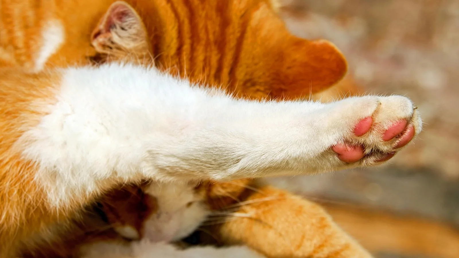
Pillow foot in cats is a skin condition in which one or more paw pads become soft, spongy, and swollen, so the normally firm pad feels like a small pillow under your finger. The medical name is plasma cell pododermatitis, and the diagnosis rests on a skin biopsy that shows a heavy infiltration of plasma cells into the pad tissue [1][2].
The classic picture is a cat with swollen pads on several paws, often the front paws, with little or no pain and no obvious lameness. Many of these cats are otherwise healthy and eating normally. That quiet presentation is exactly why pillow foot is easy to miss for weeks or months. The pads simply look and feel different, and the cat keeps walking on them without complaint.
Owner-facing triage summary:
- Soft, puffy, sponge-like paw pads on two or more paws: schedule a veterinary visit. This is not an emergency, but it needs a diagnosis.
- Pad swelling with bleeding, crusting, open sores, or a foul smell: book a visit within a few days. Secondary infection is common once the pad surface breaks down.
- Limping, refusing to walk, crying when the paws are touched, or bleeding that will not stop: treat this as urgent. Chronic hemorrhage from ulcerated pads can drop a cat's red blood cell count low enough to need intervention [3].
- Swelling that also involves the nose bridge, face, or other skin areas: still consistent with this disease pattern and worth mentioning to your veterinarian [4].
- Any cat with pillow foot should be tested for feline leukemia virus (FeLV) and feline immunodeficiency virus (FIV). Both have been documented alongside this condition [5].
This article is educational and is not a substitute for veterinary diagnosis or treatment.
At a Glance: Pillow Foot Decision Table
| What you observe | What it points toward | Next step | Urgency |
|---|---|---|---|
| Soft, doughy swelling of two or more paw pads, cat walks normally | Classic plasma cell pododermatitis | Veterinary exam, FeLV/FIV test, urinalysis, pad biopsy | Routine, within days |
| Same swelling plus ulceration, crusts, or intermittent bleeding | Plasma cell pododermatitis with surface breakdown or secondary infection | Exam plus biopsy, blood work including packed cell volume | Within 1 to 2 days |
| Swelling plus lameness or refusal to bear weight | Advanced pad disease, deep infection, or another cause | Same-day or next-day exam | Prompt |
| Bleeding that soaks a bandage or pale gums | Significant blood loss anemia | Emergency visit | Immediate |
| Swelling plus nose bridge thickening | Reaction pattern with nasal involvement | Exam, biopsy of both sites | Routine to prompt |
| Pad lumps in an older cat that do not improve | Neoplasm such as squamous cell carcinoma | Biopsy before any treatment trial | Prompt |
| Pads that look inflamed only after contact with a new litter or floor cleaner | Contact or irritant pododermatitis | Remove the trigger, exam if it persists | Routine |
What Pillow Foot Looks Like on a Cat
Owners usually describe the pads as "puffy," "squishy," or "like a marshmallow." The change is in the pad itself, not the fur or the skin between the toes. The pad loses its firm, slightly rough texture and gains volume. In classic cases the swelling is diffuse across the whole pad rather than a single lump.
The condition usually affects multiple pads. In one published report, a cat developed swelling, softening, and erythematous to violaceous discoloration across several metacarpal, metatarsal, and digital pads at once [5]. Another case described soft diffuse swelling of the pads on several feet together with a severely swollen nose bridge and mild itching [4]. A small case study of eight cats found no particular preference for sex, breed, age, or which paw was affected, but the disease typically started as pad swelling and later progressed to ulceration in some cats [1].
Front paws often draw attention first because owners see them when the cat kneads or stretches. That said, a study of pad biopsies found lesions were especially common in the front paws when silica (cat litter) material was also embedded in the tissue, which may simply reflect which paws contact the litter box most directly [6].
The single most reassuring feature for owners is the lack of pain. Plasma cell pododermatitis is characteristically non-painful, especially early. A cat with dramatically swollen pads may still jump, land, and use the scratching post without hesitation. Pain or lameness usually signals a complication such as ulceration, secondary bacterial infection, or a different diagnosis.
The Pad Anatomy That Explains the Signs
A cat's paw pad is a thick, specialized cushion of fat and connective tissue covered by a tough, hairless epidermis. It is built to absorb impact and insulate the foot from hot and cold surfaces. The pad's dermis and subcutis are the layers where the inflammatory infiltrate of plasma cell pododermatitis collects [7].
Plasma cells are immune cells that produce antibodies. When large numbers of them gather in the pad, they release inflammatory signals that loosen the normal architecture of the cushion. The result is the soft, spongy texture that gives pillow foot its nickname. The large number of plasma cells in these lesions is what first suggested an immunological basis for the disease [7].
Because the pad is a weight-bearing structure, any change in its thickness or resilience alters the way the cat stands. Mild cases compensate easily. Severe cases, especially with ulceration, can develop bleeding that is difficult to control because the pad has a rich blood supply and takes constant pressure with every step.
Causes, Risk Factors, and the Immune Question
The cause of plasma cell pododermatitis has not been determined [3]. What veterinarians know is that the lesion represents a reaction pattern rather than a single disease with a single trigger. Several lines of evidence point in different directions, and they are not mutually exclusive.
An Immune or Hypersensitivity Reaction Pattern
The dominant theory is immune-mediated or allergic. The dense plasma cell infiltrate, the response to corticosteroids in many cats, and the occasional involvement of other body sites all fit a hypersensitivity-associated reaction pattern [3][4]. A 2026 case report described a cat with plasma cell infiltration in the paw pads and a lymphoplasmacytic dermatitis on the nose bridge, and the authors argued this supports the idea that plasma cell pododermatitis is a hypersensitivity-associated reaction pattern rather than a strictly separate disease entity [4].
By the same logic, a cat can develop a plasma cell reaction elsewhere without ever having pillow foot. One report described a firm, haired, rounded swelling on the bridge of a cat's nose, and biopsy showed a plasma-cell-rich infiltrate. That cat tested negative for feline immunodeficiency virus, was treated for an upper respiratory infection, and the nasal lesion resolved within a month [8]. The lesson for owners is that plasma cell inflammation in cats is a pattern, not a fixed diagnosis with a fixed outcome.
Retrovirus Association
Feline leukemia virus and feline immunodeficiency virus have been reported alongside plasma cell pododermatitis [5]. In one detailed case, a 2-year-old intact male cat was seropositive for both FIV and FeLV, and immunohistochemistry on the pad biopsies was positive for FeLV and negative for FIV [5]. The authors highlighted retrovirus infection as a potential factor associated with the disease and called for further investigation [5].
This finding is why FeLV and FIV testing belongs in the standard workup for any cat with pillow foot. A positive test does not prove the virus caused the pad disease, but it changes the conversation about prognosis, monitoring, and household management. Retrovirus status also matters for cats living with other cats.
Concurrent Kidney Disease
A large case series of 25 cats with plasma cell pododermatitis and concurrent glomerular disease found that affected cats were most often young (under 5 years old) and castrated males [9]. Immune complex glomerulonephritis was present in 14 of 17 cats that had kidney biopsies, and urine testing consistently showed evidence of glomerular dysfunction [9]. The authors proposed that cats with plasma cell pododermatitis may be predisposed to glomerular disease [9].
This connection is important for owners because kidney changes can be silent. A cat with beautiful but puffy pads may have protein leaking through the kidney filters without any outward sign. Testing urine for protein is cheap, noninvasive, and worth doing at the first visit.
Environmental and Contact Triggers
Anything that irritates or damages the pad surface can start a chain of inflammation. A 2026 pathology study of 13 cat paw biopsies found silica-containing granulomas in the pads, with the silica material matching the Raman spectra of several brands of clay-based cat litter [6]. Ten of those 13 biopsies also showed evidence of plasma cell pododermatitis [6].
The interpretation matters. Silica granulomas are a separate finding caused by litter particles embedded in damaged pad tissue. The authors suggested that underlying pad disease may predispose a cat to litter implantation, and that the resulting granulomatous inflammation can obscure the original problem and make the clinical disease worse [6]. In practical terms, damaged pads plus abrasive or dusty litter can compound each other.
Does Age or Sex Predict Anything?
The evidence is mixed and depends on the population studied. A small histology study of eight cats found no sex, breed, or age predisposition [1]. The larger series of 25 cats with concurrent kidney disease found a cluster of young, castrated males [9]. That difference likely reflects referral patterns and the specific subgroup studied rather than a true breed or sex rule. Take the signal from your individual cat, not from a stereotype.
Differential Diagnoses: Why Biopsy Is Not Optional
Swollen paw pads have a long list of possible causes. Feline pododermatitis is a general term for pad inflammation, and plasma cell pododermatitis is only one subtype within it. Because the treatments are completely different, guessing is not safe.
Infection
Bacterial pododermatitis, fungal infections, and deep pyoderma can all swell pads. Bacterial infection often produces pus, draining tracts, odor, and pain, which plasma cell pododermatitis usually does not. Fungal disease such as sporotrichosis or cryptococcosis is less common but must be ruled out through culture or biopsy.
Allergy and Irritant Contact Reactions
Allergic dermatitis from food, fleas, or environmental allergens can affect the feet, but it usually involves the skin between the toes and the haired skin of the paw, not just the pad cushion. Contact irritation from floor cleaners, litter, or topical products can inflame pads but typically improves when the trigger is removed. Immune-mediated skin diseases in cats frequently mimic hypersensitivity and infections, which is why biopsy and culture are used together [10].
Neoplasia
Squamous cell carcinoma and other tumors of the pad are a serious concern, especially in older cats or when only one pad is affected. Malignant pad tumors often ulcerate, bleed, and destroy the normal pad contour. They do not respond to antibiotics or steroids. A pad that keeps growing despite treatment, or a single pad that looks different from the rest, needs a biopsy before any prolonged treatment trial.
Silica Granuloma
Embedded cat litter particles create granulomatous inflammation that mimics and can coexist with plasma cell pododermatitis [6]. This is detected on biopsy by examining the tissue under polarized light, which reveals birefringent crystalline material [6]. Because routine polarization is recommended for cat paw biopsies, a pathologist can identify both conditions in the same sample [6].
Why the Diagnosis Requires a Biopsy
No blood test, X-ray, or visual examination can confirm plasma cell pododermatitis. The diagnosis depends on histopathology, and in published cases it has been confirmed by examining pad tissue for the characteristic plasma cell infiltrate [5][1]. Hematoxylin and eosin staining shows the dense plasma cell population, and special stains such as methyl green-pyronin or immunohistochemistry for immunoglobulin light chains can highlight the plasma cells further [1].
The biopsy changes management. If the pathologist finds plasma cells, immunosuppressive or immunomodulatory treatment makes sense. If the pathologist finds bacteria, fungus, silica, or a tumor, the plan changes completely. A biopsy is the single most valuable test you can authorize for a cat with swollen pads.
Diagnostic Steps Your Veterinarian Will Take
A thorough workup for pillow foot follows a logical sequence. The goal is to confirm the diagnosis, identify triggers and complications, and screen for the conditions that travel with this disease.
1. History and Physical Examination
Your veterinarian will ask when the swelling started, whether it is getting worse, whether the cat limps, and whether there has been any bleeding. They will examine every paw pad, not just the ones you noticed, and will check the nose, ears, and skin for other plasma cell reactions [4].
They will also palpate the pads to grade softness and look for ulceration, crusting, and discharge. Photographing the pads at each visit is a practical way to track change over time, since pad contours are hard to remember.
2. Blood Work and Retrovirus Testing
A complete blood count and serum biochemistry panel establish baseline health and can reveal anemia from chronic pad hemorrhage [3]. Serum protein electrophoresis may show hyperglobulinemia, which is consistent with a plasma cell-driven process [8].
FeLV and FIV testing is standard because both viruses have been reported with this disease [5]. Testing is done by immunoassay, and confirmatory polymerase chain reaction testing for proviral DNA may be used [5].
3. Urinalysis and Protein Assessment
Given the link to glomerular disease, a urine sample should be collected and evaluated for protein [9]. A urine protein-to-creatinine ratio quantifies protein loss, and in the 25-cat case series these ratios varied widely between patients [9]. Urine protein electrophoresis using sodium dodecyl sulfate-polyacrylamide gel electrophoresis can identify a glomerular pattern of protein loss, which is a sensitive clue to kidney involvement [9]. When protein loss is confirmed, blood pressure measurement and sometimes a kidney biopsy are the next steps.
4. Imaging
Abdominal ultrasound is typically performed when kidney disease is suspected, but findings are often nonspecific in the early stages [9]. Chest imaging may be appropriate if a tumor or systemic disease is on the differential list. Imaging does not diagnose pillow foot, but it helps rule in or out the conditions that accompany it.
5. Pad Biopsy
The biopsy is the confirmatory test. Your veterinarian will typically take a full-thickness wedge or punch sample from an affected pad under local or general anesthesia, then close the site or allow it to heal by second intention. The sample goes to a pathologist who examines it for plasma cell infiltration, perivascular patterns, binucleated plasma cells, mitotic figures, and any foreign material [1].
Pathologists should polarize the sample routinely to look for silica crystals, because those crystals can be present alongside plasma cell pododermatitis and can change how the case is managed [6]. If infection is a concern, part of the sample can be submitted for culture.
6. Monitoring After Diagnosis
Once the diagnosis is confirmed, the follow-up plan usually includes recheck visits to assess pad size, repeat blood work to monitor for anemia or drug effects, and periodic urine testing if kidney disease is present. A case series of cats with both conditions found that immunosuppressive therapy was the primary treatment for the kidney disease but was often delayed because glomerular dysfunction was hard to recognize [9]. Early urine testing avoids that delay.
Treatment of Pillow Foot in Cats
Talking about pillow foot in cats treatment means talking about a condition that is unpredictable. Some cats improve without any medication. Others need months of therapy. Setting realistic expectations up front makes the process easier for everyone.
First-Line Treatment: Doxycycline
Doxycycline is the standard first-line treatment for plasma cell pododermatitis. It belongs to the tetracycline class and has anti-inflammatory and immunomodulatory effects that are separate from its antibiotic activity. This is why it can help even when no infection is identified.
Doxycycline is a prescription medication. Dose depends on your cat's weight and the formulation your veterinarian stocks, and it must be given with water or a small amount of food followed by a water flush to reduce the risk of esophageal stricture, a known complication of doxycycline tablets in cats. Never give a tablet dry. If your cat will not take pills, ask about a compounded liquid or another option. Follow the label directions exactly and do not adjust the dose on your own.
Corticosteroids and Other Immunosuppressants
Corticosteroids are the mainstay for cats that do not respond to doxycycline or that have severe disease. In one early report, two cats with pad enlargement and chronic hemorrhage were treated with immunosuppressive doses of corticosteroids after their ulcers were debrided, and both had considerable reduction in pad size after three to four weeks [3]. Another small study found total remission of lesions after glucocorticoid therapy in four cats [1].
Steroids are not harmless. They can raise blood sugar, suppress the immune system, and worsen heart disease. Cats on steroids need monitoring blood work, and the dose should be tapered rather than stopped suddenly. If steroids alone are not enough, veterinarians may consider other immunosuppressive drugs or immunomodulators. These are decisions for your veterinarian based on the severity of the disease and the response to initial therapy.
Spontaneous Remission
Some cats improve without treatment. One report described a cat with a plasma-cell-rich nasal lesion, no pad involvement, and a negative FIV test, whose lesion resolved on its own within a month after the respiratory infection was treated [8]. Other case reports note that the condition can fluctuate, and some cats have long periods of normal pads between flare-ups. Spontaneous remission is real, but it cannot be predicted, and waiting to see if it happens carries the risk that pad disease will progress.
What the Treatment Table Looks Like
| Drug | Class and mechanism | What it does in this disease | Monitoring | Expected response |
|---|---|---|---|---|
| Doxycycline | Tetracycline antibiotic with anti-inflammatory and immunomodulatory effects | First-line treatment for swollen pads, works even without confirmed infection | Appetite, vomiting, esophageal signs (drooling, gagging after pills), recheck exam in 3 to 4 weeks | Gradual softening of pads over weeks, partial or full response in many cats |
| Corticosteroids (such as prednisolone) | Anti-inflammatory and immunosuppressive steroid | Used when doxycycline fails, or when disease is severe or involves other sites such as the nose | Blood glucose, complete blood count and chemistry, weight, behavior changes, taper plan | Pad size reduction often visible in 3 to 4 weeks [3][1] |
| Other immunosuppressants or immunomodulators | Varies by drug, generally suppress specific immune pathways | Reserved for cats that do not respond to doxycycline and steroids, or that cannot tolerate steroids | Drug-specific blood work, typically starting at 2 to 4 weeks and then periodically | Variable. Some cats respond well, others need combination therapy |
| Supportive care: pad protection, wound cleaning, soft bedding | Not a drug. Reduces trauma and secondary infection | Helps ulcerated or bleeding pads heal between medical treatments | Daily visual check for bleeding, odor, or worsening | Improved comfort and reduced bleeding |
Any change in medication should be made by your veterinarian. Do not start, stop, or change doses on your own, and do not give your cat any human medication for swelling or pain unless your veterinarian prescribes it.
What You Can Do at Home While Treatment Is Starting
Your role is to reduce stress on the pads and report changes accurately.
- Keep the litter box clean and consider a softer, less dusty litter while the pads are fragile. Damaged pads can trap litter particles, and embedded silica can worsen inflammation [6].
- Trim the fur between the toes if it is trapping debris or moisture, but do not cut the pad itself.
- Inspect the pads daily. Note color, size, discharge, and any bleeding. Take photos with your phone in consistent lighting.
- Limit jumping from high places if the cat is willing, and provide soft bedding. This reduces trauma to swollen pads.
- Do not apply human antibiotic ointments, essential oils, or home remedies to the pads. Cats groom their feet and will ingest anything you apply.
- Give medications exactly as prescribed and finish the full course unless your veterinarian says otherwise.
- Bring your cat back for rechecks even if things look better. Pad appearance lags behind tissue healing.
Prevention and Risk Reduction
There is no proven way to prevent plasma cell pododermatitis, because the cause is not known [3]. The practical goal is to reduce known triggers and catch flares early.
- Manage retrovirus status. Test any new cat entering the household, and keep positive cats indoors to reduce transmission and stress.
- Screen for kidney protein loss if your cat has this diagnosis. Early detection allows earlier treatment [9].
- Use gentle, dust-controlled litter and avoid harsh floor cleaners. Silica from clay litter has been found in inflamed cat paw tissue [6].
- Address other allergic or immune conditions with your veterinarian so the immune system is not chronically activated.
- Keep routine wellness visits so pad changes are noticed before they become severe.
Unsafe Home Remedies and Common Mistakes
Wrong treatment can delay the correct diagnosis and cause real harm.
- Giving leftover antibiotics or human steroids. Doses, drugs, and durations are different in cats, and some human medications are toxic to cats.
- Assuming a swollen pad is "just a bruise." Plasma cell pododermatitis, infection, and tumors can all look like a bruise at first.
- Skipping the biopsy because the pads look better. Improvement can be temporary, and a tumor can be masked by a short course of steroids.
- Bandaging the paw tightly without veterinary guidance. A tight bandage on a cat's foot can cut off circulation.
- Letting a bleeding pad continue untreated. Chronic hemorrhage has caused severe anemia in a published case [3].
- Using human pain relievers. Many common human pain medications are dangerous for cats.
Prognosis and Long-Term Outlook
The outlook for plasma cell pododermatitis is generally favorable, but it depends on what else is going on. Cats that respond to doxycycline or corticosteroids usually improve over three to four weeks, and some achieve total remission [3][1]. Some cats need ongoing or repeated treatment. Others improve on their own [8].
The presence of concurrent kidney disease changes the picture. In the 25-cat series, immunosuppressive therapy was the main treatment for glomerular disease, but diagnosis was often delayed [9]. Cats with both conditions need closer monitoring and more frequent veterinary visits. Immune-mediated skin diseases in cats can occasionally be severe enough to reduce quality of life, which is why early diagnosis and consistent follow-up matter [10][2].
Emergency Red Flags
Go to a veterinarian immediately if you see any of these:
- Bleeding from a pad that does not stop within a few minutes of gentle pressure.
- Pale gums, weakness, collapse, or a fast heartbeat, which can indicate significant blood loss anemia [3].
- Refusal to eat or drink for more than 24 hours.
- A pad that is hot, draining pus, or has a foul smell.
- Sudden severe lameness or a cat that will not put a foot down at all.
- Vomiting, drooling, or gagging after doxycycline tablets, which can indicate esophageal irritation.
- Any new swelling of the face, nose, or throat that affects breathing.
Limitations and When to Contact a Veterinarian
This article describes the general pattern of plasma cell pododermatitis, but individual cats vary widely. Contact your veterinarian if:
- Your cat has soft or swollen pads that have not been evaluated, even if your cat seems comfortable.
- Pad swelling is getting worse after two weeks of treatment.
- New pads become involved, or the nose and face start to swell [4].
- Your cat develops lameness, pain, or bleeding.
- Your cat is on steroids or doxycycline and develops vomiting, diarrhea, increased thirst, increased urination, or changes in behavior.
- Your cat has a known retrovirus infection and develops any new pad or skin change [5].
- Your cat was diagnosed with pillow foot and has not had urine protein testing, especially if the cat is young or male [9].
Your veterinarian can adjust the plan based on your cat's response. Do not extend or repeat a prescription without guidance.
Frequently Asked Questions
Is pillow foot in cats painful?
Usually not in the early stages. Plasma cell pododermatitis is characteristically non-painful, and many cats walk normally despite visibly swollen pads [1]. Pain or lameness usually appears when a pad ulcerates or becomes infected, which is why newlimping is a reason to call your veterinarian.
What does doxycycline do for plasma cell pododermatitis in cats?
Doxycycline reduces pad inflammation through anti-inflammatory and immunomodulatory effects that go beyond its antibiotic action. It is the standard first-line treatment, and your veterinarian will set the dose and duration based on your cat's weight and response.
Can pillow foot in cats go away on its own?
Yes, spontaneous improvement has been reported in cats with plasma cell reaction patterns. One cat with a plasma-cell-rich nasal lesion and no pad involvement improved without specific treatment after about a month [8]. Waiting is a decision to make with your veterinarian, since some cats progress to ulceration and bleeding.
Does pillow foot in cats mean my cat has FeLV or FIV?
No. Retrovirus infection is associated with the condition in some cats, and one case report described a cat with both FeLV and FIV [5]. However, many cats with plasma cell pododermatitis test negative. Testing is still recommended because it changes monitoring and household management.
Why does my cat need a biopsy instead of just antibiotics?
A biopsy is the only way to confirm plasma cell pododermatitis and to rule out infection, silica granuloma, and cancer [5][1][6]. Treating without a diagnosis risks missing a tumor or an infection, and it can delay effective treatment.
How long does treatment for plasma cell pododermatitis cats take?
Many cats show improvement in three to four weeks, based on published reports of steroid treatment [3]. Some cats need longer courses or ongoing management, and some improve with doxycycline alone. Your veterinarian will set recheck intervals based on your cat's response.
Are plasma cell pododermatitis cats at risk for kidney disease?
There is a documented association. A series of 25 cats with this diagnosis and concurrent glomerular disease found immune complex glomerulonephritis in most cats that had kidney biopsies [9]. Ask your veterinarian about urine protein testing at the time of diagnosis.
Can I use human medicine on my cat's swollen paws?
No. Human medications, including pain relievers and steroid creams, can be toxic to cats or cause serious side effects at the wrong dose. Do not apply anything to the pads or give any oral medication unless your veterinarian prescribes it.
Related Articles
- Yeast Infection Pododermatitis Dog Paw Home Remedy
- Rabbit Care: Sore hock ulcerative pododermatitis treatment guide
- Equine Hoof and Foot Conditions: Diagnosis and Treatment
- Dog Licking Paws Treatment
- Bovine Foot Rot: Diagnosis, Treatment, Recurrence, and Pasture Risk Control
- Sheep and Goat Foot Rot: Lesion Scoring, Treatment, and Eradication Planning
- 3 Parts of Cell Theory: Explained Simply
- Cell Image Guide: Labeled Diagrams of Cell Structures
Sources
- Feline plasma cell pododermatitis: a study of 8 cases.
- Feline immune-mediated skin disorders: Part 2.
- Plasma cell pododermatitis with chronic footpad hemorrhage in two cats.
- [[Feline plasma cell pododermatitis with nasal involvement suggesting a hypersensitivity associated reaction pattern - a case report and review of the literature].](https://pubmed.ncbi.nlm.nih.gov/42624129/)
- Plasma Cell Pododermatitis Associated With Feline Leukemia Virus (FeLV) and Concomitant Feline Immunodeficiency Virus (FIV) Infection in a Cat.
- Cutaneous silica-associated (cat litter) granulomas in the paws of 13 domestic cats.
- Plasma cell pododermatitis in a cat.
- Nasal swelling due to plasma cell infiltrate in a cat without plasma cell pododermatitis.
- Feline plasma cell pododermatitis with concurrent glomerular disease: a case series of 25 cats.
- Feline immune-mediated skin disorders: Part 1.