Cell Image Guide: Labeled Diagrams of Cell Structures
By Dr. Zubair Khalid, DVM, MS, PhD ·
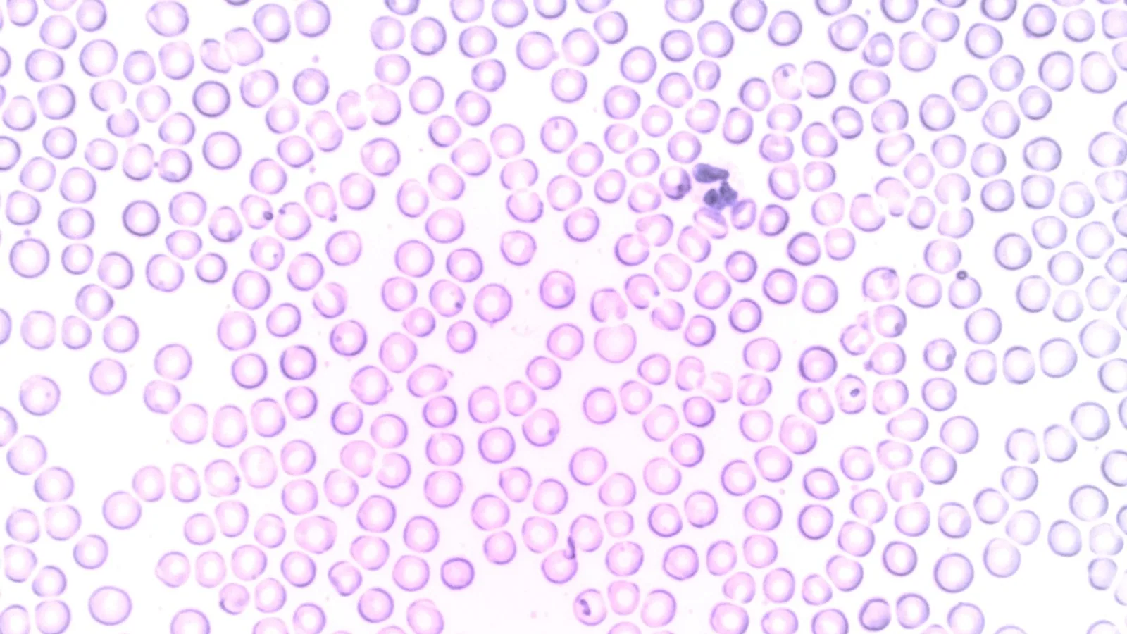
A labeled cell image is the fastest way to learn cell biology, because every organelle you can point to has a job you can remember. This guide gives you the three canonical cell pictures (animal, plant, and bacterial), a table that maps each structure to its function and to the cell types that contain it, and the practical rules for reading any cellular image without confusing one structure for another.
When you look at a pic of a cell, you are looking at a system in which compartmentalization is the whole point. Eukaryotic cells (animal and plant) wrap their chemistry inside membrane-bound compartments. Prokaryotic cells (bacteria) do not. That single difference explains most of what you see and do not see in a standard cell picture.
What You Are Actually Looking At
The resolution problem
Light microscopes resolve objects down to roughly 200 nm. A mitochondrion is about 500 nm to 1 µm wide, so it appears as a faint filament or dot. Ribosomes are about 25 nm and are invisible by light microscopy, which is why they do not appear in a routine cell picture but must be added to a labeled diagram. Transmission electron microscopy (TEM) resolves to the nanometer scale and reveals membranes, cristae, and even individual ribosomes. The classic cell structure reference images used in histology teaching are TEM preparations [1].
This matters for honesty in diagrams. A textbook animal cell image is a composite. Nobody sees the endoplasmic reticulum, the Golgi, and the lysosomes in their true relative positions in one light micrograph. You see them by fluorescence tagging or electron microscopy, one or two structures at a time.
Why every diagram looks slightly different
Illustrators normalize sizes so the labels fit. In a real cell, the nucleus is not small and centered. In an adipocyte, a lipid droplet pushes the nucleus to the rim. In a skeletal muscle fiber, hundreds of nuclei sit under the plasma membrane. Plant cells are frequently dominated by one enormous vacuole, so the cytoplasm is a thin rind against the wall [2]. Bacterial cells, at 0.5 to 5 µm, are smaller than most animal cell nuclei.
The Animal Cell: What Each Label Means
An animal cell is a eukaryotic cell with a plasma membrane, a membrane-bound nucleus, and a full set of membrane-bound organelles. It has no cell wall. Instead, animal cells sit in an extracellular matrix and use the actin cytoskeleton, integrins, and cadherins for shape and adhesion.
Plasma membrane
A phospholipid bilayer with embedded proteins, cholesterol, and glycolipids. It is the selective barrier. Because animal cells lack a wall, the membrane also carries most of the mechanical signaling load.
Nucleus, nucleolus, and nuclear envelope
The nucleus holds the DNA. The nuclear envelope is a double membrane perforated by nuclear pores that control traffic of RNA and proteins. The nucleolus is the dense region where ribosomal RNA is transcribed and pre-ribosomal subunits are assembled. Nucleoli are visible in almost every cellular image of an animal cell because they are dense enough to contrast.
Endoplasmic reticulum (ER)
The rough ER is studded with ribosomes and is the entry point for secreted and membrane proteins. The smooth ER synthesizes lipids and, in liver cells, detoxifies drugs. The two regions are continuous.
Golgi apparatus
A stack of flattened cisternae that modifies, sorts, and packages proteins and lipids arriving from the ER. Every secreted protein in your body passed through a Golgi stack.
Mitochondria
The ATP-producing organelle. Mitochondria have their own circular DNA and a double membrane, with the inner membrane folded into cristae to increase surface area. Mitochondria are one of the structures that separate eukaryotic cells from bacteria in a labeled diagram.
Lysosomes and peroxisomes
Lysosomes are acidic vesicles (around pH 5) packed with hydrolytic enzymes. They digest material delivered by endocytosis and autophagy. This degradation route is a real experimental constraint in targeted therapy, because plant-derived type I ribosome-inactivating proteins such as gelonin are vulnerable to lysosomal breakdown, which limits cytosolic delivery and antitumor efficacy [3]. Peroxisomes handle fatty acid oxidation and hydrogen peroxide metabolism.
Ribosomes
The protein synthesis machines. Eukaryotic cytoplasmic ribosomes are 80S (a 60S large subunit plus a 40S small subunit). They are either free in the cytosol or bound to the rough ER.
Cytoskeleton
Microtubules, actin filaments, and intermediate filaments. They set cell shape, move cargo, and drive division. Cortical microtubule arrays are especially prominent in plant cells, where they organize under the plasma membrane rather than radiating from a central centrosome [4].
Centrioles and centrosome
A pair of centrioles forms the core of the centrosome, the main microtubule-organizing center of animal cells. Plant cells lack centrioles and organize microtubules from dispersed cortical and nuclear sites instead [4]. This is one of the cleanest ways to tell an animal cell from a plant cell in a labeled diagram.
Membrane-bound organelles absent in bacteria
The nucleus, mitochondria, ER, Golgi, lysosomes, peroxisomes, and centrioles have no prokaryotic counterparts. If you see any of them labeled, you are looking at a eukaryotic cell image.
The Plant Cell: What Each Label Means
A plant cell is a eukaryotic cell with three structures an animal cell does not have: a cell wall, chloroplasts, and a large central vacuole. It also lacks centrioles [4].
Cell wall
A rigid wall outside the plasma membrane, built mainly of cellulose microfibrils cross-linked in a pectin and hemicellulose matrix. It provides tensile strength and resists turgor pressure. The plant cell wall and the bacterial peptidoglycan sacculus are chemically unrelated, and they are not homologous even though both are called "wall" in casual speech.
Chloroplasts
Double-membrane plastids containing thylakoid membranes and chlorophyll. They carry out photosynthesis and have their own circular DNA like mitochondria. Chloroplasts are the label that most reliably identifies a plant cell image, along with the wall and central vacuole.
Central vacuole
A large, single membrane-bound compartment (the tonoplast encloses it) that occupies most of a mature plant cell's volume. It stores water, ions, pigments, and metabolites, and it generates turgor pressure. In some plant species, storage tissue is dominated by parenchyma cells with abundant polysaccharides, and phenolic compounds concentrate in thick cell walls, which is visible histochemically [5].
Plasmodesmata
Cytoplasmic channels through the wall that connect adjacent plant cells. They are the plant equivalent of gap junctions.
Plant microtubule arrays
Plant cells do not have centrosomes. Microtubules assemble at the cell cortex into ordered arrays whose patterns depend on assembly dynamics and microtubule-associated proteins [4]. Computer models reproduce these cortical patterns as self-organized structures, which is why plant cell images often show a band of microtubules hugging the membrane.
What is shared with animal cells
Nucleus, nucleolus, ER, Golgi, mitochondria, peroxisomes, ribosomes (80S), and a plasma membrane. Plant cells also contain lysosome-like lytic vacuoles rather than discrete lysosomes, which is a nuance worth knowing because "lysosome" is often omitted from plant diagrams on purpose.
Microscopy of plant tissue
Structural and histochemical studies of plant stems describe an ordered arrangement of cell types and deposits, from calcium oxalate raphide bundles in the cortex to phenolic compounds in thick walls, and they rely on schematic diagrams to capture that organization [5]. A plant cell image is therefore often a tissue-level picture, not an isolated cell, and interpretation requires knowing which tissue you sectioned.
The Bacterial Cell: What Each Label Means
Bacteria are prokaryotes. They have no membrane-bound organelles, no nucleus, and no cytoskeleton organized into the same filament systems you see in eukaryotes.
Nucleoid
The nucleoid is the region containing the circular chromosome. It is not enclosed by a membrane. Chromosomal DNA is compacted by supercoiling and nucleoid-associated proteins, and it is the structure that replaces the eukaryotic nucleus in every bacterial cell image.
Plasmids
Small circular extrachromosomal DNA molecules that replicate independently. Plasmids often carry antibiotic resistance genes or metabolic genes. Many bacterial cells carry several plasmids at once.
Ribosomes (70S)
Bacterial ribosomes are 70S, made of a 50S and a 30S subunit. They are structurally and functionally distinct from eukaryotic 80S ribosomes, which is why many antibiotics target the 70S ribosome selectively. In an electron micrograph of bacterial cytoplasm, ribosomes fill much of the internal volume.
Cell envelope
Bacteria have a plasma membrane, and most also have a rigid sacculus of peptidoglycan outside it. Gram-positive bacteria have a thick peptidoglycan layer and no outer membrane. Gram-negative bacteria have a thin peptidoglycan layer plus an outer membrane containing lipopolysaccharide. This envelope is the target of many antimicrobial peptides. Bombyx mori cecropin A, for example, binds and permeabilizes bacterial membranes in a detergent-like manner, then damages DNA, and its C-terminal amidation and the serine-lysine-glycine motif are required for those activities [6].
Flagella, pili, and capsules
Flagella drive swimming motility, pili mediate adhesion and horizontal gene transfer, and capsules provide a protective polysaccharide coat. None of these are membrane-bound organelles.
What is missing
No nucleus, no mitochondria, no ER, no Golgi, no lysosomes, no peroxisomes, no chloroplasts, and no centrioles. If a diagram of a cell has any of these, it is not a bacterium.
Eukaryotic Versus Prokaryotic Cells: The Defining Difference
The single defining difference is the nucleated compartment. Eukaryotes (Greek for "true kernel") sequester DNA in a membrane-bound nucleus and run oxidative metabolism in mitochondria. Prokaryotes ("before kernel") keep DNA in a nucleoid and run metabolism in the cytoplasm or at the membrane.
| Feature | Animal cell | Plant cell | Bacterial cell |
|---|---|---|---|
| Cell type | Eukaryotic | Eukaryotic | Prokaryotic |
| Nucleus | Yes | Yes | No (nucleoid) |
| Nucleolus | Yes | Yes | No |
| Plasma membrane | Yes | Yes | Yes |
| Cell wall | No | Yes (cellulose) | Yes (peptidoglycan) in most |
| Mitochondria | Yes | Yes | No |
| Chloroplasts | No | Yes | No |
| Central vacuole | No | Yes | No |
| Endoplasmic reticulum | Yes | Yes | No |
| Golgi apparatus | Yes | Yes | No |
| Lysosomes | Yes | Rare (lytic vacuoles) | No |
| Peroxisomes | Yes | Yes | No |
| Centrioles / centrosome | Yes | No | No |
| Ribosomes | 80S | 80S | 70S |
| Plasmids | Rare | Rare (in some plastids) | Common |
| Cytoskeleton | Actin, microtubules, intermediate filaments | Actin, microtubules, no intermediate filaments of the same type | Bacterial cytoskeletal homologs (MreB, FtsZ) |
| Typical size | 10 to 30 µm | 10 to 100 µm | 0.5 to 5 µm |
This table is the fastest answer for the common study question: is this structure in animal, plant, or bacterial cells?
Table of Organelle Function
| Structure | Function | Animal | Plant | Bacteria |
|---|---|---|---|---|
| Plasma membrane | Selective barrier, signaling | Yes | Yes | Yes |
| Nucleus | Stores DNA, transcription | Yes | Yes | No |
| Nucleolus | rRNA synthesis, ribosome assembly | Yes | Yes | No |
| Nucleoid | Compacts and houses chromosome | No | No | Yes |
| Ribosome | Translation of mRNA to protein | 80S | 80S | 70S |
| Rough ER | Secreted and membrane protein synthesis | Yes | Yes | No |
| Smooth ER | Lipid synthesis, detoxification | Yes | Yes | No |
| Golgi | Protein and lipid sorting, modification | Yes | Yes | No |
| Mitochondrion | ATP synthesis, oxidative metabolism | Yes | Yes | No |
| Chloroplast | Photosynthesis | No | Yes | No |
| Lysosome | Acidic degradation of macromolecules | Yes | No | No |
| Peroxisome | Fatty acid oxidation, H2O2 handling | Yes | Yes | No |
| Central vacuole | Storage, turgor, pH control | No | Yes | No |
| Cell wall | Rigidity, turgor resistance | No | Cellulose | Peptidoglycan |
| Centriole | Microtubule organization, spindle poles | Yes | No | No |
| Plasmid | Accessory genes, e.g. resistance | Rare | Rare | Common |
| Cytoskeleton | Shape, transport, division | Yes | Yes | Homologs |
Use this table as a quick-reference card. When you look at a cellular picture and a structure is labeled but you cannot remember what it does, match the label to the row.
How to Read a Cell Picture Without Getting Confused
Match scale to method
An electron micrograph at 50,000x shows membranes as thin dark lines and mitochondria with visible cristae. A phase-contrast micrograph of a live culture shows flattened cells with a clear nucleus and a bright halo. If the image shows individual organelles as distinct outlined bodies with clean shapes, it is almost certainly an illustration, not a photograph, so it is schematic.
Recognize the four giveaway structures
- A rigid outer wall plus chloroplasts plus a large central vacuole: plant cell [5].
- An irregular cell with a nucleus, no wall, and visible centrioles: animal cell.
- A small cell with a nucleoid, no nucleus, and a thick envelope: bacterium, and the envelope is peptidoglycan [6].
- A cell with a nucleus and mitochondria but no chloroplast, no wall, and no centriole: plant or animal, and you need another clue such as a cell plate during division or a cortical microtubule band [4].
Distinguish structure from cytoplasm texture
Ribosomes and glycogen granules both appear as dense particles in TEM. Only the rough ER is a membrane system, not a particulate field. A dense cytoplasmic ground substance in a good TEM cell image is largely ribosome-studded cytosol [1].
Do not overread tissue-level images
A stem cross-section with labeled cell walls and deposits is a tissue structure, not a single-cell anatomy [5]. The histochemistry identifies classes of molecules (polysaccharides, saponins, phenolics), so interpret it at that level.
Interpret functional experiments carefully
A bacterial cell picture with a labeled envelope is often used to explain permeabilization mechanisms. Cecropin A is a good example because it binds in a detergent-like way and damages DNA, and its activity depends on specific residues and C-terminal amidation [6]. A plant cell image used for defense signaling is usually a diagram of a network rather than a photograph of a single organelle arrangement [7].
Practical Implications for Students and Bench Workers
For exam purposes
The highest-yield facts are: animal cells have centrioles and lysosomes but no cell wall, plant cells have chloroplasts, a central vacuole, and a cellulose wall, and bacterial cells are prokaryotic with a nucleoid, 70S ribosomes, and often plasmids. Ribosomal size is a classic distinguishing number: 80S in eukaryotic cytoplasm versus 70S in bacteria. This is the basis for selective antibiotic action, and it is worth memorizing as a pair.
For microcopy on the bench
If you are preparing a cell culture image of an animal line, fix and permeabilize, then counterstain nuclei with DAPI and visualize the plasma membrane with a lipophilic dye. If you image a plant cell, remember that the cortical microtubule array reorients in response to growth and stress [4], so the pattern is dynamic, not fixed. If you image a bacterium, the envelope is the surface that determines uptake of dyes and antimicrobial peptides [6].
For understanding drug delivery
Endosomal escape and lysosomal degradation are the main barriers for toxins and biologics delivered into eukaryotic cells [3]. When you look at an animal cell image, the lysosome label is not just a textbook object, it is the organelle that limits efficacy of targeted therapies such as ribosome-inactivating proteins.
For understanding variability
Cells in a population are not identical. Even in a highly regulated differentiation process such as trichome patterning, cell-to-cell variability disturbs the geometric pattern, and the concentrations of key protein complexes determine how much variation appears [8]. When you compare a cell image of one cell to another, expect differences in size, organelle number, and vacuole volume.
Common Mistakes and Limitations
Calling the plant cell wall and the bacterial wall the same thing. The plant wall is cellulose-based, while the bacterial wall is peptidoglycan-based [6]. They perform a similar mechanical job but they are chemically unrelated. Never transfer conclusions about one to the other.
Assuming bacteria are just small animal cells. They lack every membrane-bound organelle, including the nucleus. The nucleoid is not a nucleus, and the 70S ribosome is not an 80S ribosome. If a diagram assigns a nucleus to a bacterium, the diagram is wrong.
Treating one labeled drawing as a universal cell. No single cell image represents all cell types. Plant cells vary from thin-walled cortical cells to heavily lignified fibers, and animal cells range from keratinocytes to neurons. Structural studies of plants, for example, show that cell types and deposits differ across developmental stages of the same organ [5].
Forgetting that image interpretation depends on method. TEM, fluorescence, phase-contrast, and schematic illustration each emphasizes different structures. The classic TEM cell structure reference shows details that are invisible by light microscopy [1]. If you compare a TEM image to a brightfield image, expect apparent contradictions.
Overgeneralizing from cultured cells. A cultured animal cell is flattened and adapted to plastic, so it displays less elaborate organelle geometry than the same cell type in tissue. Plant cells in culture often lose their large central vacuole and cortical microtubule pattern.
A note on scope: this guide covers structure and function, not individual clinical or diagnostic questions. For those, the interpretation of a specific specimen requires professional evaluation.
Frequently Asked Questions
What is the easiest way to tell an animal cell from a plant cell in a labeled image?
Look for a rigid cell wall, chloroplasts, and one large central vacuole. Plant cells have all three, and animal cells have none of them. Animal cells also have centrioles and lysosomes, while plant cells generally do not.
Do bacteria have a nucleus?
No. Bacteria are prokaryotes, so their DNA sits in a nucleoid that has no surrounding membrane. Any diagram that shows a bacterial nucleus is incorrect.
Are plant cell walls made of the same material as bacterial cell walls?
No. Plant cell walls are built from cellulose, while bacterial cell walls are built from peptidoglycan. They are structurally analogous but chemically unrelated.
Why do bacterial ribosomes matter in medicine?
Bacterial ribosomes are 70S, whereas eukaryotic cytoplasmic ribosomes are 80S. That difference lets many antibiotics bind the bacterial ribosome selectively. The 50S and 30S subunits are the classic drug targets.
What is the difference between a nucleoid and a nucleus?
A nucleus is bounded by a double membrane with pores and holds linear chromosomes. A nucleoid is an unbounded region that holds a circular chromosome. Only eukaryotes have a nucleus.
Do animal cells have a cell wall?
No. Animal cells rely on the plasma membrane, the extracellular matrix, and the actin cytoskeleton for shape and adhesion. The absence of a wall is one reason animal cells can migrate and change shape.
Can a cell picture be used to identify what an organelle actually does?
Not from the image alone. Structure suggests function (folded membranes suggest surface area for chemical reactions), but function is confirmed by experiments, not by appearance. Use the image to learn the map and the table to learn the jobs.
Why do plant cells lack centrioles?
Plant cells organize microtubules from dispersed cortical and nuclear sites rather than from a single centrosome, so centrioles are not needed [4]. Their cortical microtubule arrays self-organize into patterns that depend on assembly dynamics and microtubule-associated proteins.
Related Articles
- Ribosome Diagram: Structure, Function, and Labeled Parts
- DNA Drawing & Structure Diagram: Labeled Double Helix Step-by-Step
- Protein Synthesis Diagram Labeled
- Diagram of Yeast Cell: Structure, Function, and Labeling Guide
- Microscope Parts and Functions: A Visual Guide to Labeled Diagrams
- Nucleosome Diagram: Structure, Function, and How to Draw It
- Nephron Diagram: Labeled Structure and Function
- Chigger Bite Pictures: What Chiggers Look Like
- Muscle Cell: Structure, Types, and Function
Sources
- Cell Structure (TEM) - The Cell
- Differential growth and shape formation in plant organs.
- A Spectroscopic Study on Secondary Structure and Thermal Unfolding of the Plant Toxin Gelonin Confirms Some Typical Structural Characteristics and Unravels the Sequence of Thermal Unfolding Events.
- Understanding phase behavior of plant cell cortex microtubule organization.
- Structure and histochemistry of the stem of Dracaena cambodiana Pierre ex Gagnep.
- Antibacterial mechanism and structure-activity relationships of Bombyx mori cecropin A.
- Signalling network construction for modelling plant defence response.
- Influence of cell-to-cell variability on spatial pattern formation.