Lump on a Dog's Hind Quarter: What It Could Be
By Dr. Zubair Khalid, DVM, MS, PhD ·
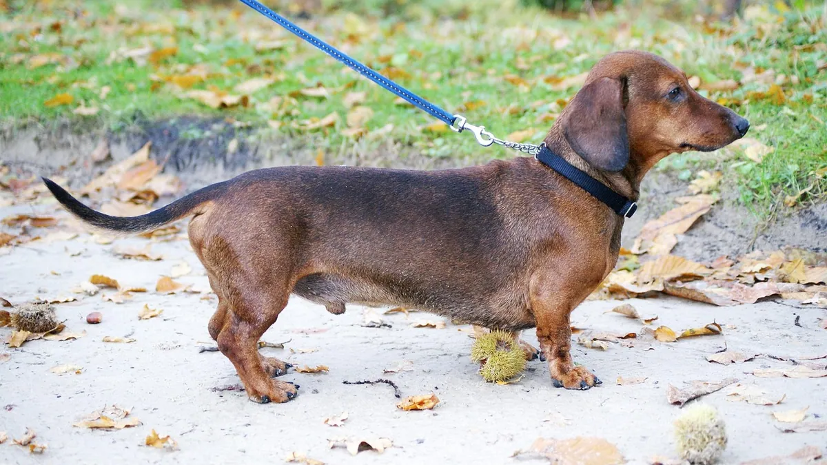
A hard lump on a dog's hind quarter is often a benign fatty tumor, but it can also be a cyst, an injury-related swelling, or a malignant growth. Because the hind quarter sits over major muscles and joints, the location changes how your veterinarian examines and tests the lump. This article is educational and is not a substitute for veterinary diagnosis or treatment.
Quick Answer
- Any new lump deserves a veterinary exam, even if it feels small and your dog seems fine.
- Firmness alone does not tell you whether a lump is benign or malignant. Only cell or tissue testing can do that [1].
- Lumps near the hip, knee, or ankle need extra attention because they can affect walking and lameness.
- Fine needle aspiration is a quick, low-stress test that collects cells for evaluation, and it is often the first step [1].
- Watch for rapid growth, pain, redness, discharge, or limping, and call your vet promptly if any appear.
What a Lump on the Hind Quarter Looks Like and What Is Normal
Dogs get lumps. Older dogs get more of them. A lump on the hind quarter can sit in the skin, in the fat under the skin, in a muscle, or near a joint. It may be pea-sized or as large as a golf ball or bigger.
Some lumps are soft and move freely under the skin. Others feel hard and fixed in place. Some have normal hair over them. Others are hairless, discolored, or ulcerated [1]. Skin tumors in dogs are diverse, and they can show up as small bumps, patches, rashes, or nonhealing sores [1].
Normal findings in an older dog include soft, movable lumps under the skin that have been stable for months. These are often benign fatty tumors called lipomas. Adipose tumors are common in older dogs, and most present as benign subcutaneous lipomas [2].
That said, "normal for an older dog" is not the same as "safe to ignore." A lump that changes in size, firmness, or color needs a fresh look. So does a lump that was never checked in the first place.
Main Causes of a Lump on a Dog's Hind Quarter
The list below covers the most common possibilities. Keep in mind that a physical exam cannot confirm a diagnosis. Distinguishing a benign tumor from a cancerous one requires specialized knowledge and laboratory equipment [1].
| Cause | Typical clues | How urgent |
|---|---|---|
| Lipoma (benign fatty tumor) | Soft, movable, under the skin, slow growing, common in older dogs [2] | Routine visit |
| Sebaceous cyst or follicular cyst | Round, smooth, may drain a thick material, often in the skin itself | Routine visit |
| Histiocytoma | Red, button-like, often in young dogs, may regress on its own | Routine visit, monitor closely |
| Mast cell tumor | Often raised, may swell or redden when touched, can change size quickly | Prompt visit |
| Soft tissue sarcoma | Firm, deep, fixed to tissue beneath, slow but progressive growth | Prompt visit |
| Joint or synovial tumor | Near a joint, causes lameness, may mimic arthritis on x-rays [3] | Prompt visit |
| Muscle or tendon injury | Swelling at a muscle origin, hindlimb lameness, often in active dogs [4] | Prompt visit |
| Abscess or infection | Warm, painful, may drain, often follows a wound or bite | Same-day visit |
| Bone tumor | Firm, painful, deep, may cause limping that worsens | Same-day visit |
Lipomas and Other Fatty Tumors
Lipomas are the most common benign lump in older dogs. They usually feel soft and squishy, and they slide under your fingers. Most stay under the skin and cause no problems [2].
Rarely, fatty tumors grow inside the body. A perirenal lipoma, for example, sits near the kidney and can cause weakness, urinary changes, or organ displacement [2]. These internal masses are uncommon and are found with imaging, not by touch.
Cysts and Skin Growths
Cysts form when a gland or hair follicle gets blocked. They often feel round and firm, and they may rupture and drain. Skin tumors can also look like cysts, rashes, or nonhealing ulcers, which is why a vet should identify them [1].
Histiocytomas
Histiocytomas are common in young dogs. They often look like a red, raised button on the skin. Many regress without treatment, but a vet should confirm the diagnosis first. You can read more about this in Histiocytoma in Dogs: The Red Button Lump That Often Regresses on Its Own.
Mast Cell Tumors and Other Malignant Growths
Mast cell tumors are one of the most common skin cancers in dogs. They can look like almost anything, including a harmless bump. They may swell, redden, or change size after you touch them.
Other malignant tumors include soft tissue sarcomas, which tend to feel firm and deep. Merkel cell carcinoma is a rare malignant tumor of small round cells that can be confused with other skin growths, and it has been reported in a young dog [5].
Joint and Muscle Problems
The hind quarter includes the hip, knee, and the muscles that attach around them. A mass near a joint can be a synovial tumor. One case report described a synovial myxosarcoma in a dog's shoulder joint that caused a year of intermittent lameness and did not respond well to anti-inflammatory drugs [3]. Joint tumors can even mimic septic arthritis on x-rays [3].
Muscle and tendon injuries can also create a firm swelling. A study of dogs with proximal gastrocnemius musculotendinopathy found chronic hindlimb lameness as the main sign, with soft tissue swelling and contrast enhancement at the muscle origin on CT [4]. Border Collies made up a third of the cases in that study [4].
Abscesses and Infections
An abscess is a pocket of pus, often from a bite, scratch, or foreign object. It feels warm and painful, and your dog may limp or resist touch. Abscesses need drainage and often antibiotics. If your dog has a draining wound plus a fever or refuses to eat, call your vet the same day.
Red Flags That Make It an Emergency
Call an emergency clinic right away if you see any of these:
- The lump grew noticeably in a few days or hours.
- The lump is hot, red, oozing pus, or bleeding without stopping.
- Your dog cannot bear weight on the leg or cries when you touch the area.
- Your dog has a fever, vomiting, or seems dull and weak.
- The lump ruptured and your dog is licking it constantly.
- Your dog has trouble breathing or collapses.
These signs can point to infection, a rapidly growing tumor, or another serious problem. Do not wait for a routine appointment if any of them appear.
Safe Things You Can Do at Home While You Wait
You cannot diagnose the lump at home. You can, however, gather useful information and avoid making things worse.
- Take a photo with a ruler or coin next to the lump for scale.
- Measure the lump with a soft tape and write down the date.
- Note whether it feels soft, firm, or hard, and whether it moves under the skin.
- Check for pain by gently pressing nearby, not on the lump itself.
- Keep your dog from licking or chewing the area. A cone or a light bandage can help.
- Do not squeeze, poke, or try to drain the lump. You can cause bleeding or infection.
- Do not apply human creams, ointments, or essential oils. Dogs can react badly or lick them off.
- Do not give any medication unless your veterinarian prescribes it. Your vet calculates the dose from your dog's weight and health.
If your dog is limping, limit running, jumping, and stairs until the exam. Rest helps many muscle and joint problems, and it prevents a small issue from getting worse.
What the Veterinarian Will Check and Test
Your vet will start with a full physical exam. They will check the lump's size, firmness, and mobility. They will also check the nearby lymph nodes, the hip and knee joints, and your dog's gait.
Fine Needle Aspiration
Fine needle aspiration is often the first test. The vet inserts a thin needle into the lump, collects cells, and looks at them under a microscope [1]. It takes seconds and usually needs no sedation. Results can guide the next step, though cytology and histopathology do not always agree. In one study of splenic masses, cytology and histopathology agreed in 55% of dogs, and disagreement occurred in 24% [6]. That study looked at the spleen, not the skin, but it shows why a single test is not always the final word.
Biopsy
A biopsy removes a small piece of tissue for evaluation [1]. It gives a more complete picture than cytology alone. Your vet may recommend a biopsy if the aspiration results are unclear or if the lump looks suspicious.
Imaging
X-rays can show whether a mass involves bone or a joint [3]. Ultrasound helps evaluate lumps that sit deeper in the body. CT gives detailed images of soft tissue and joint involvement, and it has been used to identify muscle and tendon problems in the hind limb [4]. Your vet will choose the imaging that fits your dog's case.
Blood Work and Other Tests
Blood work checks your dog's overall health before any procedure. It also helps rule out infection or other systemic problems. If the lump is near a joint, your vet may test joint fluid to rule out infection or inflammation.
Referral
Some cases need a specialist. A veterinary oncologist or surgeon can plan surgery, staging, or other treatment. Ask for a referral if you want a second opinion or if the diagnosis is unclear.
Prevention and Monitoring
You cannot prevent most lumps. You can catch problems early.
- Do a monthly lump check. Run your hands over your dog's whole body, including the hind quarters, armpits, belly, and between the toes.
- Record the size and location of every lump you find. A simple notebook or phone note works.
- Take photos every few months for comparison.
- Keep your dog at a healthy weight. Extra weight adds stress to hips and knees.
- Schedule wellness exams at least once a year, and twice a year for senior dogs.
- Ask your vet to check any lump that is new, changing, or painful.
If your dog has had a lump removed before, ask your vet how often to recheck. Some tumors can recur, and early detection makes treatment easier.
For a broader look at lumps in dogs, see Dog Skin Bumps and Lumps: When to See the Vet and Why Is My Dog having lumps on skin? Lipoma vs Mast Cell vs Cyst Diagnosis.
Limitations and When to Contact a Veterinarian
No article can tell you what your dog's lump is. Only a veterinarian who examines your dog and tests the lump can do that. Individual cases need a veterinarian.
Contact your vet the same day if the lump is new, growing, painful, or draining. Go to an emergency clinic now if your dog cannot walk on the leg, has a fever, is bleeding heavily, or seems weak or collapsed.
Schedule a routine visit if the lump is small, soft, and stable but has never been checked. A routine visit is also right if your dog has several lumps and you want a baseline.
If your dog has a lump plus other symptoms, such as vomiting, diarrhea, or weight loss, mention all of them at the visit. If your dog ate something that could be stuck, read Dog Ate a Foreign Object: Signs of Obstruction and When to Go to the Vet. If you are worried about parasites, see How To Tell If A Dog Has Roundworms Or Hookworms.
Frequently Asked Questions
Can a hard lump on my dog's hind quarter be harmless?
Yes, a hard lump can still be benign. Lipomas, cysts, and old injuries can all feel firm. Firmness alone does not prove cancer, and only testing can tell the difference [1].
Does a lump near the hip or knee mean something different?
It can. Lumps near joints may involve the joint lining, tendons, or muscles, and they often cause lameness. Joint tumors can even look like arthritis on x-rays, so imaging and testing matter more in these spots [3].
How long does it take to get results from a fine needle aspiration?
Many vets can look at the slides in the clinic the same day. If the sample goes to a lab, results often come back within a few days. Your vet will tell you what to expect for your dog.
Should I wait and see if the lump goes away?
Only wait if your veterinarian tells you to. Some lumps, like histiocytomas in young dogs, can regress on their own. Others grow or spread. A short monitoring plan with a set recheck date is safer than waiting without a plan.
Can I prevent lumps on my dog's hind quarter?
There is no proven way to prevent most lumps. Monthly home checks, regular vet visits, and prompt evaluation of any change are the best tools you have. Early detection gives you more options.
Related Articles
- Dog Skin Bumps and Lumps: When to See the Vet
- Histiocytoma in Dogs: The Red Button Lump That Often Regresses on Its Own
- Why Is My Dog having lumps on skin? Lipoma vs Mast Cell vs Cyst Diagnosis
- Dog Ate a Foreign Object: Signs of Obstruction and When to Go to the Vet
- How To Tell If A Dog Has Roundworms Or Hookworms
- Dog Skin Rash with Bumps: What Could It Be?
- Lump on Your Dog's Leg, Knee, or Ankle
- Lumps on Your Dog's Legs, Elbows, and Armpits: What They Mean
- Lump on Your Dog's Side or Abdomen
References
- Tumors of the Skin in Dogs - Dog Owners. Merck Veterinary Manual.
- Perirenal lipoma in a 14-year-old spayed female dog: A rare diagnosis. The Canadian veterinary journal = La revue veterinaire canadienne, 2026.
- Synovial myxosarcoma in the scapulohumeral joint of a dog: a case report. Veterinary research communications, 2024.
- Proximal Gastrocnemius Musculotendinopathy in Dogs: A Retrospective Computed Tomography-Based Study. Veterinary and comparative orthopaedics and traumatology : V.C.O.T, 2026.
- Diagnosis and treatment of Merkel cell carcinoma in a puppy: A comparative oncology approach. The Canadian veterinary journal = La revue veterinaire canadienne, 2026.
- Cytology and histopathology have poor to fair agreement for determination of neoplastic or nonneoplastic lesions in dogs with splenic masses or nodules. Journal of the American Veterinary Medical Association, 2026.