Lumps on Your Dog's Legs, Elbows, and Armpits: What They Mean
By Dr. Zubair Khalid, DVM, MS, PhD ·
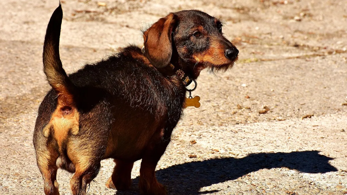
A dog bump on the leg is usually a benign growth such as a hygroma, lipoma, or cyst, though some lumps on the legs, elbows, armpits, and hips can be malignant and need treatment. The location, texture, and how fast a lump grows all help point toward a cause. This article is educational and is not a substitute for veterinary diagnosis or treatment.
Quick Answer
- Most soft, movable lumps under the skin are benign fatty tumors called lipomas, which are very common in dogs [1].
- Firm, fluid-filled swellings over the elbow or hip are often hygromas, pressure sores that form where a dog lies on hard surfaces.
- A lump that grows quickly, feels fixed to deeper tissue, or sits in the armpit deserves a veterinary exam soon.
- Your veterinarian can usually identify a lump with a simple needle sample (fine needle aspirate) in the exam room.
- Never squeeze, poke, or try to drain a lump at home, and never give human medications without veterinary guidance.
What a Dog Bump on the Leg Looks Like and What Is Normal
Lumps on a dog's legs come in many forms. Some are flat and barely raised. Others are round, pea-sized, or as large as a golf ball. Some sit right on the skin surface, and others hide in the fat beneath it. Many are hairless, and some feel firm and rubbery while others feel soft and mushy [2].
It helps to know what is normal for your own dog. Dogs commonly develop small skin tags, warts, and harmless bumps as they age. A lump that has been the same size and texture for months is less worrisome than one that appeared last week and doubled in size.
Location matters too. The legs, head, and neck are common sites for certain benign fibrous lumps, partly because these areas get bumped and scraped more often [2]. The elbows and hips take constant pressure when a dog lies down, so they are prime spots for fluid-filled swellings.
Common Causes of Lumps on Dog Legs, Elbows, Armpits, and Hips
The table below covers the lumps owners ask about most. It is a starting point, not a diagnosis. Only your veterinarian can tell you what a specific lump is.
| Cause | Typical clues | How urgent |
|---|---|---|
| Hygroma | Soft, fluid-filled swelling over the elbow or hip, often in large or heavy dogs that lie on hard floors | Routine visit, sooner if it is hot, painful, or draining |
| Lipoma | Soft, movable, squishy lump under the skin, slow growing, common in middle-aged and older dogs [1] | Routine visit |
| Sebaceous cyst | Round, firm lump that may have a pore or a cheesy discharge if it ruptures | Routine visit |
| Collagenous nevus | Flat to raised lump in the skin or fat, often on the legs or areas prone to trauma, common in middle-aged and older dogs [2] | Routine visit |
| Fibroma | Isolated, often raised, hairless lump under the skin, firm and rubbery or soft and mushy, most common in older dogs [2] | Routine visit, sooner if it changes or grows large |
| Soft tissue sarcoma | Firm lump with poorly defined edges that feels attached to deeper tissue, more common in medium and large breeds and older dogs [3] | Prompt visit, within days |
| Merkel cell carcinoma | Solitary, rapidly growing skin mass, can occur even in young dogs [4] | Prompt visit, within days |
Hygromas on the Elbow
A hygroma is a pocket of fluid that forms over a bony pressure point. It happens when a dog repeatedly lies on a hard surface, so the elbow or hip rubs against the floor. The result is a soft, squishy swelling that is usually not painful at first.
Small hygromas often improve with soft bedding and pressure relief. Large ones, or ones that become infected, may need drainage or other treatment. Your veterinarian can tell the difference between a hygroma and a tumor that happens to sit over the elbow.
Lipomas and Fatty Lumps
Lipomas are benign fatty tumors and are among the most common lumps in dogs [1]. They feel soft, move easily under the skin, and grow slowly. Most lipomas do not need removal unless they interfere with walking, sit in a spot that gets rubbed, or keep getting bigger.
A rare variant called a spindle cell lipoma has been described in dogs, and it can look similar to a plain lipoma on a needle sample [1]. This is one reason a veterinarian may recommend lab testing instead of judging by feel alone.
Cysts and Benign Fibrous Growths
Cysts are sacs of oil or other material that form under the skin. They are usually round and firm, and they may rupture and drain a thick, cheesy material. Do not squeeze them, because that can cause infection.
Collagenous nevi are benign collections of fibrous protein that form flat to raised lumps, most often on the legs, head, and neck in middle-aged or older dogs [2]. Fibromas are similar benign lumps that feel firm and rubbery or soft and mushy, and they are seen most often in aged dogs, with Doberman Pinschers, Boxers, and Golden Retrievers at higher risk [2]. Treatment for fibromas is optional unless the lump changes appearance or grows large [2].
Malignant Lumps That Can Look Harmless
Some cancers start as a lump that seems minor. Soft tissue sarcomas are malignant tumors of connective tissue that are common in dogs, especially medium and large breeds and older animals [3]. They have poorly defined margins, which makes them invasive and prone to coming back in the same spot, though they spread to other parts of the body at low rates [3].
Merkel cell carcinoma is a malignant tumor that can appear as a solitary, rapidly growing skin mass, and it has been reported even in a young dog [4]. It can be confused with other skin tumors, so tissue testing matters [4].
Cancer is not the only serious possibility. A soft tissue sarcoma has been reported invading the humerus bone through a small opening in a dog with front leg lameness and a mass near the shoulder [5]. That case shows why a lump that causes limping or pain needs imaging, not just a quick feel.
If you want a broader look at lumps anywhere on the body, see Dog Skin Bumps and Lumps: When to See the Vet and Why Is My Dog having lumps on skin? Lipoma vs Mast Cell vs Cyst Diagnosis.
Red Flags That Make a Dog Lump an Emergency
Most lumps can wait for a scheduled visit. Some cannot. Get emergency care right away if you see any of these:
- The lump is bleeding heavily and will not stop.
- The lump ruptured and the area looks black, foul-smelling, or is spreading fast.
- Your dog is collapsing, struggling to breathe, or has pale gums along with the lump.
- Your dog cannot bear weight on the leg and the limb looks swollen, hot, and painful.
- The lump sits in the armpit or groin and is growing by the day.
Call your veterinarian the same day for these signs:
- A lump that appeared suddenly and is growing quickly.
- A lump that is firm, fixed in place, and feels attached to bone or muscle.
- New limping, stiffness, or reluctance to climb stairs along with the lump.
- A hygroma that becomes warm, red, or starts to drain.
- Any lump that changes color, texture, or size over a few days.
What You Can Safely Do at Home While You Wait
You cannot diagnose a lump at home, but you can gather useful information for your veterinarian.
- Measure the lump with a soft tape or compare it to a coin, and write down the size.
- Take a photo next to a ruler every week so you can see change over time.
- Note whether the lump is soft or firm, movable or stuck, and whether it is warm or cool.
- Check for hair loss, redness, discharge, or a bad smell.
- Watch your dog for limping, licking, chewing, or reluctance to lie on that side.
- Switch to soft, padded bedding to reduce pressure on elbow and hip lumps.
- Keep the area clean and dry, and cover a draining lump with a clean bandage if your veterinarian advises it.
Do not squeeze, pierce, or try to drain a lump. Do not apply human creams, essential oils, or over-the-counter medications. Do not give any pain medication unless your veterinarian prescribes it, because the dose depends on your dog's weight and health, and some human pain relievers are toxic to dogs.
What Your Veterinarian Will Check and Test
Your veterinarian will start with a physical exam. They will feel the lump, measure it, check whether it moves freely, and look at the surrounding skin. They will also check the nearby lymph nodes and watch your dog walk.
The most common next step is a fine needle aspirate. A thin needle collects cells from the lump, and those cells are examined under a microscope. This test is quick, usually well tolerated, and often gives an answer the same day.
If the aspirate is unclear, your veterinarian may recommend a biopsy, which removes a piece of the lump for lab analysis. Blood work and imaging such as X-rays or ultrasound may be needed if the lump is deep, attached to bone, or if your dog is limping [5]. In some cases, advanced imaging helps distinguish a benign mass from a malignant one, since even benign tissue can look aggressive on a scan [6].
Treatment depends on the diagnosis. Benign lumps like collagenous nevi and fibromas can often be removed surgically with good results, though very large ones may be hard to remove completely [2]. Soft tissue sarcomas are usually treated with surgery, and they respond poorly to chemotherapy [3]. Some tumors, including Merkel cell carcinoma, can have a good outcome when removed completely with clean margins [4].
For a deeper reference on lumps under the skin, see Lump Under Dogs Skin: Comprehensive Veterinary Reference Guide.
Prevention and Monitoring
You cannot prevent every lump, but you can catch problems early.
- Do a monthly lump check from nose to tail, including the armpits, groin, and between the toes.
- Keep a simple log with the date, location, and size of each lump you find.
- Use padded bedding, especially for large or heavy dogs prone to elbow hygromas.
- Keep your dog at a healthy weight, since extra body weight adds pressure to joints and elbows.
- Schedule routine wellness exams so your veterinarian can track any changes.
- Ask about removal for any lump that keeps getting rubbed, irritated, or infected.
Limitations and When to Contact a Veterinarian
No article can tell you what your dog's lump is. Lumps that feel identical can have very different causes, and only testing can sort them out. Your veterinarian needs to examine your dog in person.
Contact a veterinarian the same day if a lump is growing fast, is firm and fixed in place, causes limping, or sits in the armpit or groin. Go to an emergency clinic now if a lump is bleeding heavily, your dog is having trouble breathing, or your dog cannot use the leg. Schedule a routine visit for any new lump that is small, soft, and not changing, so it can be measured and tracked over time.
Frequently Asked Questions
Are lumps on a dog's legs usually cancer?
No, most lumps on the legs are benign. Lipomas, cysts, hygromas, collagenous nevi, and fibromas are all common and noncancerous. That said, malignant tumors like soft tissue sarcomas also occur on the legs, so any new lump should be checked by a veterinarian.
Should I worry about a soft lump on my dog's elbow?
A soft lump on the elbow is often a hygroma, which is a fluid pocket caused by pressure from lying on hard surfaces. These usually improve with soft bedding and pressure relief. If the lump becomes warm, red, painful, or starts to drain, have it examined.
Can a lump in my dog's armpit be removed?
Yes, most armpit lumps can be surgically removed. The armpit is a sensitive area with major blood vessels and nerves nearby, so your veterinarian will plan the surgery carefully. Whether removal is needed depends on what testing shows the lump to be.
How fast can a malignant lump grow on a dog?
Some malignant lumps grow quickly, sometimes within weeks. A rapidly growing skin mass in a young dog has been reported as a malignant tumor, so speed of growth is a sign that deserves prompt attention. Any lump that doubles in size over a short period should be seen soon.
Does my dog need surgery for every lump?
No, many benign lumps can be monitored without surgery. Fibromas, for example, often need no treatment unless they change appearance or grow large. Your veterinarian will weigh the lump's type, location, growth rate, and your dog's overall health before recommending surgery.
Related Articles
- Dog Skin Bumps and Lumps: When to See the Vet
- Why Is My Dog having lumps on skin? Lipoma vs Mast Cell vs Cyst Diagnosis
- Dog Skin Rash with Bumps: What Could It Be?
- Dog Skin Bumps: Comprehensive Veterinary Reference Guide
- Lump Under Dogs Skin: Comprehensive Veterinary Reference Guide
- Histiocytoma in Dogs: The Red Button Lump That Often Regresses on Its Own
- Lump on Your Dog's Leg, Knee, or Ankle
- Lump in Your Dog's Armpit: What It Could Be
- Dog Lump on Back, Neck, or Leg: Location Guide
References
- Cytology and Differential Diagnosis of Canine Spindle Cell Lipoma. Veterinary medicine and science, 2025.
- Tumors of the Skin in Dogs - Dog Owners. Merck Veterinary Manual.
- Canine cutaneous and subcutaneous soft tissue sarcoma in dogs: a consensus report from the Brazilian association of veterinary oncology. Frontiers in veterinary science, 2026.
- Diagnosis and treatment of Merkel cell carcinoma in a puppy: A comparative oncology approach. The Canadian veterinary journal = La revue veterinaire canadienne, 2026.
- Soft tissue sarcoma invasion of the humerus via the nutrient foramen in a dog. The Canadian veterinary journal = La revue veterinaire canadienne, 2026.
- Ectopic Splenic Tissue Mimicking Malignant Neoplasia in a Dog. Veterinary radiology & ultrasound : the official journal of the American College of Veterinary Radiology and the International Veterinary Radiology Association, 2026.