Histiocytoma in Dogs: The Red Button Lump That Often Regresses on Its Own
By Dr. Zubair Khalid, DVM, MS, PhD ·
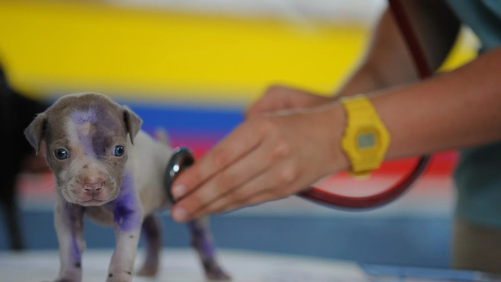
A histiocytoma in dogs is a benign skin tumor that most often shows up as a single, firm, hairless, red raised lump that looks like a button pressed into the skin. It grows quickly over one to four weeks, sits still for a while, and then, in most young dogs, shrinks and disappears on its own within roughly three months. The classic patient is a dog younger than three years old with one new lump on the head, ear flap, or leg. The lump is not cancer in the dangerous sense. It is a harmless overgrowth of a specific immune cell in the skin, and it is one of the most common skin tumors diagnosed in young dogs worldwide [1][2][3].
The catch is that a histiocytoma can look almost identical to a mast cell tumor, which is a potentially serious cancer that requires treatment. You cannot tell them apart by appearance alone. A fine needle aspiration with a pathologist's review is the standard way to sort this out, and it is the single most important step in managing a new button-like lump on your dog. This article explains what a histiocytoma is, how veterinarians confirm the diagnosis, how to tell a histiocytoma from a mast cell tumor, when waiting is safe, and when surgical removal makes sense.
At a Glance
| Feature | Typical Histiocytoma | What It Means for You |
|---|---|---|
| Typical age | Under 3 years, with nearly half of cases in dogs 2 years or younger [4] | A new red button lump in a young dog is most likely a histiocytoma |
| Typical breeds | French Bulldogs and Boxers have the highest risk, followed by other breeds [4] | Breed raises suspicion but does not confirm the diagnosis |
| Appearance | Solitary, round, hairless, red, button-shaped, rapidly growing [5] | Take a photo and measure it so you can track change |
| Common locations | Head, ear flap, and limbs, with breed-specific patterns [4] | Ear and face lumps are common and still need testing |
| Growth pattern | Fast growth over weeks, then a plateau, then shrinkage [6][5] | Rapid growth alone does not mean cancer |
| Regression | Most regress spontaneously within about three months [5] | Waiting is a legitimate plan when the diagnosis is confirmed |
| Key test | Fine needle aspiration with cytology to rule out mast cell tumor [7] | This test separates a harmless lump from a serious one |
| Removal | Reasonable when the lump is not regressing, is ulcerated, infected, or bothersome [5][8] | Surgery is straightforward and usually curative |
| Prognosis | Excellent for typical solitary lesions [5] | Most dogs do not need any treatment at all |
What a Histiocytoma Actually Is
A histiocytoma is a tumor made of Langerhans cells. Langerhans cells are a type of histiocyte, which is a family of immune cells that live in the skin and act as sentinels. They grab material from the skin surface, process it, and carry it to lymph nodes so the rest of the immune system can respond. A cutaneous histiocytoma is a clonal overgrowth of these Langerhans cells, which is why pathologists classify it as a benign Langerhans cell tumor [4][6][9].
The word tumor frightens owners, and that reaction is understandable. In this case, the word describes a lump of cells that multiplied too much, not a cancer that spreads and destroys tissue. Histiocytomas are benign. They grow, they attract an immune response, and then they are dismantled from within. The Swiss Canine Cancer Registry found histiocytomas made up about 12 percent of all canine skin tumors, and a large German pathology dataset ranked them as the fourth most common skin tumor overall at about 7 percent of cases [2][3]. In young dogs, they are the most common neoplastic skin condition [1].
The Immunology in One Short Paragraph
The reason a histiocytoma disappears is that the immune system eventually recognizes the tumor cells as abnormal and attacks them. As lymphocytes move into the lump, the tumor cells mature and change shape, the local blood supply shifts, and immune checkpoint molecules that were suppressing the response get rebalanced [6][10][9][11][12]. The result is a self-limited inflammatory event that clears the tumor without any treatment. This process is unusual among tumors and is the reason veterinarians are comfortable watching a confirmed histiocytoma instead of cutting it out immediately.
What a Histiocytoma Looks Like (the Button Tumor)
Owners often describe the lump as a red button. It is usually a single, well-defined, dome-shaped or button-shaped nodule that is hairless on top. The surface is often bright red or pink, and it can look shiny, raw, or ulcerated. Some histiocytomas develop a small crust or a collar of thickened skin around the base. The classic button tumor dog presentation is one new lump on a young dog, growing fast, with no other signs of illness.
The lump often ulcerates and becomes secondarily infected, which can make it look worse than it is [13]. A bleeding or crusted button lump is still usually a histiocytoma, but ulceration is also common in mast cell tumors, so appearance alone never settles the question.
Locations follow patterns. A study of 359 confirmed cases found that histiocytomas cluster in specific body sites, and those sites differ by breed and sex [4]. The head, ear flap, and limbs are common. Ear canal histiocytomas are reported and can behave more aggressively than skin surface lesions, which is one reason ear lumps deserve a proper diagnosis rather than a wait-and-see approach [8].
What dog histiocytoma pictures show
If you search for dog histiocytoma pictures, you will see a consistent theme. The lesions are round, raised, hairless, and red, often with a smooth or slightly ulcerated surface. They frequently sit alone. They do not usually have the angry, spreading, irregular border of an infection, and they do not look like the flat, scaly patches of many allergic skin diseases. Photos are useful for recognizing the pattern, but they cannot distinguish a histiocytoma from a mast cell tumor. Only cytology or biopsy can do that.
How Common Is It, and Who Gets It?
Histiocytomas are common. The Swiss registry reported an incidence rate of 44.6 per 100,000 dogs per year, and histiocytomas accounted for 12.1 percent of all skin tumors in that dataset [2]. A German study of more than 109,000 tissue samples found histiocytoma in 7.0 percent of all neoplasms [3]. A South African pathology survey found histiocytoma as the second most frequent skin tumor at 9.4 percent [14]. These numbers vary by region and by which dogs get biopsies, but the message is consistent. This is a routine diagnosis in general practice.
Age patterns
Age is the strongest clue. In a Portuguese case series, 48.8 percent of confirmed histiocytomas occurred in dogs 2 years old or younger [4]. The same study identified three age bands with different behavior, roughly 1.5 years and under, 1.5 to 3.5 years, and over 3.5 years [4]. Another study described histiocytomas as predominantly affecting dogs under four years of age [1]. A separate report confirmed that histiocytomas are common benign skin tumors in young dogs [5].
Older dogs can still develop histiocytomas. A 12-year-old dog was reported with a large histiocytoma on the front of the face, and it was successfully removed [5]. Age shifts the probability, it does not close the door.
Breed and sex patterns
Breed matters. French Bulldogs had a relative risk of 11.5 and Boxers had a relative risk of 8.3 compared with mixed-breed dogs in one population study [4]. Boxers also appear repeatedly in breed predisposition data for skin tumors generally [3][14]. Males were overrepresented at 58.5 percent of cases in one series, and the incidence rate was higher in males than females [4]. Breed and sex raise or lower suspicion. They never replace a diagnostic test.
Why the Lump Appears and What Else It Could Be
The direct cause of a histiocytoma is not known. Investigators have looked for infectious triggers. One study tested histiocytoma tissue for Bartonella species, bacteria that can live inside histiocytes and are transmitted by arthropod bites and scratches, and found that Bartonella DNA could be amplified from these tumors [13]. That finding shows an association worth studying. It does not prove that Bartonella causes histiocytomas, and it does not change how a histiocytoma is managed.
Leishmania organisms have also been found inside a cutaneous histiocytoma in an older dog, another example of an infectious agent appearing within these immune-cell tumors [15]. These reports point to the biology of histiocytes, which sample and carry material from the skin. They do not establish a preventable cause.
The differential diagnosis list
A new button lump on a dog can be several things. The most important to rule out is a mast cell tumor, because mast cell tumors can be malignant and can spread. Other possibilities include histiocytic sarcoma, plasmacytoma, lymphoma, transmissible venereal tumor, and non-cancerous inflammatory nodules such as pyogranulomas [12][7][16][17]. The Merck Veterinary Manual groups histiocytomas with other round cell and histiocytic skin tumors, and notes that they are benign and often regress [16][18].
Because these tumors can look alike, the diagnostic path is the same for all of them. Sample the lump, look at the cells, and let the cell type drive the plan.
Histiocytoma vs Mast Cell Tumor: Why the Test Matters
This is the central decision in every new button lump. A histiocytoma and a mast cell tumor can both be solitary, raised, hairless, red, and fast-growing. They can both ulcerate. They can both appear on the head, limbs, or trunk. Owners and even experienced veterinarians cannot reliably separate them by eye or by touch.
The difference in outcome is large. A histiocytoma usually regresses without treatment. A mast cell tumor may need surgery, and depending on grade and stage, may need additional treatment. Missing a mast cell tumor because it looked like a benign button lump is the main risk of a wait-and-see approach without a diagnosis.
Fine needle aspiration is the first-line test
Fine needle aspiration is quick, inexpensive, and minimally invasive. A veterinarian inserts a small needle into the lump, collects cells, spreads them on a slide, and sends the slide to a clinical pathologist. The pathologist identifies the cell type. In a study of round cell tumors that included histiocytoma, mast cell tumor, lymphoma, plasmacytoma, and transmissible venereal tumor, diagnostic agreement between cytology and histopathology was excellent for some tumor types when good images were available, and cytology was confirmed as a practical first-line method for round cell neoplasms [7].
If cytology is inconclusive, the next step is a tissue biopsy. Biopsy gives the pathologist the full architecture of the lump, which allows a definitive diagnosis. Immunohistochemistry can be added when the cell type is ambiguous. Histiocytomas express markers such as CD18 and, in many cases, MUM1/IRF-4, while lacking lymphocyte and macrophage markers, and these stains help pathologists separate histiocytoma from plasma cell tumors and histiocytic sarcoma [19][20].
What the pathologist sees
Under the microscope, a histiocytoma is a dense sheet of round to oval cells with abundant cytoplasm, often with a rim of small lymphocytes around the edges. As regression advances, lymphocyte infiltration increases, tumor cells enlarge and change shape, and the nuclear-to-cytoplasmic ratio falls [12]. Pathologists use these features to stage the regression and confirm the diagnosis. In one series, most tumors showed epidermal invasion and frequent mitotic figures, and necrosis was present in more than half of cases, findings that can look alarming but are expected in a tumor that is being destroyed by the immune system [1].
The Natural Course: Growth, Plateau, and Regression
The typical timeline has three phases.
The growth phase lasts one to four weeks. The lump appears and enlarges quickly. This rapid growth is one of the most frightening features for owners, and it is also one of the most characteristic features of a histiocytoma [6][5].
The plateau phase follows. The lump stops growing and may ulcerate, bleed, or develop a crust. Secondary infection is common [13]. The lump may look worse during this phase even though the biology is moving toward resolution.
The regression phase is when the lump shrinks. Most histiocytomas regress spontaneously within a few months, and the commonly quoted window is about three months [5]. As the lump shrinks, it often flattens, changes color, and eventually disappears, sometimes leaving a small area of hairless or slightly thickened skin.
Regression is not passive. It is an active immune event. Studies show that immune checkpoint molecules, co-stimulatory molecules such as CD80 and CD86, regulatory T cells, dendritic cells, and angiogenic factors all shift during regression [6][10][9][11]. One study found that angiogenesis, the growth of new blood vessels, appears compromised in early histiocytomas, which may help trigger the regression process [9]. Another found that CD80 expression density was negatively correlated with regression, meaning the immune balance shifts as the tumor resolves [6]. These details matter to researchers. For owners, the practical takeaway is that regression is expected and normal.
Diagnosis: What Your Veterinarian Will Do
A typical workup for a suspected histiocytoma includes several steps.
History and physical examination come first. Your veterinarian will ask when the lump appeared, how fast it grew, whether it has changed, and whether your dog has other lumps or symptoms. They will measure the lump and record its location.
Fine needle aspiration follows. This is the key test. Cells are collected and evaluated by a pathologist. The goal is to identify the cell type and rule out mast cell tumor and other round cell tumors [7].
Imaging is not routine for a typical skin histiocytoma. It becomes relevant when a lump is large, fixed to deeper tissue, or in an unusual location. In one case of a large facial histiocytoma, radiographs confirmed a subcutaneous mass with no evidence of lymph node metastasis before surgery [5].
Biopsy is used when cytology is inconclusive or when the lump is being removed anyway. Excisional biopsy provides both diagnosis and treatment in one step.
Lymph node evaluation is considered when the lump is large, when the draining lymph node is enlarged, or when the location is unusual. Lymph node metastasis has been documented in a small number of solitary cutaneous histiocytomas, so an enlarged draining node deserves attention [19]. This is uncommon, and most solitary histiocytomas never spread.
Management: Watching, Treating Symptoms, or Removing
Management depends on the diagnosis, the location, the size, and how the lump is behaving.
Watchful waiting
Watchful waiting is appropriate when a histiocytoma has been confirmed by cytology, the lump is small, the dog is comfortable, and the location allows monitoring. The plan is simple. Measure the lump, photograph it, and recheck it with your veterinarian at a set interval, often every two to four weeks. Most histiocytomas regress within about three months [5]. If the lump is shrinking, continue monitoring. If it is growing after the expected window, or if it is causing problems, move to treatment.
Supportive care during regression
Ulcerated histiocytomas can become secondarily infected [13]. Your veterinarian may recommend cleaning the area, preventing your dog from licking or scratching it, and treating infection if it develops. Do not apply human antibiotic ointments or other products without veterinary guidance, because some are toxic to dogs if licked off.
When histiocytoma removal is reasonable
Surgical removal is a reasonable choice in several situations.
The lump is not regressing after a reasonable observation period. If a confirmed histiocytoma is still growing or unchanged after about three months, removal ends the uncertainty [5].
The lump is ulcerated, infected, bleeding, or painful. A lump that will not heal or that bothers your dog is better removed.
The location is problematic. Lumps on the eyelid, in the ear canal, on the paw, or near the mouth can interfere with function. Ear canal histiocytomas can behave more aggressively and may require more than simple excision [8].
The diagnosis is uncertain. If cytology cannot rule out a mast cell tumor, removal provides a definitive answer and treatment at the same time.
The owner prefers a definitive solution. Waiting requires rechecks and monitoring. Some owners prefer to remove a confirmed histiocytoma and be done with it. That is a legitimate choice, and surgery for a typical histiocytoma is usually straightforward.
Removal is typically a simple excisional procedure under anesthesia or heavy sedation, depending on the size and location. In one reported case of a large facial histiocytoma, en bloc excision resulted in an intact dog with no recurrence at a six-month recheck [5]. Most solitary histiocytomas are cured by complete excision.
What about aggressive histiocytomas?
A small number of cutaneous histiocytomas behave aggressively. Lymph node metastasis has been documented in eight dogs with solitary cutaneous histiocytomas, and an ear canal histiocytoma in a Rottweiler was locally invasive and required total ear canal ablation plus chemotherapy [19][8]. These are exceptions. They are the reason a lump that keeps growing, a lump in an unusual location, or an enlarged draining lymph node deserves prompt veterinary attention rather than prolonged waiting.
What Not to Do at Home
Do not try to remove or drain the lump yourself. Do not apply human medications, essential oils, or home remedies to the lump. Do not use a rubber band, string, or any ligature around the base. These approaches cause pain, infection, and tissue damage, and they do not treat the underlying problem.
Do not assume a new lump is a histiocytoma because your dog is young. Young dogs get mast cell tumors too. The age pattern makes a histiocytoma more likely, not certain.
Do not skip rechecks. A confirmed histiocytoma that is being monitored needs scheduled examinations to confirm it is actually shrinking. A lump that is unchanged or growing at the three-month mark needs a new plan.
Prevention
There is no known way to prevent a histiocytoma. The tumor arises from the dog's own immune cells, and no diet, supplement, vaccine, or grooming practice has been shown to prevent it. The infectious associations reported in the literature, including Bartonella and Leishmania, are research findings and do not translate into prevention recommendations for histiocytomas [13][15].
General skin care still matters. Routine veterinary examinations help catch new lumps early. Monthly monitoring at home, sometimes called a lump check, helps you notice changes. If you find a new lump, have it evaluated rather than waiting to see what happens.
Prognosis
The prognosis for a typical solitary histiocytoma is excellent. Most regress without treatment within about three months [5]. Those that are removed surgically are usually cured by complete excision [5]. The small subset with lymph node metastasis or locally invasive behavior has a more guarded outlook and needs individualized treatment [19][8].
Emergency Red Flags
Contact a veterinarian promptly if you see any of the following.
The lump is growing rapidly after the initial growth phase, especially after three months of observation.
The lump is bleeding heavily, has a foul odor, or is deeply ulcerated and not healing.
Your dog is painful when the lump is touched, or the lump interferes with eating, drinking, blinking, walking, or hearing.
The draining lymph node near the lump is enlarged, firm, or growing.
New lumps appear elsewhere on the body.
Your dog has weight loss, reduced appetite, lethargy, vomiting, or diarrhea along with the lump.
Limitations and When to Contact a Veterinarian
This article is educational and is not a substitute for veterinary diagnosis or treatment. Any new lump on a dog should be checked by a veterinarian, because a histiocytoma cannot be distinguished from a mast cell tumor by appearance alone.
This article describes general patterns. Your dog is an individual, and a lump that looks like a histiocytoma may not be one. Contact a veterinarian if a new lump appears, if a known lump changes in size, color, or texture, if a lump fails to shrink after about three months, or if your dog shows any of the red flags listed above. A fine needle aspiration is the fastest way to get a clear answer, and it is worth doing before you commit to any plan. If your dog has a confirmed histiocytoma that is being monitored, keep every recheck appointment, because the decision to wait or remove depends on what the lump does over time.
Frequently Asked Questions
What does a histiocytoma look like on a dog?
A histiocytoma usually looks like a single, round, hairless, red or pink raised lump that resembles a button pressed into the skin. It often grows quickly over one to four weeks and may ulcerate or develop a crust. The classic location is the head, ear flap, or a limb, and the classic patient is a dog under three years old [4][5].
Is a histiocytoma in dogs cancer?
A histiocytoma is a benign tumor, which means it is a growth of cells but not a spreading cancer. It is derived from Langerhans cells in the skin and typically regresses on its own [4][6]. The word tumor can be alarming, but a histiocytoma behaves very differently from a malignant skin cancer such as a mast cell tumor.
How long does a histiocytoma take to go away?
Most histiocytomas regress spontaneously within a few months, and about three months is the commonly expected window [5]. The lump usually grows first, then plateaus, then shrinks. If a confirmed histiocytoma is still growing or unchanged after about three months, your veterinarian may recommend removal.
How do you tell a histiocytoma from a mast cell tumor?
You cannot tell them apart by appearance. A fine needle aspiration with cytology is the standard first step, and it allows a pathologist to identify the cell type [7]. If cytology is inconclusive, a tissue biopsy provides a definitive diagnosis. This distinction matters because mast cell tumors can be malignant and may need more aggressive treatment.
Does a histiocytoma need to be removed?
Not always. A confirmed histiocytoma that is small, comfortable, and in a monitorable location can be watched, because most regress on their own [5]. Removal is reasonable when the lump is not regressing after about three months, is ulcerated or infected, is in a troublesome location such as the ear canal or eyelid, or when the diagnosis is uncertain [5][8].
Can a histiocytoma spread to lymph nodes?
It can, but this is uncommon. Lymph node metastasis has been documented in a small number of dogs with solitary cutaneous histiocytomas [19]. An enlarged draining lymph node near a button lump should be evaluated by a veterinarian rather than watched at home.
Are certain dog breeds more likely to get histiocytomas?
Yes. French Bulldogs and Boxers have the highest reported risk compared with mixed-breed dogs, and other breeds also appear in predisposition data [4]. Breed raises suspicion, but it does not confirm the diagnosis. Any breed can develop a histiocytoma, and any new lump still needs cytology to rule out a mast cell tumor.
What should I do if my dog has a new red button lump?
Schedule a veterinary visit and ask for a fine needle aspiration. Do not wait to see if it goes away on its own, because a mast cell tumor can look identical to a histiocytoma [7]. Once the diagnosis is confirmed, your veterinarian can recommend monitoring, supportive care, or removal based on the size, location, and behavior of the lump.
Related Articles
- Lump Under Dogs Skin - Comprehensive Veterinary Reference Guide
- Why Is My Dog having lumps on skin? Lipoma vs Mast Cell vs Cyst Diagnosis
- Dog Skin Bumps and Lumps - When to See the Vet
- Boxer Health Problems - Dilated Cardiomyopathy Mast Cell and Aortic Stenosis
- Mast Cell Tumors in Dogs - Histopathological Grading and Treatment Guide
- Warts On Dogs - Comprehensive Veterinary Reference Guide
Sources
- Histopathological and Ultrastructural Study of a Canine Langerhans Cell Tumour (Canine Cutaneous Histiocytoma).
- Cutaneous Tumors in Swiss Dogs: Retrospective Data From the Swiss Canine Cancer Registry, 2008-2013.
- Tumour Incidence in Dogs in Germany: a Retrospective Analysis of 109,616 Histopathological Diagnoses (2014-2019).
- Epidemiology of Canine Cutaneous Histiocytoma: Insights From Age and Topographic Clustering.
- A case report of massive cutaneous histiocytoma in a 12-year-old intact bitch: Cytologic, radiologic, pathologic investigation.
- Immune checkpoint regulation is critically involved in canine cutaneous histiocytoma regression.
- Assessment of diagnostic accuracy and inter-pathologist agreement in telecytology performed for canine round cell tumours using images captured by a smartphone camera.
- Invasive histiocytoma in the ear canal of a dog.
- A Role for Angiogenesis in Canine Cutaneous Histiocytoma Regression: Insights into an Old Clinical Enigma.
- Immuno-oncologic profiling by stage-dependent transcriptome and proteome analyses of spontaneously regressing canine cutaneous histiocytoma.
- FOXP3, CD208, and CD206 Expression in Canine Cutaneous Histiocytoma.
- Cytomorphometry of canine cutaneous histiocytoma.
- Prevalence of Bartonella spp. in Canine Cutaneous Histiocytoma.
- Breed and anatomical predisposition for canine cutaneous neoplasia in South Africa during 2013.
- Leishmania spp. in a cutaneous histiocytoma of an old dog.
- Lymphocytic, Histiocytic, and Related Cutaneous Tumors in Animals (Merck Veterinary Manual)
- Round Cell Tumors in Dogs (VCA Animal Hospitals)
- Tumors of the Skin in Dogs (Merck Veterinary Manual)
- Retrospective characterisation of solitary cutaneous histiocytoma with lymph node metastasis in eight dogs.
- Immunohistochemical Labeling of Multiple Myeloma Oncogene 1/Interferon Regulatory Factor 4 (MUM1/IRF-4) in Canine Cutaneous Histiocytoma.