Lump on Your Dog's Head or Chin: Common Causes Explained
By Dr. Zubair Khalid, DVM, MS, PhD ·
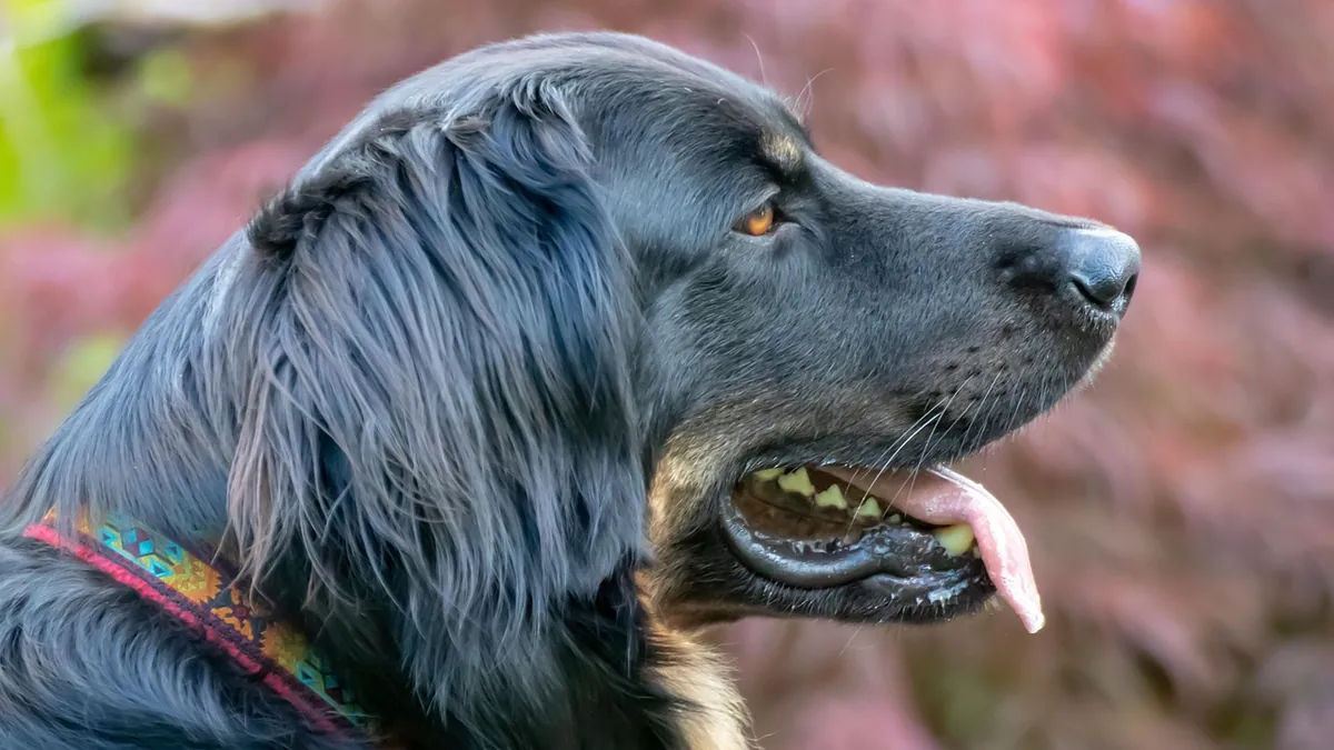
A new dog growth on head or chin is common, and most turn out to be harmless. The tricky part is that benign lumps and serious tumors can look almost identical from the outside. Size, texture, and how fast it changes are your best clues, but only a veterinarian can tell you what it really is.
This article is educational and is not a substitute for veterinary diagnosis or treatment.
Quick Answer
- Most lumps on a dog's head or chin are benign, including cysts, warts, and histiocytomas, a benign tumor that often shrinks on its own [1][2].
- Fast growth over days to weeks, redness, ulceration, or pain raises concern for a mast cell tumor or another malignancy.
- Any lump that is new, changing, or bothering your dog should be examined, and many will be sampled with a fine needle.
- A fine needle aspirate is quick, inexpensive, and often gives an answer without surgery.
- Never squeeze, poke, or try to drain a lump at home. You can cause bleeding, infection, or spread of cells.
What a Lump on the Head or Chin Looks Like
Lumps on the head and chin come in many forms. Some are pea-sized and firm. Some are soft and squishy. Some sit in the skin and move freely when you touch them. Others feel attached to deeper tissue and do not move.
Location matters. The chin, muzzle, eyelids, and ears have thin skin and little padding, so even small lumps are easy to spot. A lump on the top of the head may go unnoticed for weeks under fur.
What Is Normal
Not every bump is a problem. Dogs commonly develop small skin tags, warts (papillomas), and harmless fatty lumps as they age. A lump that has been the same size and texture for months is usually less concerning than one that appeared last week and doubled in size.
It also helps to know your dog's baseline. Run your hands over the head, chin, neck, and ears once a month. You will notice changes faster, and your veterinarian will have a better history to work with.
Common Causes of a Dog Growth on the Head or Chin
The table below groups the most common causes by what you are likely to see and feel, plus how quickly you should act.
| Cause | Typical clues | How urgent |
|---|---|---|
| Sebaceous cyst | Soft to firm, round, often with a dark center or "pore." May drain a thick, white, cheesy material. Common on the head and eyelids. | Routine visit. Watch for infection or sudden enlargement. |
| Histiocytoma | Firm, pink, dome-shaped, often called a "red button." Most common in young dogs, often under 2 years old [1]. Frequently regresses on its own [2]. | Routine visit to confirm. Fast growth can mimic cancer. |
| Wart (papilloma) | Cauliflower-like, often on the muzzle or chin. Common in young dogs and older dogs with weakened immunity. | Routine visit. |
| Lipoma | Soft, movable, under the skin. Usually not on the head, more common on the trunk and legs. | Routine visit. |
| Mast cell tumor | Firm or soft, may swell and shrink, may be itchy or red. Can appear anywhere, including the head. | See a vet promptly. These need sampling. |
| Basal cell tumor | Small, firm, often on the head and eyelids. Benign version is a basal cell tumor, malignant version is a basal cell carcinoma [3]. | Routine to prompt visit. |
| Abscess or infected bite wound | Warm, painful, swollen, may drain pus. Often follows a scratch or bite. | Same-day visit. |
| Congenital cyst | Fluid-filled, nonpainful mass in the neck or head region. Rare, present from birth but may enlarge later [4]. | Routine visit with imaging. |
| Odontoma | Bony-hard mass near the upper jaw or cheek in a young dog, sometimes with a missing or unerupted tooth [5]. | Prompt visit. Needs imaging. |
| Lymphoma or other cancer | Rapidly growing mass, sometimes with enlarged lymph nodes or eye changes [6]. | Urgent. Same-day or next-day visit. |
Sebaceous Cysts
Sebaceous cysts are blocked oil glands. They are one of the most common reasons for a lump on a dog's head or chin. They often feel like a small rubber ball under the skin and may have a tiny dark dot on top.
Many sebaceous cysts can be watched. If one becomes red, painful, or starts draining, it may be infected and need treatment. Your veterinarian may drain it, flush it, or remove it if it keeps coming back.
Histiocytomas
Histiocytomas are benign tumors of a type of immune cell in the skin [1]. They are most common in young dogs, with about half of cases occurring in dogs 2 years old or younger [1]. They often look like a raised, hairless, red button on the head, ear, or leg.
The good news is that most histiocytomas shrink and disappear on their own through an immune response [2]. The catch is that they can grow quickly at first, which makes them look alarming. A veterinarian can usually identify one with a needle sample.
Mast Cell Tumors
Mast cell tumors are the lump that veterinarians worry about most on the head and elsewhere. They are malignant tumors of mast cells, a type of immune cell. They can be firm or soft, and they sometimes swell after being touched or irritated.
Mast cell tumors vary a lot in behavior. Some grow slowly for years. Others spread quickly to lymph nodes and internal organs. Because they can look like almost anything, any lump that changes or grows should be sampled.
Other Causes
Basal cell tumors arise from cells at the base of the top layer of skin [3]. The benign form is a basal cell tumor, and the malignant form is a basal cell carcinoma [3]. They are often small, firm, and slow-growing.
Congenital cysts of the head and neck are rare [4]. They come from leftover tissue from the embryo and are usually fluid-filled and nonpainful [4]. They may enlarge later in life, sometimes after a respiratory infection [4].
Odontomas are rare benign tumors made of tooth-like tissue. One report described a 3-month-old German shepherd with a 3 cm round mass near the eye and an unerupted tooth [5]. Surgery was curative, and the dog was free of signs one year later [5].
Red Flags That Mean See a Vet Now
Some lumps need attention the same day. Others can wait for a routine visit. Use these signs to decide.
- The lump appeared suddenly and is growing fast (doubling in days).
- The lump is red, ulcerated, bleeding, or draining pus.
- Your dog is painful when you touch the area, or is pawing at the head.
- Your dog has trouble eating, breathing, or swallowing.
- The lump is firm and fixed to the bone or deep tissue.
- Your dog has swollen lymph nodes under the jaw or in front of the shoulder.
- Your dog has lost weight, is vomiting, or seems tired.
- The lump is near the eye and causing swelling, discharge, or vision changes.
A rapidly growing mass near the eye or jaw can be serious. One report described a dog with an eye mass that later invaded the face and sinuses, and it was diagnosed as lymphoma [6]. Early evaluation gives you more options.
What You Can Safely Do at Home
While you wait for your appointment, keep things simple and safe.
- Take a photo with a ruler or coin next to the lump for scale.
- Measure the lump with a soft tape measure and write down the date.
- Check it once a week, not every day, so you do not irritate it.
- Keep your dog from scratching or rubbing the area. A soft cone may help.
- Do not squeeze, poke, or try to pop the lump.
- Do not apply human creams, oils, or essential oils. Dogs lick these off and can get sick.
- Keep the area clean with plain water and a soft cloth.
If the lump starts bleeding, cover it with a clean gauze pad and apply gentle pressure. If bleeding does not stop in a few minutes, call your veterinarian or an emergency clinic.
What Your Veterinarian Will Check
Your veterinarian will start with a full physical exam. They will feel the lump, measure it, and check the lymph nodes under the jaw and around the shoulders. They will also look in the mouth and ears if the lump is near those areas.
Fine Needle Aspirate
This is the most common first test. Your veterinarian inserts a thin needle into the lump, pulls out a few cells, and looks at them under a microscope. It takes a minute or two and rarely needs sedation. It can identify cysts, lipomas, mast cell tumors, and histiocytomas in many cases.
Biopsy
If the needle sample is unclear, your veterinarian may recommend a biopsy. This means removing a small piece of tissue or the whole lump. The tissue is sent to a lab for a histopathology test, which confirms the tumor type and checks the margins [3].
Margins tell you whether tumor cells were left behind. "Dirty" margins mean tumor cells reach the edge of the removed tissue, so some cells likely remain in your dog [3]. "Narrow" margins mean cells are close to the edge but not at it [3]. "Wide" or "clean" margins mean cells are far from the edge, so it is unlikely any remain [3]. Clean margins are the goal for malignant tumors.
Imaging and Other Tests
If cancer is suspected, your veterinarian may recommend X-rays, ultrasound, or a CT scan. Blood work and urinalysis help check overall health before anesthesia or chemotherapy. In one lymphoma case, CT showed the mass invading the frontal sinus, which changed the treatment plan [6].
Treatment Options
Treatment depends on the diagnosis. Benign lumps may need no treatment at all. Infected cysts may need antibiotics and drainage. Malignant tumors often need surgery, and some need chemotherapy or radiation. In one report, a puppy with a malignant Merkel cell carcinoma was cured with wide surgery and had no recurrence for at least 20 months [7].
Monitoring and Prevention
You cannot prevent most lumps. You can catch them early.
- Do a monthly head-to-tail check with your hands.
- Note the size, location, and texture of any lump you find.
- Take photos with a ruler for reference.
- Keep up with routine wellness visits.
- Ask your veterinarian to check any lump at every visit.
Some lumps are more common in certain breeds and ages. Histiocytomas cluster in young dogs, with about half of cases in dogs 2 years or younger [1]. If you have a young dog with a new red button lump, mention it at your next visit. If you have an older dog with a new lump, get it checked sooner.
For a broader look at lumps anywhere on the body, see Dog Skin Bumps and Lumps: When to See the Vet. If you are trying to sort out whether a lump is a lipoma, cyst, or mast cell tumor, this guide walks through the differences: Why Is My Dog having lumps on skin? Lipoma vs Mast Cell vs Cyst Diagnosis. And if your dog also has hair loss around the lump, see Why Is My Dog Losing Hair in Patches? Common Causes and Next Steps.
Limitations and When to Contact a Veterinarian
This article cannot tell you what your dog's lump is. Only a veterinarian who examines it can do that. Individual cases vary, and some lumps that look harmless turn out to be serious.
Contact your veterinarian the same day if the lump is growing fast, bleeding, ulcerated, or painful. Go to an emergency clinic now if your dog has trouble breathing, cannot swallow, has a swollen face or throat, or is collapsing. Call for a routine visit if the lump is small, slow-growing, and not bothering your dog, but do not wait months to have it checked.
Frequently Asked Questions
Should I worry about a lump on my dog's chin?
Most chin lumps are benign cysts, warts, or histiocytomas, but mast cell tumors can also appear there. Because chin lumps are easy to see and often get licked, they can become irritated or infected. Have your veterinarian check any new chin lump, especially if it is growing or changing.
Can a dog lump go away on its own?
Some lumps do go away on their own. Histiocytomas often shrink and disappear through an immune response, especially in young dogs [2]. Cysts may drain and flatten, then refill later. Other lumps, including mast cell tumors, do not go away and need treatment.
How fast do cancerous lumps grow on a dog's head?
Growth speed varies widely. Some malignant tumors grow slowly for months, while others double in size in days. A lump that grows quickly is more concerning and should be sampled promptly. There is no reliable way to tell cancer from a benign lump by look or feel alone.
Will my vet remove the lump right away?
Not always. Your veterinarian will usually start with a fine needle aspirate to identify the cells. If the lump is benign and not bothering your dog, you may choose to monitor it. If it is malignant or causing problems, surgery is often the next step, and the tissue is sent for a histopathology test to check the margins [3].
Can I put antibiotic ointment on my dog's lump?
No. Human antibiotic ointments are not made for dogs, and your dog can lick them off and swallow them. Some ingredients are toxic to dogs. Keep the area clean with plain water and let your veterinarian decide on any medication.
Related Articles
- Dog Skin Bumps and Lumps: When to See the Vet
- Why Is My Dog having lumps on skin? Lipoma vs Mast Cell vs Cyst Diagnosis
- Why Is My Dog Losing Hair in Patches? Common Causes and Next Steps
- Histiocytoma in Dogs: The Red Button Lump That Often Regresses on Its Own
- Dog Shaking Head Causes
- Dog Skin Rash with Bumps: What Could It Be?
- Bump on Your Dog's Chin or Jaw: Causes and Next Steps
- Bump on Your Dog's Head, Nose, or Snout: Causes and Next Steps
- Lump on Your Dog's Head or Neck: Cancer or Benign?
References
- Epidemiology of Canine Cutaneous Histiocytoma: Insights From Age and Topographic Clustering. Veterinary dermatology, 2026.
- Age distribution and regression-associated immunophenotypic features in canine cutaneous histiocytoma. The Journal of veterinary medical science, 2026.
- Tumors of the Skin in Dogs - Dog Owners. Merck Veterinary Manual.
- Congenital Cysts of the Head and Neck in Animals - Digestive System. Merck Veterinary Manual.
- Compound maxillary odontoma in a young German shepherd dog. The Journal of small animal practice, 2005.
- Presumed solitary ocular lymphoma of large B-cell origin with Mott cell change in a dog. The Canadian veterinary journal = La revue veterinaire canadienne, 2024.
- Diagnosis and treatment of Merkel cell carcinoma in a puppy: A comparative oncology approach. The Canadian veterinary journal = La revue veterinaire canadienne, 2026.