Wolf Teeth in Horses: Extraction and Management
By Dr. Zubair Khalid, DVM, MS, PhD ·
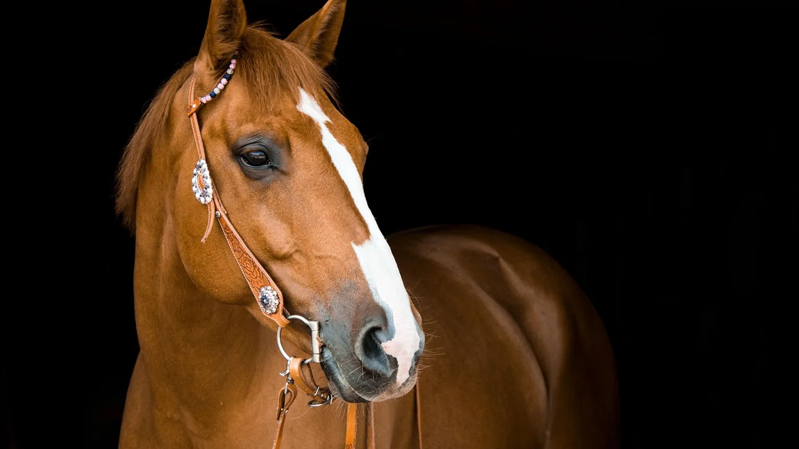
Wolf teeth in horses are small, vestigial first premolars that sit just in front of the large cheek teeth. They usually erupt in the upper jaw, they often have nothing to oppose them in the lower jaw, and in many horses they never cause a problem at all. When a wolf tooth sits under the bit, grows into an awkward position, or becomes fractured or infected, it can produce genuine bitting resistance, head tossing, and evasive behavior under saddle. The standard fix is a short surgical procedure performed under sedation with local anesthesia, and most horses return to work within a few days.
This article is educational and is not a substitute for veterinary diagnosis or treatment.
Quick Triage: What Owners Should Do First
If your horse is suddenly fussy in the bridle, tosses its head, or refuses to move forward, do not assume the problem is behavioral. Oral pain is a common and frequently missed cause of ridden resistance. Book a dental examination with a veterinarian who uses a full mouth speculum, and describe exactly when the behavior appears (at the walk, on contact, on one rein, or only with a bit in place).
If you can see a small tooth in front of the cheek teeth, that is likely a wolf tooth. Do not attempt to remove it yourself. Do not use a screwdriver, pliers, or any improvised tool. Do not ask a non-veterinary lay dentist to extract it. Extraction requires sedation, local anesthesia, and equipment designed for the job, and the tooth can fracture during removal, leaving a root fragment behind [1].
Call a veterinarian promptly if you see any of the following:
- A fractured tooth with exposed pulp or a visible cavity
- Facial or jaw swelling, especially over the maxillary region
- Nasal discharge from one nostril, particularly if it smells abnormal
- Dropping feed, quidding, or weight loss
- Bleeding from the mouth that does not stop
- Severe head shaking that interferes with eating or handling
At a Glance: Tooth Types and Their Management
| Tooth type | Triadan position | Typical location | Typical management |
|---|---|---|---|
| Wolf tooth (vestigial first premolar) | 05 (105, 205, 305, 405) | Just rostral to the cheek teeth, most often the upper arcade | Extract when it interferes with the bit, is displaced or enlarged, sits in the mandible, or is fractured or diseased [2] |
| Canine tooth | 04 (104, 204, 304, 404) | Usually the lower arcade, with a smaller upper counterpart, more common in males | Leave in place unless diseased, displaced, or fractured, or unless calculus becomes a problem [2] |
| Cheek teeth (premolars and molars) | 06 through 11 | The long grinding arcades on each side | Routine floating, and extraction only for specific disease [2] |
| Blind or displaced wolf tooth | 05 | Under the gingiva or in an abnormal position | Extract when it causes pain, bitting interference, or cyst formation [3] |
| Supernumerary wolf tooth | 05 (extra) | Adjacent to a normal first premolar | Surgical removal, especially when associated with a cystic structure [3] |
What Are Wolf Teeth in Horses?
Wolf teeth are the vestigial first premolars of the horse. In the Triadan numbering system used across equine dentistry, they occupy the 05 position. That means the upper right wolf tooth is 105, the upper left is 205, the lower right is 305, and the lower left is 405. They sit immediately in front of the second premolar (the 06), which is the first of the large grinding cheek teeth.
These teeth are called vestigial because they no longer serve a meaningful function. The horse's modern dentition is built for grinding fibrous forage across the cheek teeth, and the wolf tooth contributes nothing to that process. It is a remnant of a more complete premolar row. Because it is vestigial, its size and shape vary enormously from horse to horse.
How Common Are Wolf Teeth?
Roughly 15 to 25 percent of horses have at least one wolf tooth, and they appear far more often in the upper arcade than the lower. When they are present, they are usually small, measuring about 5 to 18 mm in the visible clinical crown. The variation is real. In a study of 65 extracted first premolar teeth, total tooth length ranged from 12 to 34 mm, with a median of 21 mm. Root length ranged from 0 to 19.6 mm with a median of 13.2 mm, and crown height ranged from 2 to 20 mm with a median of 7 mm [4].
That size range matters clinically. A wolf tooth with a long root is a more demanding extraction than one with a short, rounded root. The same study found that root length exceeded crown height in 61 of 65 teeth, and that the visible crown is a poor predictor of how much root sits beneath the gumline [4]. A veterinarian cannot judge the difficulty of extraction simply by looking at how much tooth is showing.
Why Horses Have Them at All
The wolf tooth is a normal anatomical structure, not a disease. It is a first premolar that has lost its function over evolutionary time. Archaeological evidence from equine skeletal remains spanning the past 3,200 years shows that nomadic horsemen in northeast Asia practiced removal of problematic deciduous teeth and vestigial first premolars, and that first premolar removal appears in the record around 750 BCE, coinciding with the archaeological appearance of jointed bronze and iron bits [5]. In other words, the problem of the wolf tooth under the bit is at least as old as the bit itself.
Anatomy: Where Wolf Teeth Sit and Why Position Matters
The wolf tooth sits in the diastema region, the gap between the incisors at the front of the mouth and the cheek teeth at the back. The bit rests in this same general area. That shared real estate is the entire reason wolf teeth become a clinical issue.
When a horse accepts the bit, the bit sits across the bars of the mouth and the tongue, and the lips and cheeks close around it. A wolf tooth that protrudes from the upper arcade can be pressed by the bit, by the rider's hands, or by the horse's own tongue and cheek movements. The result is discomfort that shows up as resistance rather than as an obvious wound.
Upper Versus Lower Wolf Teeth
Upper wolf teeth are far more common, and they are also the ones most likely to interfere with the bit because of how the bit sits against the upper arcade. Lower wolf teeth are uncommon. When they do occur, they are considered an indication for extraction in their own right, because they sit directly in the path of the bit and are difficult to protect [2].
Blind and Displaced Wolf Teeth
A blind wolf tooth is one that has not erupted through the gingiva and remains covered by soft tissue. You cannot see it, but it can still be present and can still cause pain when the bit presses over it. A displaced wolf tooth sits in an abnormal position, sometimes rostral or lateral to where it should be. Displaced or enlarged wolf teeth are among the main indications for extraction [2].
Supernumerary wolf teeth also occur. A case report describes a 3-year-old Quarter Horse mare with a supernumerary first premolar at Triadan 105 that was associated with a dentigerous cyst, a cystic structure containing dental material. The cyst required surgical removal along with the extra tooth [3]. This is uncommon, but it is a reminder that an extra or abnormally positioned tooth is not always a harmless finding.
Wolf Teeth Versus Canine Teeth: A Common Confusion
Owners frequently mix up wolf teeth and canine teeth. They are different teeth in different positions with different management.
Canine teeth sit at the 04 Triadan position. They are typically found in the lower arcade, with a smaller upper counterpart, and they are much more common in males than females. Canines are not vestigial in the same way wolf teeth are, and they are generally left in place. The main disorder of canine teeth is the development of calculus on the lower canines, along with occasional developmental displacements and traumatic fractures [2].
Wolf teeth sit at the 05 position, just in front of the cheek teeth. They are usually upper, they are small, and they are the teeth most often extracted for bitting reasons. If you are unsure which tooth you are looking at, a veterinarian can identify it by position and by radiography if needed. Do not guess, and do not let anyone extract a tooth based on a guess.
| Feature | Wolf tooth (05) | Canine tooth (04) |
|---|---|---|
| Position | Just in front of cheek teeth | Further rostral, near the incisor region |
| Most common arcade | Upper | Lower |
| Sex predilection | Both sexes | More common in males |
| Typical action | Extract when problematic | Usually leave in place |
| Main concern | Bit interference, displacement, fracture | Calculus, displacement, fracture [2] |
Why Wolf Teeth Cause Problems Under Saddle
A wolf tooth does not have to be large to cause trouble. It only has to be in the wrong place relative to the bit. The clinical signs owners notice are usually behavioral, which is why wolf teeth are sometimes mistaken for training problems.
Common signs attributed to problematic wolf teeth include:
- Head tossing or head shaking under saddle
- Bracing or leaning on one rein
- Refusing to move forward or stopping abruptly
- Opening the mouth, tongue lolling, or grabbing the bit
- Evasion when the bit is first placed or when contact is taken up
- Reluctance to accept the bridle
These signs are not specific to wolf teeth. They overlap with many other causes of bitting resistance, including cheek tooth overgrowths, sharp enamel points, oral ulcers, ill-fitting tack, and musculoskeletal pain. That overlap is exactly why a proper examination matters. Removing a wolf tooth will not fix a horse whose real problem is a sharp cheek tooth or a sore back.
The Bit as a Mechanical Trigger
The bit applies pressure in the diastema, and a wolf tooth that protrudes into that space becomes a pressure point. The horse cannot avoid the pressure without changing its head position, and so it does. Over time, that avoidance becomes a trained response, and the horse may continue to resist even after the tooth is gone. This is one reason early evaluation is valuable. The longer a horse practices an evasion, the more likely it is to persist as a habit.
When Extraction Is Indicated
Extraction is not automatic. Many horses have wolf teeth and never have a problem. The decision to extract rests on specific findings.
The main indications for extraction of wolf teeth are the presence of displaced or enlarged wolf teeth, or their presence in the mandible [2]. To that list, add teeth that interfere with the bit, teeth that cause bitting resistance, and teeth that are fractured or diseased.
A practical way to think about it:
- Bit interference. The tooth sits where the bit contacts the mouth and the horse shows resistance.
- Displacement or enlargement. The tooth is not where it should be, or it is unusually large [2].
- Mandibular location. Lower wolf teeth are an indication for extraction [2].
- Fracture or disease. The tooth is broken, infected, or associated with a cystic structure [3].
- Blind tooth with clinical signs. The tooth has not erupted but is causing pain.
When Extraction May Not Be Needed
A small, well-positioned upper wolf tooth in a horse that works comfortably in a bit may never need to come out. Juvenile dental care includes management of wolf teeth, but management does not always mean extraction [6]. The decision should be made by a veterinarian after examining the whole mouth, not by a rule applied to every horse.
If your horse has a wolf tooth and no clinical signs, ask your veterinarian whether monitoring is reasonable. If your horse has a wolf tooth and bitting resistance, the tooth becomes a much stronger candidate for removal.
Diagnosing the Problem: Examination and Imaging
A thorough clinical examination using a full mouth speculum is a prerequisite to performing any equine dental procedure [2]. That standard applies directly to wolf teeth. You cannot properly assess the mouth without opening it and looking.
The Oral Examination
The veterinarian will sedate the horse, place a full mouth speculum, and examine the oral cavity systematically. This includes the incisors, the canines, the diastema, the wolf tooth region, and the cheek teeth on both sides. The examination looks for sharp enamel points, overgrowths, ulcers, fractured teeth, and retained deciduous teeth, any of which could explain the horse's signs better than the wolf tooth does.
The wolf tooth itself is assessed for size, position, mobility, and whether it has erupted or remains blind. The veterinarian will also palpate the region and check for swelling or discharge.
Diagnostic Imaging
Radiography is useful when the tooth is blind, displaced, or unusually large, and when a root fragment is suspected. Because the visible crown does not predict root length [4], imaging helps the veterinarian plan the extraction and anticipate difficulty. Imaging is also important when a cystic structure or supernumerary tooth is suspected [3].
Ruling Out Other Causes
Because bitting resistance has many causes, the examination should include the whole horse, not just the mouth. Cheek tooth disorders are common and can produce the same signs. Developmental abnormalities of the cheek teeth include rostral positioning of the upper rows relative to the lower rows, with resulting focal overgrowths on the upper 06s and lower 11s, and displaced cheek teeth develop overgrowths on unopposed aspects [2]. A horse with a normal wolf tooth and a significant cheek tooth overgrowth needs the overgrowth addressed, not the wolf tooth removed.
The Extraction Procedure: Sedation and Local Anesthesia
Sedation plus local anesthesia is the standard approach for wolf tooth extraction. This is not a procedure to attempt on a standing, unsedated horse with hand tools.
Step by Step
- Physical examination and risk assessment. The veterinarian confirms the horse is fit for sedation and identifies any concurrent problems.
- Sedation. A sedative protocol is chosen to keep the horse calm and still while maintaining safety for the horse and the handler.
- Local anesthesia. Local anesthetic is infiltrated around the tooth to block sensation in the region.
- Extraction. The tooth is elevated and removed using dental instruments designed for the purpose. The veterinarian works carefully to avoid fracturing the tooth and to avoid damaging adjacent structures.
- Inspection of the socket. The socket is checked for retained fragments and for evidence of disease.
- Aftercare instructions. The owner receives specific guidance on feeding, riding, and monitoring.
Why the Procedure Is Done Under Anesthesia
The diastema is a sensitive region. Extraction without adequate anesthesia would be painful and would put the horse and the handler at risk. Sedation also allows the veterinarian to work precisely, which reduces the chance of fracturing the tooth or damaging nearby tissue. The vast majority of equine oral procedures are dental-related, and unless great care is taken, almost all such procedures have the potential to cause marked short- or long-term damage to other oral structures [1]. Precision is not optional.
Complications: Fracture and Root Retention
The most common complication of wolf tooth extraction is fracture of the tooth, which can leave a root fragment in the socket. This is a recognized complication of oral surgery in this region [1]. The risk is higher when the root is long or curved, when the tooth is blind, and when the tooth is diseased.
A retained root fragment is not always a disaster. Small fragments may be left in place if they are not infected and not causing signs, and the veterinarian will make that judgment based on the individual case. Larger fragments, infected fragments, or fragments associated with a cystic structure usually need to be removed, sometimes surgically [3].
Other Complications
Oral surgery in the wolf tooth region can be associated with complications beyond fracture. Salivary duct disruption and dental sinusitis are described among the complications related to procedures of the wolf teeth and cheek teeth [1]. Maxillary and mandibular fractures are also discussed in the surgical literature, though these are uncommon with routine wolf tooth extraction [1].
Signs that a complication may have occurred include:
- Persistent swelling at the extraction site
- Nasal discharge, especially from one nostril
- Foul odor from the mouth or nose
- Continued pain or resistance after an appropriate healing period
- Bleeding that does not resolve
Report any of these to your veterinarian rather than waiting.
Post-Procedure Care: A Step-by-Step Plan
Post-procedure care is straightforward, but it matters. The goal is to let the socket heal without disturbing the clot and without introducing contamination.
The First 24 Hours
- Follow your veterinarian's instructions exactly on feeding and turnout.
- Offer soft or soaked feed if instructed.
- Avoid putting a bit in the mouth.
- Keep the horse in a clean environment.
- Monitor for swelling, bleeding, and changes in attitude.
The First Week
- Continue to avoid bitting the horse.
- Inspect the mouth if your veterinarian asks you to, using a clean technique.
- Watch for nasal discharge, facial swelling, or reluctance to eat.
- Do not poke at the extraction site or attempt to clean it with instruments.
Return to Work
Most horses can return to work after a short rest period, but the exact timeline depends on the difficulty of the extraction and the healing of the socket. Your veterinarian will give you a specific plan. Do not put a bit back in the mouth before you are cleared to do so, because pressure on a healing socket can cause pain and delay healing.
Feeding During Recovery
Follow your veterinarian's feeding instructions. In many cases, horses can continue eating hay and pasture, but if the extraction was complicated, a short period of softened feed may be recommended. Do not add supplements, medications, or topical products to the mouth unless your veterinarian prescribes them.
What Not to Do: Unsafe Home Remedies
There is no safe home extraction method for a wolf tooth. The following are unsafe and should be avoided:
- Using pliers, forceps, screwdrivers, or any household tool to pull the tooth
- Asking a lay person or non-veterinary "dentist" to remove the tooth
- Applying caustic or numbing products to the gums
- Sedating the horse with a product not prescribed by a veterinarian
- Ignoring a fractured tooth and hoping it resolves
- Continuing to ride a horse with clear bitting pain
The reason these are unsafe is not just pain. A fractured tooth can leave a root fragment, and a retained fragment can become infected or form the nidus for a cystic structure [3][1]. A tooth that could have been removed cleanly in one procedure becomes a more complicated surgical problem.
Prevention and Routine Dental Care
You cannot prevent a wolf tooth. It is a normal anatomical variant, present or absent at birth. What you can do is detect problems early and manage them before they become entrenched behaviors.
Routine Examination
Regular dental examination with a full mouth speculum is the foundation of equine dental care [2]. For young horses, juvenile dental care includes oral examination, management of sharp enamel points, management of deciduous teeth, and management of wolf teeth [6]. This is the right time to identify a wolf tooth and decide whether it needs to come out.
Watch the Bit
If your horse's way of going changes, pay attention. A sudden change in acceptance of the bit, a new head toss, or a new reluctance to move forward deserves an oral examination. Do not write it off as attitude.
Do Not Extract Prophylactically Without Reason
Removing every wolf tooth in every horse is not necessary. The indications are specific: displacement, enlargement, mandibular location, bit interference, fracture, or disease [2]. A small, well-positioned tooth in a comfortable horse can be monitored.
Prognosis
The prognosis for a horse with a problematic wolf tooth is generally good when the tooth is identified and removed appropriately. Most horses recover from routine extraction without lasting effects. The prognosis is also good when the wolf tooth is not the primary problem, provided the real issue is found and treated.
The prognosis is more guarded when complications occur. A retained root fragment, a salivary duct injury, or dental sinusitis can require additional treatment and a longer recovery [1]. Early reporting of post-procedure signs improves the outcome.
Emergency Red Flags
Seek veterinary care promptly if you observe any of the following after an extraction or in a horse with a suspected wolf tooth problem:
- Bleeding from the mouth that does not stop
- Rapidly progressive facial or jaw swelling
- Difficulty breathing or noisy breathing
- Nasal discharge that is bloody, foul-smelling, or from one nostril only
- Inability to eat or drink
- Severe head shaking that prevents handling
- Signs of colic, including pawing, flank watching, or rolling
- Fever or depression
These signs may indicate a complication that needs treatment, and they should not wait for a routine appointment.
Limitations and When to Contact a Veterinarian
This article describes general principles of wolf tooth anatomy, diagnosis, extraction, and aftercare. It cannot tell you whether your individual horse needs an extraction, and it cannot replace an oral examination. Every horse's mouth is different, and the decision to extract depends on findings that only a veterinarian can make in person.
Contact a veterinarian if:
- Your horse shows new bitting resistance, head tossing, or evasions under saddle
- You can see a tooth in front of the cheek teeth and your horse has oral signs
- Your horse has a fractured tooth, oral swelling, or nasal discharge
- Your horse has had a wolf tooth extracted and is not healing as expected
- You are unsure whether a tooth you see is a wolf tooth or a canine tooth
- Your horse is due for a routine dental examination
If you are in doubt, book the examination. An oral examination is low risk, and it answers the question directly.
Frequently Asked Questions
What are wolf teeth in horses?
Wolf teeth are vestigial first premolars located at the Triadan 05 position, just in front of the cheek teeth. They are present in roughly 15 to 25 percent of horses, more often in the upper arcade, and they usually measure about 5 to 18 mm.
Do all horses need their wolf teeth removed?
No. Many horses have wolf teeth and never have a problem. Extraction is indicated when the tooth interferes with the bit, is displaced or enlarged, sits in the mandible, or is fractured or diseased [2].
How do I know if my horse's wolf tooth is causing a problem?
The most common sign is bitting resistance, including head tossing, bracing on one rein, and reluctance to move forward. These signs overlap with other oral problems, so a full oral examination is needed to confirm the cause.
Are wolf teeth the same as canine teeth?
No. Wolf teeth are at the 05 position and are usually in the upper arcade. Canine teeth are at the 04 position, are usually in the lower arcade, and are more common in males. Canines are generally left in place unless diseased [2].
How are wolf teeth removed?
The standard approach is sedation plus local anesthesia, followed by elevation and extraction with dental instruments. The socket is then inspected for retained fragments. The procedure is performed by a veterinarian.
Can a wolf tooth break during extraction?
Yes. Fracture with retention of a root fragment is a recognized complication [1]. A retained fragment may be left in place if it is small and not infected, or it may need surgical removal.
How long does recovery take after wolf tooth extraction?
Recovery is usually short, but the timeline depends on the difficulty of the extraction. Your veterinarian will give you a specific plan for rest, feeding, and return to bitting.
What should I watch for after the procedure?
Watch for swelling, nasal discharge, bleeding, foul odor, and reluctance to eat. Report any of these to your veterinarian promptly, because they may indicate a complication that needs treatment [1].
Related Articles
- Horse Dental Care and Floating Teeth
- How To Brush a Dog's Teeth Safely
- How To Brush a Cat's Teeth Safely
- How to Brush Your Dog's Teeth: A Step-by-Step Guide
- How to Brush Your Cat's Teeth: A Step-by-Step Guide for Seniors
- Do Frogs Have Teeth? What It Means for Feeding and Handling
- Primary Dentition: Deciduous Teeth Explained
- Teratoma: Tumors with Hair and Teeth
- Teeth Diagram: Types, Numbers and Animal Comparison