Horse Teeth and Dentition: Care and Common Problems
By Dr. Zubair Khalid, DVM, MS, PhD ·
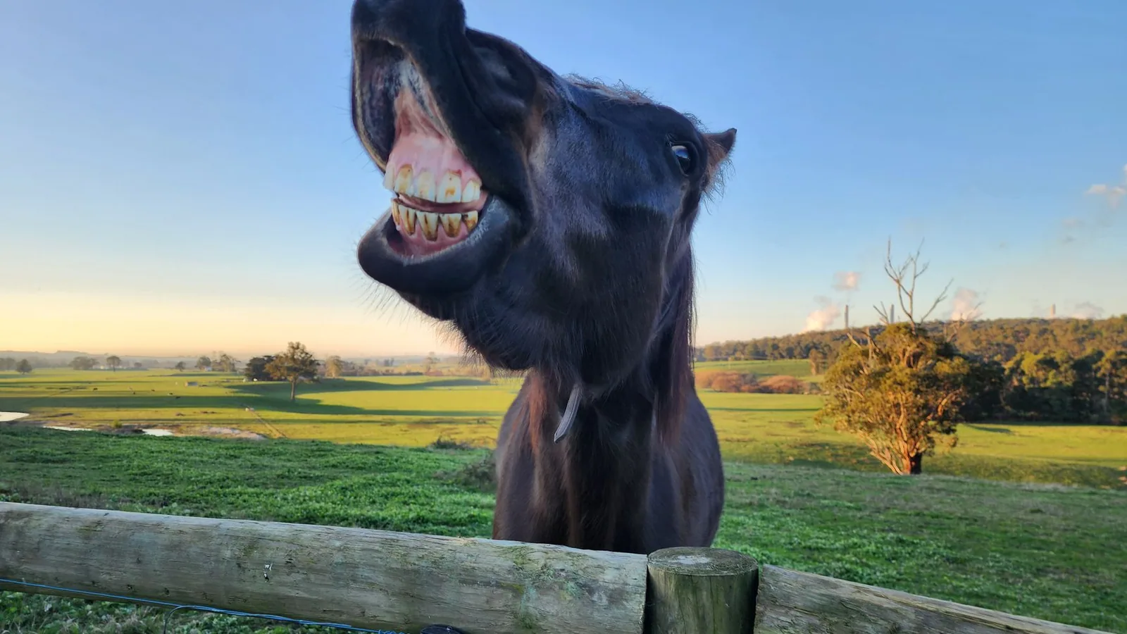
Horse dentition is built for a lifetime of grinding. Equine teeth have long reserve crowns that sit inside the jaw and erupt slowly as the exposed chewing surface wears away, which is why horses can process tough forage for decades without replacing their teeth [1]. That same design explains most of what goes wrong. Dental problems in horses are usually problems of wear, alignment, or the supporting tissues rather than simple cavities, and they develop slowly enough that owners often miss them until eating or ridden behavior changes.
The practical answer for owners is straightforward. Book a complete oral examination with a veterinarian at least once a year, expect sedation and a full mouth speculum, and treat floating as a targeted treatment based on a diagnosis rather than a routine rasping job [2][3]. The American Association of Equine Practitioners (AAEP) recommends at least an annual dental examination for adult horses, with more frequent checks for older horses or those with known disease.
This article is educational and is not a substitute for veterinary diagnosis or treatment.
At a Glance
| Task | What it involves | Timing |
|---|---|---|
| Complete oral examination | Sedation, mouth speculum, systematic inspection of both arcades, incisors, canines, gums, cheeks and tongue | At least annually for adult horses |
| Dental charting | Recording every tooth, every wear change, and every lesion so progress can be tracked | At every examination |
| Floating (odontoplasty) | Site-specific reduction of sharp enamel points, transverse ridges, or malocclusions identified on examination | Only when a specific diagnosis calls for it [2] |
| Incisor and canine check | Careful palpation and radiographs if resorption or hypercementosis is suspected | Every examination from roughly 10 years of age onward [4] |
| Imaging | Skull and dental radiographs as the first-line tool, computed tomography when radiographs are inconclusive | When a lesion is suspected or found [5] |
| Diet and management review | Forage type, turnout, feeding surface, and group feeding arrangements | Annually, and after any dental diagnosis [6] |
How Horse Dentition Works
Equine teeth are hypsodont. The visible crown is only part of the tooth, and a substantial reserve crown remains below the gumline, gradually erupting to replace what is worn away at the occlusal surface. Staszyk describes equine teeth and their occlusal surfaces as structures in continuous change, driven by age and by the high amount of occlusal wear specific to the horse [1]. That has an important clinical consequence. What looks like a variation in a young horse may be normal, and what looks normal in a 20-year-old may be the late stage of a progressive problem. Telling the two apart requires defined anatomical criteria and consistent naming [1].
The three functional groups
The incisors sit at the front of the mouth and shear forage close to the ground. The canines sit behind the incisors in the bars of the mouth and are commonly examined alongside the incisors because they share the same disease processes [7]. The cheek teeth, which comprise the premolars and the molars, do the grinding. Horse molars and premolars form a single functional arcade on each side, and most of the work of digestion depends on those surfaces meeting evenly through the full chewing cycle.
Why the occlusal surface is more than a flat plane
The chewing surface of a cheek tooth is a mixed structure of enamel, dentine, and cementum that wears at different rates. Earley and Reiswig note that an irregular occlusal surface, prominent cingula, transverse ridges, and enamel points all contribute to the function, form, and longevity of the equine cheek tooth [2]. In other words, some irregularity is normal and functional. The clinical question is not whether the surface is perfectly flat, because it never is, but whether a specific irregularity has become excessive enough to interfere with chewing or traumatize soft tissue.
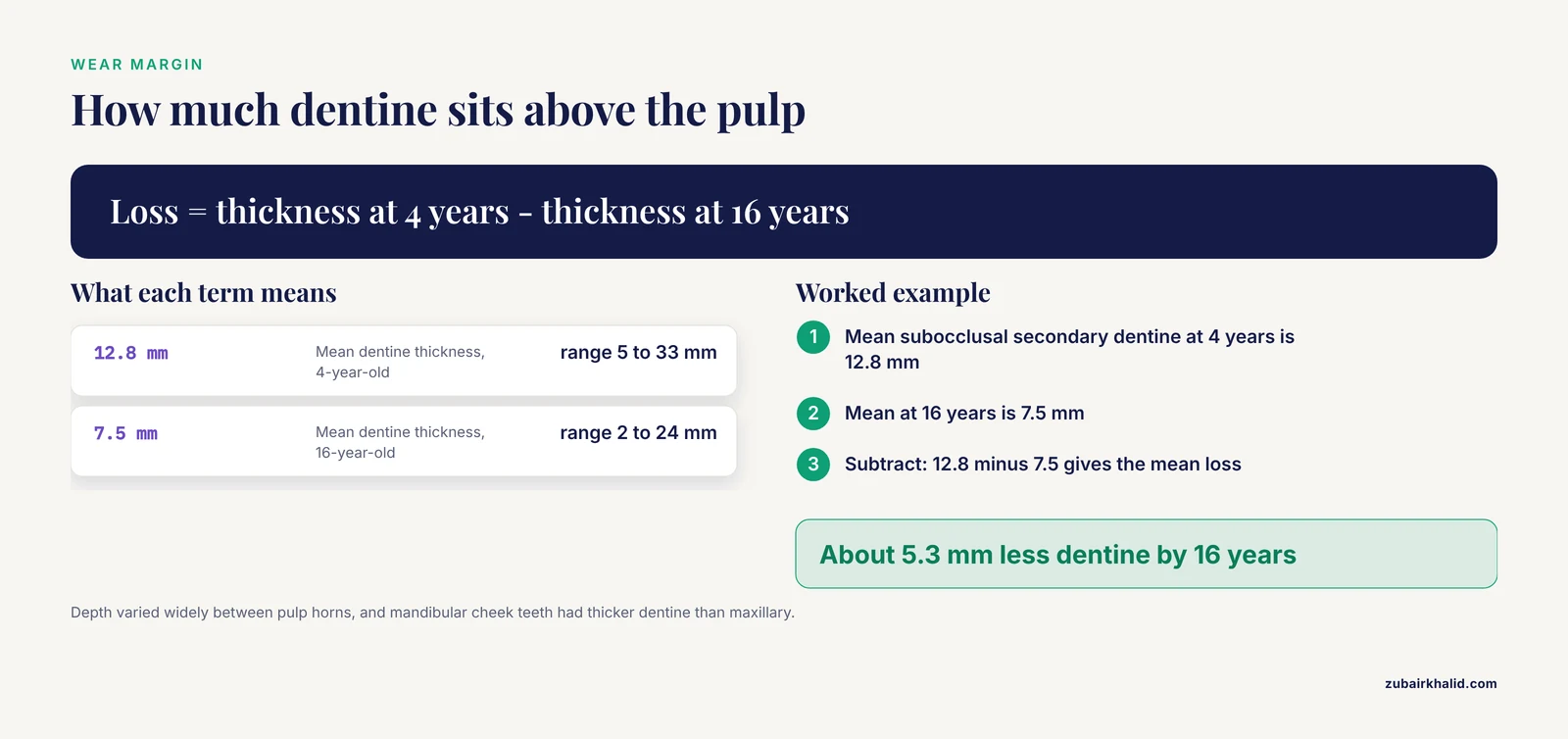
The safety margin between the surface and the pulp is smaller than many owners assume. In a study of cheek teeth from 17 horses with no history of dental disease, subocclusal secondary dentine thickness above the pulp horns ranged from a mean of 12.8 mm (range 5 to 33 mm) in a 4-year-old to 7.5 mm (range 2 to 24 mm) in a 16-year-old [8]. Depth varied widely between different pulp horns, even within the same tooth, and mandibular cheek teeth had significantly thicker subocclusal secondary dentine than maxillary cheek teeth [8].
Incisors have their own margin. High-resolution computed tomography of incisors from 13 horses found subocclusal dentine thickness between 1.5 and 11.7 mm in upper incisors and 0.7 and 6.7 mm in lower incisors, and thickness decreased with tooth age [9]. The distance from the pulp to the labial surface increased with age, ranging from 3.5 to 9.0 mm in upper incisors and 3.8 to 8.1 mm in lower incisors [9]. Older incisors therefore have less material to spare beneath the occlusal surface. Any procedure that reduces incisor height needs to account for that.
The Timeline of Horse Teeth
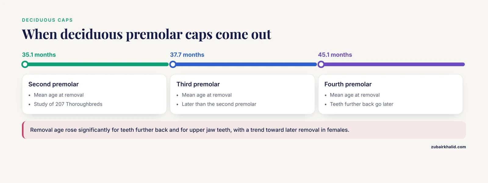
Foals are born with deciduous incisors and premolars that are gradually replaced. The most visible milestone is the loss of the deciduous premolar caps, and the timing of that event is less predictable than older textbooks suggest.
A prospective study of 508 deciduous premolar caps removed from 207 Thoroughbred racehorses found mean ages at removal of 35.1 months for the second premolar, 37.7 months for the third premolar, and 45.1 months for the fourth premolar [10]. Removal age increased significantly for teeth further back in the mouth and for teeth in the upper jaw, and there was a significant trend toward later cap removal in females, the first evidence of sexual dimorphism in permanent premolar eruption in horses [10]. The authors noted that these ages differed considerably from the ages of permanent tooth emergence widely reported in equine publications [10]. That matters to owners who are told a specific cap should be gone by a specific birthday. A retained cap is a reason to look, not a reason to panic on its own.
Age-related change continues throughout life. The occlusal surfaces migrate, the reserve crown shortens, and the proportions of enamel, dentine, and cementum at the surface shift. Staszyk's review stresses that these are normal dynamic changes, and that the examination should distinguish them from disease [1]. A veterinarian who sees a horse every year has the advantage of comparing one year to the next rather than judging a single snapshot.
Common Problems in Horse Teeth
A large survey of 206 privately owned horses found that 95% had at least one dental disorder, with disorders distributed roughly evenly between incisors and cheek teeth [11]. Malocclusions of incisor, premolar, and molar teeth were more prevalent than dental diseases across all age, gender, and breed groups [11]. Curvatures and calculus were the most common incisor findings, while sharp enamel points and caries predominated in the cheek teeth [11].
Those numbers are a useful corrective to the idea that a horse either has a dental problem or does not. Most horses have something worth recording. The clinical task is deciding which findings need treatment now, which need monitoring, and which are normal for that horse's age.
Sharp enamel points and excessive transverse ridges
Enamel points form where the outer edge of an upper cheek tooth or the inner edge of a lower cheek tooth is not worn down by an opposing surface. They are one of the components Earley and Reiswig describe as part of normal cheek tooth function, but they become a problem when they lengthen and start to lacerate the cheek or tongue [2]. Excessive transverse ridges are a separate finding, and a study comparing management systems found them more often in horses kept under intensive management [6].
Malocclusions
Malocclusion means the teeth do not meet correctly. Diagonal incisor malocclusion is a well-recognized example, and a retrospective evaluation of 204 corrections found that excursion to molar contact distances were commonly asymmetric before correction, and that prudent corrective measures usually increased symmetry [12]. The same study found that decreases in excursion to molar contact after correction were much more common than increases, and that the side with the shorter distance was more likely to stay the same [12]. It also identified patterns by quadrant, with the right side most likely to decrease in 100-300 quadrant malocclusions and the left side most likely to decrease in 200-400 quadrant malocclusions [12].
The authors were careful about prediction. Their data strongly suggested multiple causes and progressive compensatory mechanisms that complicate any precise forecast of how a given horse will respond [12]. Earley and Rawlinson place diagonal incisor malocclusion in the context of skull asymmetry and note how it commonly relates to premolar and molar occlusion [7]. That relationship is why an incisor problem is never examined in isolation.
Dorsal curvature malocclusion is another pattern recorded in practice, and it was more common in horses managed under semi-extensive systems in one comparative study [6].
Diastemata and periodontal disease
Diastema means a widened gap between adjacent cheek teeth. Food packs into the gap, and the tissue between the teeth becomes inflamed. Premolar and molar diastemata occurred more frequently in horses kept under extensive management in a comparison of three management systems [6]. Periodontal disease is a common feature of advanced dental disease in horses, characterized primarily by moderate lymphoplasmacytic inflammation in histologic samples [13]. Because diastemata and periodontal disease are often painful and progress quietly, they are a strong argument for a hands-on examination rather than a look at the front teeth.
Caries and fractures
Caries, or tooth decay, was one of the two predominant cheek tooth findings in the Polish survey, alongside sharp enamel points [11]. Fractures are classified and assessed to determine treatment options, and the same review covers oral and radiographic assessment of incisive bone fracture and incisor avulsion [7]. A fractured tooth is not always an extraction. The decision depends on which tooth, how much crown is left, whether the pulp is involved, and what the radiograph shows [7].
EOTRH
Equine odontoclastic tooth resorption and hypercementosis is a painful, progressive disease that mainly affects the incisors and canine teeth of aged horses [4]. It is probably underdiagnosed. In a German prevalence study of 142 horses aged 10 and older, 94% had at least minor radiologic changes associated with EOTRH and 62% had moderate to severe changes [4]. No horse older than 14 years was free of radiological signs, and every horse over 28 years had at least moderate changes [4]. The mean age of the study population was 21 years [4].
Histopathology explains the clinical appearance. Affected teeth show cemental hyperplasia and lysis, and in severe cases the proliferation of cementum causes bulbous enlargement of the intra-alveolar portion of the tooth [13]. Several teeth contained necrotic debris, bacteria, and plant material in areas of cemental lysis, all horses had dentinal lysis in at least one affected tooth, and endodontic disease was often present [13]. Resorption with secondary hypercementosis appears to begin on the external surface of the tooth rather than inside the pulp cavity [13]. That external origin is why radiographs are the diagnostic mainstay, and why researchers have worked on quantifying radiographic change. Studies using density standards and scaled pixel counting have shown that pixel values increase with EOTRH grade, allowing differentiation of both early and late radiographic signs [14]. Texture analysis of radiographic images has also identified features that change with disease progression [15].
Clinical signs, staging, and classification of tooth resorption in incisors and canines are covered in the equine dental literature alongside canine odontoplasty and the consequences of heavy plaque and calculus on lower canines [7].
Canine teeth
Lower canines accumulate plaque and calculus, and excessive accumulation can lead to periodontal disease and abscess formation [7]. Canine teeth are frequently overlooked because they sit back from the incisors and because many owners assume they are not part of routine dental care. They are.
Wolf teeth
Wolf teeth are small vestigial teeth that may be present in the premolar region. Dental disorders were recorded in wolf teeth in the Polish prevalence survey, though at low numbers compared with incisors and cheek teeth [11]. Whether a wolf tooth needs removal depends on its position, its size, and whether it interferes with the bit or with the cheek teeth arcade. That is a conversation for the examining veterinarian.
The occlusal angle question
Owners sometimes worry that the angle of the molar surfaces is causing poor digestion. A study of 40 pregnant mares found molar occlusal angles ranging from 6.3 to 19.3 degrees, and none of those angles was significantly associated with feed digestibility, water balance, or fecal particle size [16]. The number of dental abnormalities was also not associated with feed digestibility, and occlusal angle did not vary significantly with age [16]. A separate observational study of 230 mandibular cheek teeth from 21 horses found occlusal angle was not significantly affected by tooth position or tooth age, and that measurement of an individual tooth was predictive for the whole mouth [17]. This does not mean angle never matters. It means an angle in that range is not by itself an explanation for poor condition or loose manure.
What a Dental Examination Involves
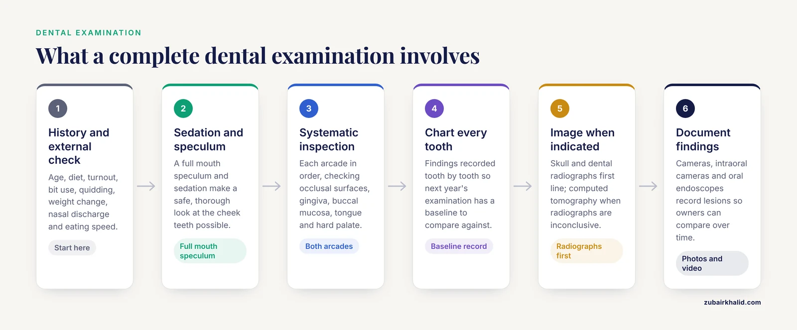
Easley makes the case plainly. Equine dentistry is not just carpentry work involving the removal of sharp enamel points. Every horse deserves a complete veterinary dental examination on a regular basis, and without that examination the practitioner cannot determine which corrective procedures are needed [3].
A complete examination usually follows a sequence.
- History and external assessment. Age, diet, turnout, work type, bit use, quidding, weight change, nasal discharge, and any change in eating speed.
- Sedation and a full mouth speculum. This is what makes a safe, thorough look possible.
- Systematic inspection. Each arcade is examined in order, with attention to the occlusal surfaces, the gingiva, the buccal mucosa, the tongue, and the hard palate.
- Charting. Findings are recorded tooth by tooth so that next year's examination has a baseline.
- Imaging when indicated. Radiography is the first-line imaging modality and is available to most equine veterinarians providing primary care [5]. Diagnostic radiographs of the skull and dentition require careful positioning and technique, and the images have known limitations that may prompt computed tomography [5].
- Documentation. Digital cameras, intraoral cameras, and oral endoscopes have become part of equine dental practice and help record what was found [18].
Galloway and Easley note that each veterinarian must choose the camera or endoscope system that fits that practice's clinical needs and budget, and they encourage research into the whole system rather than just the camera, including software compatibility, manufacturer service policies, and colleague references [18]. For owners, that translates into a simple observation. A practice that photographs or films lesions and shares the images is giving you a record you can compare over time.
Floating and Odontoplasty
Odontoplasty, commonly called floating, is the most commonly performed procedure in equine dentistry [2]. It is also the one most often misunderstood.
Earley and Reiswig set out the constraint clearly. With limited reserve crown available and an average functional life range between 18 and 25 years, removal of tooth structure should be conservative [2]. Individual tooth evaluation should lead to a specific diagnosis and treatment plan, and odontoplasty should be site-specific [2]. The authors consider a quality oral examination to be the most important dental procedure performed in the horse, more important than the floating that follows it [2].
That has direct consequences for how owners should think about routine floating.
- Floating without an examination is guesswork. If no one has looked at every tooth, no one knows which surfaces need reduction.
- Over-reduction is irreversible. Each pass of a float removes reserve crown that will not come back within the horse's working life.
- Not every irregularity needs treatment. Some surface features are functional, and reducing them removes structure without benefit [2].
- The goal is a functional occlusal surface, not a flat one.
For a horse with a specific problem such as a diagonal incisor malocclusion, corrective work may extend beyond simple point reduction. The 204-case retrospective found that correction usually improved symmetry of excursion to molar contact, but also that outcomes were difficult to predict because of multiple causes and compensatory mechanisms [12]. Follow-up timing should be set by the veterinarian who did the work, based on what was corrected and how the horse responds.
Pain Control During Dental Procedures
Dental procedures are painful, and pain control has become a larger part of equine dental practice. Intraligamentary anesthesia, in which local anesthetic is injected into the periodontal ligament, is well established in human dentistry. Teysen and colleagues adapted the technique for equine dentistry and evaluated it in a cadaver study [19]. After injection, the solution spread extensively through the periodontal ligament and alveolar bone and accumulated around the apex of the tooth, apparently through both diffusion along the periodontal ligament and infiltration into bone marrow spaces [19]. The technique is described as straightforward to perform, and the cadaver work supported the anatomical basis for desensitizing the pulp, periodontium, and surrounding tissues, though clinical studies are still needed to confirm efficacy [19]. For owners, the takeaway is that asking how pain will be managed is a reasonable question before any extraction or deep procedure.
Prevention and Everyday Care
Examination frequency
Annual examination is the foundation. The AAEP recommends at least annual dental examinations for adult horses, and more frequent checks make sense for seniors, for horses with a history of diastemata or periodontal disease, and for horses that have had a malocclusion corrected.
Management and feeding
What a horse eats and how it is kept changes what a veterinarian finds. A comparison of 69 clinically healthy adult horses kept under extensive, semi-extensive, and intensive management found that incisor problems, premolar and molar diastemata, and irregular incisors were more frequent under extensive systems, while oral mucosal ulcers and excessive transverse ridges were more common under intensive systems [6]. Overall prevalence of orodental disorders was lower under semi-extensive management than under extensive or intensive management. That last point matters. No management system is dentally free. Each produces a different pattern of problems, and the examination should be read against the horse's management.
Home checks owners can do
Owners cannot see the cheek teeth, but they can notice change. Watch for slower eating, dropping partially chewed forage, quidding, weight loss despite adequate feed, a foul smell from the mouth, swelling on the side of the face, nasal discharge from one nostril, resistance to the bit, head tossing, and reluctance to have the face handled. None of these signs confirms a dental diagnosis, and all of them justify a call to the veterinarian.
Equipment and Supplies: Selection Criteria
Owners do not need dental instruments. They do need to understand what a properly equipped examination requires, so they can ask informed questions.
- Mouth speculum. Necessary for a safe and complete look at the cheek teeth.
- Good light and a mirror. The back of the mouth cannot be assessed by direct vision alone.
- Dental picks and probes. Needed to check for diastemata, caries, and pockets.
- Floats and power equipment. Chosen by the veterinarian based on the procedure planned [3].
- Radiography. Skull and dental radiographs require positioning and technique appropriate to the region being imaged [5].
- Camera or endoscope. An optional but increasingly common documentation tool, chosen for system compatibility and support as much as for image quality [18].
A note on owner-applied rasps and floats. Removing tooth structure without a diagnosis risks removing reserve crown the horse cannot replace [2]. That is the argument against do-it-yourself floating.
Welfare and Safety Points
EOTRH is described in the literature as a painful and progressive disease [4]. Pain is not a side effect of dental disease. It is often the reason a horse stops eating normally. Recognizing and treating pain is a welfare obligation, not a refinement.
Sedation and a speculum are what make a thorough examination possible, and they are also why the procedure belongs with a veterinarian.
Conservative odontoplasty protects the horse's future. A horse whose teeth are reduced aggressively at 8 years old has less reserve crown at 20 [2].
Limitations and When to Contact a Veterinarian
Individual horses vary enough in dental anatomy, age, and management that no general article can replace an examination. Contact a veterinarian promptly if you notice any of the following.
- Dropping feed, quidding, or slow eating that persists for more than a few days.
- Weight loss or loss of body condition without an obvious nutritional reason.
- Foul breath, facial swelling, or discharge from one nostril.
- Blood in the mouth or on the bit.
- Resistance to the bit, head tossing, or a new reluctance to be bridled.
- Visible damage to an incisor, or a loose incisor.
- Reluctance to eat hard feed or to graze, particularly in a horse over 15 years old.
- Any horse over 10 years old that has never had an incisor examination for EOTRH.
Signs of advanced disease warrant a same-day call. These include inability to eat, severe facial swelling, a visibly displaced or fractured tooth after trauma, and any horse showing signs of colic alongside dental signs.
Related Guides
- Why Is My Horse Grinding or Showing Its Teeth?: Explains grinding and tooth-showing behavior that may signal dental discomfort
- Common Horse Health Problems Every Owner Should Know: Broad overview of horse health problems, including dental concerns
- Bacterial Infections in Goats and Horses: Common Pathogens and Treatment Protocols: Covers bacterial infections and pathogens relevant to periodontal disease
- Common Horse Feeding Mistakes to Avoid: Feeding management affects wear patterns and common dental problems
- Complete Horse Care Guide for New Owners: Comprehensive care guide for new owners managing horse health
- Senior Horse Care: Dental, Nutrition, and Arthritis Needs: Dental, nutrition, and arthritis care for senior horses
- Monthly Horse Care Checklist: Checklist helps schedule dental exams and routine horse care
Frequently Asked Questions
How often should a horse's teeth be checked?
At least once a year for most adult horses. The AAEP recommends annual dental examinations, and older horses or horses with known disease often need checks every six months.
What is floating a horse's teeth?
Floating is odontoplasty, the reduction of sharp enamel points and other excessive wear features on the occlusal surfaces [2]. It should follow a complete examination and target specific teeth rather than treat the whole mouth the same way.
What are the early signs of dental problems in horses?
Quidding, slow eating, dropping feed, weight loss, foul breath, facial swelling, and resistance to the bit are the signs owners most often notice. None is specific to one diagnosis, so an examination is needed.
What is EOTRH in horses?
Equine odontoclastic tooth resorption and hypercementosis is a progressive disease of the incisors and canines in older horses [4][13]. It is diagnosed primarily by radiographs and is painful, so early recognition matters.
Do horses lose baby teeth?
Yes. Foals lose their deciduous premolar caps as the permanent premolars erupt, usually between roughly 2.5 and 4 years of age depending on the tooth [10]. Retained caps should be assessed rather than assumed to be normal.
Can a horse's teeth be worn down too far?
Yes. Each tooth has limited reserve crown, and the average functional life of the dentition is between 18 and 25 years [2]. Repeated aggressive reduction removes structure the horse cannot regrow.
Are sharp enamel points painful?
They can be. Enamel points that have lengthened can lacerate the cheek or tongue, which is why a site-specific examination matters more than a routine schedule [2].
Does my horse need sedation for a dental examination?
A complete examination of the cheek teeth generally requires sedation and a mouth speculum. A brief look at the incisors can be done without sedation, but that is not a substitute for a full examination.
Sources
- [[Clinical anatomy of the horse: teeth and dentition].](https://pubmed.ncbi.nlm.nih.gov/26603675/)
- Equine Dental Floating (Crown Osontoplasty).
- Dental care and instrumentation.
- Radiological prevalence of equine odontoclastic tooth resorption and hypercementosis.
- Dental Radiography and Radiographic Signs of Equine Dental Disease.
- Semi-extensive management system drives lower prevalence of equine orodental disorders.
- A new understanding of oral and dental disorders of the equine incisor and canine teeth.
- A study of the thickness of cheek teeth subocclusal secondary dentine in horses of different ages.
- Anatomy of equine incisors: Pulp horns and subocclusal dentine thickness.
- Chronology and sequence of emergence of permanent premolar teeth in the horse: study of deciduous premolar 'cap' removal in Thoroughbred racehorses.
- Malocclusions and Dental Diseases in Privately Owned Horses in the Mazovia Region of Poland.
- A retrospective evaluation of 204 diagonal incisor malocclusion corrections in the horse.
- Equine Odontoclastic Tooth Resorption and Hypercementosis: Histopathologic Features.
- An application of the density standard and scaled-pixel-counting protocol to assess the radiodensity of equine incisor teeth affected by resorption and hypercementosis: preliminary advancement in dental radiography.
- Selection of Filtering and Image Texture Analysis in the Radiographic Images Processing of Horses' Incisor Teeth Affected by the EOTRH Syndrome.
- Effect of premolar and molar occlusal angle on feed digestibility, water balance, and fecal particle size in horses.
- Observations of the cheek tooth occlusal angle in the horse.
- Incorporating oral photography and endoscopy into the equine dental examination.
- Intraligamentary anaesthesia: a local anaesthesia technique in equine dentistry.