Endocytosis vs Exocytosis
By Dr. Zubair Khalid, DVM, MS, PhD ·

Endocytosis is the process by which a cell takes in extracellular material through invagination of its plasma membrane, and exocytosis is the process by which a cell releases intracellular material through fusion of vesicles with the plasma membrane. Both are active, membrane-remodeling processes that move cargo and membrane in opposite directions.
The balance between these two processes shapes how cells eat, listen, speak, and remember. Endocytosis brings in nutrients and signaling receptors, clears apoptotic cells, and provides entry routes for pathogens and medicines. Exocytosis releases hormones, neurotransmitters, immune mediators, and membrane proteins. The two pathways also depend on each other. In human neutrophils, blocking clathrin-mediated endocytosis prevented priming of superoxide release and inhibited exocytosis of all four granule subsets after TNFα stimulation [1]. That result is a useful warning against treating uptake and secretion as separate topics. They are coupled arms of one membrane traffic system.
Endocytosis Definition and the Core Machinery
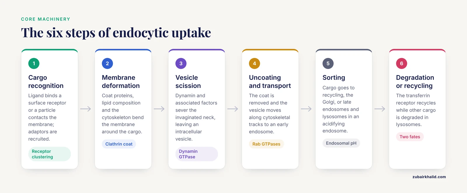
The endocytosis definition is straightforward: a cell surrounds extracellular cargo with a patch of plasma membrane, pinches off a vesicle, and delivers the cargo to an intracellular compartment. The cargo can be soluble fluid, a ligand bound to a receptor, a membrane protein, a virus, a toxin, or a particle such as an apoptotic body. The newly formed vesicle is a membrane-bound carrier whose lumen is topologically continuous with the outside of the cell. That topology matters because the inside of an endocytic vesicle is chemically similar to the extracellular space until pumps and enzymes modify it.
The core machinery includes coat proteins, adaptors, small GTPases, phosphoinositides, actin, and fission factors. Clathrin is a protein that assembles into a triskelion lattice and bends the membrane into a coated pit. Adaptor protein 2, usually called AP2, links cargo receptors to clathrin and helps select which membrane proteins enter the vesicle. Dynamin is a GTPase that forms a collar around the vesicle neck and promotes scission. Rab GTPases are molecular switches that mark vesicles and help them find the correct target membrane. Soluble N-ethylmaleimide-sensitive factor attachment protein receptors, or SNAREs, mediate fusion between vesicles and target membranes. Endosomes are sorting stations that receive endocytic cargo. Lysosomes are acidic organelles that degrade cargo. Recycling endosomes return receptors and membrane back to the plasma membrane.
The physical steps are easier to remember as a numbered cycle.
- Cargo recognition. A ligand binds a surface receptor, or a particle contacts the membrane. Receptor clustering and adaptor recruitment begin. For receptor-mediated endocytosis, a particle must approach the membrane closely enough for ligand-receptor binding to occur, sometimes within a few nanometers [2].
- Membrane deformation. Coat proteins, lipid composition, and the cytoskeleton bend the membrane around the cargo. Clathrin-coated pits are the classic example, but caveolae and other coats use different proteins.
- Vesicle scission. Dynamin and associated factors sever the invaginated neck. The result is an intracellular vesicle.
- Uncoating and transport. The coat is removed and the vesicle moves along cytoskeletal tracks toward an early endosome.
- Sorting. Cargo is sorted for recycling to the plasma membrane, transport to the Golgi, or delivery to late endosomes and lysosomes. Endosomal acidification is part of this sorting environment. In proximal tubule cells, expression of cystic fibrosis transmembrane conductance regulator (CFTR) and vacuolar H+-ATPase (V-ATPase) supports the endosomal acidification needed for efficient albumin endocytosis and degradation [3].
- Degradation or recycling. Receptors such as the transferrin receptor recycle, while other cargo is degraded in lysosomes. The choice depends on the receptor, the ligand, and the cell type.
The entry of Mycoplasma pneumoniae CARDS toxin shows how a pathogen can exploit this machinery. CARDS toxin induced rapid, transient translocation of annexin A2 (AnxA2) to the plasma membrane, peaking at 15 minutes after exposure [4]. The recruitment was phosphorylation-independent. Co-immunoprecipitation showed enhanced association between AnxA2 and clathrin heavy chain, and the toxin co-localized with AnxA2 in early endosomes. CRISPR/Cas9 knockout of AnxA2 in A549 cells reduced toxin internalization [4]. This is a clean endocytosis example with a specific host protein, a specific cargo, and a specific uptake route.
Endocytosis Types: A Practical Map
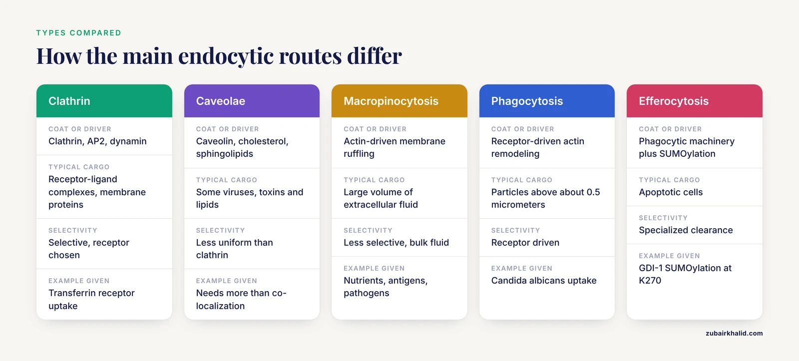
Endocytosis is not one pathway. The endocytosis types differ in coat, cargo size, membrane shape, and regulation. A single cell can use several of them at the same time.
Clathrin-Mediated Endocytosis
Clathrin-mediated endocytosis uses clathrin-coated pits and vesicles. It is the best-characterized route for receptor-ligand uptake and for internalization of membrane proteins. The pathway can be constitutive or triggered by ligand binding. In hippocampal neurons, constitutive endocytosis of AMPA-type glutamate receptors was not dependent on clathrin and dynamin, while endocytosis triggered by long-term depression-inducing stimulation was clathrin- and dynamin-dependent [5]. That finding is a reminder that the same receptor can use different routes depending on context.
Clathrin-mediated endocytosis also regulates signaling. ROBO1, a roundabout guidance receptor for SLIT ligands, undergoes constitutive endocytosis that is enhanced by NSLIT2 binding [6]. Pharmacologic inhibitors and molecular techniques showed that clathrin-mediated endocytosis is necessary for NSLIT2-induced inhibition of cell spreading. Adenomatous polyposis coli (APC), a multifunctional tumor suppressor, is in close proximity to ROBO1 but dissociates after NSLIT2 binding, and that dissociation is needed for clathrin-mediated ROBO1 endocytosis [6]. The pathway is therefore part of the signal, not just a way to remove the receptor.
Receptor-Mediated Endocytosis
Receptor-mediated endocytosis is a broad term for uptake that depends on a cell-surface receptor recognizing a ligand. It is often used as a synonym for clathrin-mediated endocytosis, but the terms are not identical. Some receptor-mediated pathways use caveolae, lipid rafts, or adaptors other than AP2.
Examples span nutrients, proteins, and drugs. The transferrin receptor takes up iron-loaded transferrin. The low-density lipoprotein receptor clears cholesterol-carrying particles. In the kidney, proximal tubule cells reabsorb albumin through receptor-mediated endocytosis and then degrade it. High albumin overload in LLC-PK1 proximal tubule cells reduced CFTR and V-ATPase expression and impaired albumin endocytosis and degradation [3]. That result connects a membrane transport pathway to a disease-relevant stress response.
Therapeutic delivery often exploits receptor-mediated endocytosis. Ctx-ETAC is a degrader-antibody conjugate in which the anti-EGFR antibody cetuximab delivers an engineered proteolysis-targeting chimera, or PROTAC, into EGFR-expressing tumor cells and activated cancer-associated fibroblasts through receptor-mediated endocytosis [7]. The payload is a bifunctional molecule that recruits a target protein to an E3 ubiquitin ligase for degradation. The delivery route matters because the conjugate must enter the right cells before the PROTAC can act.
Chemokine receptor trafficking adds another nuance. XCR1, a chemokine receptor selectively expressed on mature conventional type 1 dendritic cells, undergoes constitutive internalization through a β-arrestin-independent but AP2-dependent pathway [8]. β-arrestin is a scaffolding protein long associated with receptor desensitization and internalization. The XCR1 result shows that AP2-dependent endocytosis can proceed without that canonical β-arrestin step. Receptor-mediated endocytosis therefore has more than one molecular grammar.
Caveolae and Lipid Raft Uptake
Caveolae are flask-shaped membrane invaginations enriched in caveolin proteins, cholesterol, and sphingolipids. They participate in uptake of some viruses, toxins, and lipids, and they organize signaling at the membrane. Lipid rafts are ordered membrane domains that can concentrate receptors and signaling lipids. Membrane lipid organization, fluidity, glycocalyx architecture, and membrane protein identity all shape recognition, adhesion, endocytosis, and intracellular delivery [9]. Caveolar uptake is less uniform than clathrin-mediated uptake, and assigning a cargo to caveolae requires more than a co-localization image.
Macropinocytosis
Macropinocytosis is an actin-driven process in which the plasma membrane ruffles and engulfs a large volume of extracellular fluid. It is less selective than receptor-mediated uptake and can deliver soluble nutrients, antigens, and pathogens. Macropinosomes are large vesicles that mature and fuse with endosomal compartments. The pathway is prominent in some immune cells and cancer cells, where it can support nutrient acquisition and antigen sampling.
Phagocytosis and Efferocytosis
Phagocytosis is the receptor-driven uptake of large particles, typically greater than about 0.5 micrometers. It requires actin polymerization and coordination of signaling around the particle. In innate immunity, host receptors recognize Candida albicans cell wall components such as β-glucan, chitin, and mannan and trigger phagocytosis, reactive oxygen species, and inflammatory cytokine production [10]. Antibody-dependent cellular phagocytosis, or ADCP, is another route. CD47 on tumor cells suppresses Fcγ receptor-mediated ADCP through binding to signal regulatory protein α (SIRPα) on macrophages, and blockade of that interaction enhances antibody-dependent phagocytosis [11].
Efferocytosis is the specialized phagocytosis of apoptotic cells. It clears dying cells before their contents leak and provoke inflammation. In C. elegans, SUMOylation of GDP dissociation inhibitor GDI-1 at K270 triggered phagosomal degradation, and the same conserved site contributed to efferocytosis regulation in mammals [12]. The pathway connects phagosome maturation to the SUMO conjugation system. Efferocytosis is not the same as macropinocytosis or clathrin-mediated endocytosis, and mixing the terms obscures the receptor and cytoskeletal requirements.
Constitutive and Triggered Endocytosis
Constitutive endocytosis runs continuously and captures receptors, nutrients, and membrane for recycling. Triggered endocytosis begins after a specific stimulus, such as ligand binding, synaptic activity, or immune activation. The AMPA receptor and XCR1 examples show why the distinction matters [5][8]. A pathway that looks constitutive in one assay may be a triggered pathway that is already active at baseline. Experimental design has to separate basal uptake from stimulus-dependent uptake.
Exocytosis Definition and the Secretory Cycle
Exocytosis is the fusion of an intracellular vesicle with the plasma membrane, which releases the vesicle lumen contents outside the cell and delivers vesicle membrane proteins and lipids to the cell surface. It is the opposite of endocytosis in direction, but it uses overlapping machinery. Rab GTPases, tethering factors, and SNAREs participate in both pathways.
The secretory cycle begins with protein synthesis in the endoplasmic reticulum and processing in the Golgi apparatus. Cargo is packaged into transport vesicles or storage granules. Vesicles move along microtubules and actin filaments toward the plasma membrane. Tethering factors and Rab GTPases bring the vesicle close to the membrane. SNARE proteins on the vesicle and target membrane form a complex that pulls the two bilayers together. In regulated exocytosis, a calcium signal triggers fusion. After fusion, membrane and some proteins are retrieved by endocytosis and recycled.
Constitutive exocytosis runs continuously in many cells. It delivers newly made membrane proteins, releases serum proteins, and supplies lipids to the plasma membrane. Regulated exocytosis is triggered. Neurons release neurotransmitters from synaptic vesicles after calcium entry. Endocrine cells release insulin or other hormones from storage granules. Immune cells release granule enzymes, antimicrobial peptides, and cytokines. Human neutrophils use exocytosis of granule subsets during priming, and clathrin-mediated endocytosis is required for that response [1]. The coupling is tight enough that blocking uptake changes what the cell secretes.
Secretion can also reshape the tissue environment. In clear cell renal cell carcinoma, FOXA2 recruits histone acetyltransferase HAT1 to upregulate growth differentiation factor 15 (GDF15) [13]. Tumor cell-secreted GDF15 activates GFRAL/RET-mediated STAT3 signaling and the epithelial-to-mesenchymal transition, and it facilitates CD8+ T cell exhaustion in the tumor microenvironment [13]. The exocytotic release of GDF15 is part of a signaling axis that affects metastasis and immune evasion. Exocytosis is therefore not a passive export step. It is a regulated signaling event with local and systemic consequences.
Endocytosis vs Exocytosis: The Differences That Matter
The core endocytosis and exocytosis differences are summarized in the table below. The table is a comparison of normal cell physiology, not a list of mutually exclusive rules. Some cargoes use mixed pathways, and some vesicles recycle between the two arms.
| Feature | Endocytosis | Exocytosis |
|---|---|---|
| , - | , - | , - |
| Direction of cargo movement | Cargo enters the cell | Cargo leaves the cell or is delivered to the cell surface |
| Membrane event | Invagination and vesicle scission | Vesicle docking and fusion |
| Net membrane effect | Removes membrane from the surface before recycling | Adds membrane to the surface |
| Typical cargo | Nutrients, ligands, receptors, pathogens, apoptotic bodies | Secreted proteins, neurotransmitters, hormones, granule contents, membrane proteins |
| Vesicle path | Plasma membrane to endosome | Golgi or storage granule to plasma membrane |
| Energy and core machinery | ATP, actin, dynamin, coat proteins, Rab GTPases, SNAREs | ATP, calcium, Rab GTPases, tethering factors, SNAREs |
| Common examples | Transferrin uptake, albumin reabsorption, AMPA receptor internalization, phagocytosis | Insulin release, neurotransmitter release, neutrophil granule release, GDF15 secretion |
| Typical readouts | Fluorescent cargo uptake, surface receptor loss, endosome co-localization | Secreted cargo ELISA, surface exposure, granule marker release, pHluorin flashes |
Direction is the first difference. Endocytosis moves membrane and cargo inward. Exocytosis moves membrane and cargo outward. The net membrane effect is the second difference. Endocytosis removes plasma membrane, while exocytosis adds it. Cells balance the two to keep surface area stable during secretion and during cycles of receptor uptake. If exocytosis outpaces endocytosis, the surface expands. If endocytosis outpaces exocytosis, the surface shrinks or membrane is stored internally.
Cargo selection is the third difference. Endocytosis is often selective because receptors and adaptors choose cargo. It can also be bulk uptake through macropinocytosis. Exocytosis is usually selective because cargo is sorted into vesicles in the endoplasmic reticulum and Golgi. Regulated exocytosis adds a trigger, which allows a cell to store cargo and release it only after a signal.
The fourth difference is the relationship to signaling. Endocytosis can terminate a signal by removing receptors from the surface, but it can also sustain a signal from an endosomal platform. ROBO1 endocytosis is required for NSLIT2-induced changes in cell spreading [6]. XCR1 internalization is constitutive and AP2-dependent, which affects how dendritic cells present antigen and respond to chemokines [8]. Exocytosis releases signals such as GDF15 into the extracellular space [13]. The same cell can use endocytosis to tune a receptor and exocytosis to release a ligand.
The fifth difference is the assay. Endocytosis is often measured as cargo uptake or surface receptor loss. Exocytosis is often measured as cargo release or surface appearance of a vesicle marker. The neutrophil priming study measured both arms: endocytosis inhibitors, granule component release by ELISA or chemiluminescence, and superoxide release as reduction of ferrocytochrome c [1]. That combination is stronger than either assay alone because it tests the coupling between uptake and secretion.
How to Read an Endocytosis Diagram
A useful endocytosis diagram, whether in a textbook or a teaching animation, should show the topology clearly. Use the following checklist to evaluate one.
- The extracellular space and the cytosol are labeled. The plasma membrane is drawn as a bilayer.
- A cargo molecule or ligand is shown binding a surface receptor.
- The membrane invaginates, and the extracellular face of the membrane remains facing the lumen of the newly formed vesicle.
- Coat proteins are shown on the cytosolic face. Clathrin, AP2, caveolin, or another coat should match the pathway being taught.
- A fission event separates the vesicle from the plasma membrane. Dynamin is a common label at the neck.
- The vesicle is shown delivering cargo to an early endosome, with recycling and lysosomal routes separated.
- Exocytosis is drawn as a separate fusion event, not as endocytosis run backward.
- The diagram does not imply that every endocytic vesicle goes directly to a lysosome. Recycling is a major route.
A diagram that omits recycling or labels all endocytosis as clathrin-mediated teaches the wrong mental model. A diagram that shows a vesicle floating through the cytosol without membrane continuity also hides the topology. Students should be able to point to the extracellular side of the membrane before and after vesicle formation and explain why the vesicle lumen is not the same as the cytosol.
Endocytosis Examples Across Cell Types
The fastest way to learn endocytosis vs exocytosis is to attach each term to a concrete cell. The examples below are not isolated trivia. They show how the same molecular machinery is adapted to different cargo and different physiological needs.
Nutrient Uptake by Transferrin Receptor
Transferrin binds iron and binds the transferrin receptor at the cell surface. The receptor-ligand complex enters through clathrin-coated pits. After the vesicle is uncoated, the iron is released in the acidic endosome. The transferrin receptor and transferrin are then recycled to the surface. This is a classic receptor-mediated endocytosis example because the receptor is reused, not degraded. The pathway connects iron supply to cell growth and is used in many cultured cells as a marker of clathrin-mediated uptake.
Albumin Reabsorption in the Kidney
Proximal tubule cells reabsorb filtered albumin and degrade it. That process depends on receptor-mediated endocytosis and endosomal acidification. In LLC-PK1 cells, high albumin overload reduced CFTR and V-ATPase expression and impaired albumin endocytosis and degradation [3]. The result has two lessons. First, endocytosis is a physiological clearance route for a plasma protein. Second, the degradative arm depends on the endosomal environment, not just on the uptake receptor. A defect in acidification can change the fate of the cargo after it has already entered the cell.
AMPA Receptor Endocytosis in Synaptic Plasticity
AMPA-type glutamate receptors mediate fast excitatory transmission. Long-term depression is expressed in part by a decrease in postsynaptic AMPA receptors, and endocytosis is required for that decrease [5]. In the PSLM preparation, constitutive endocytosis of AMPA receptors did not depend on clathrin and dynamin, in contrast to transferrin receptor endocytosis. Endocytosis triggered by long-term depression-inducing stimulation did depend on clathrin and dynamin [5]. The example matters because it shows that an endocytosis defect can alter synaptic strength, learning-related plasticity, and the surface distribution of a neurotransmitter receptor.
Pathogen Entry by CARDS Toxin
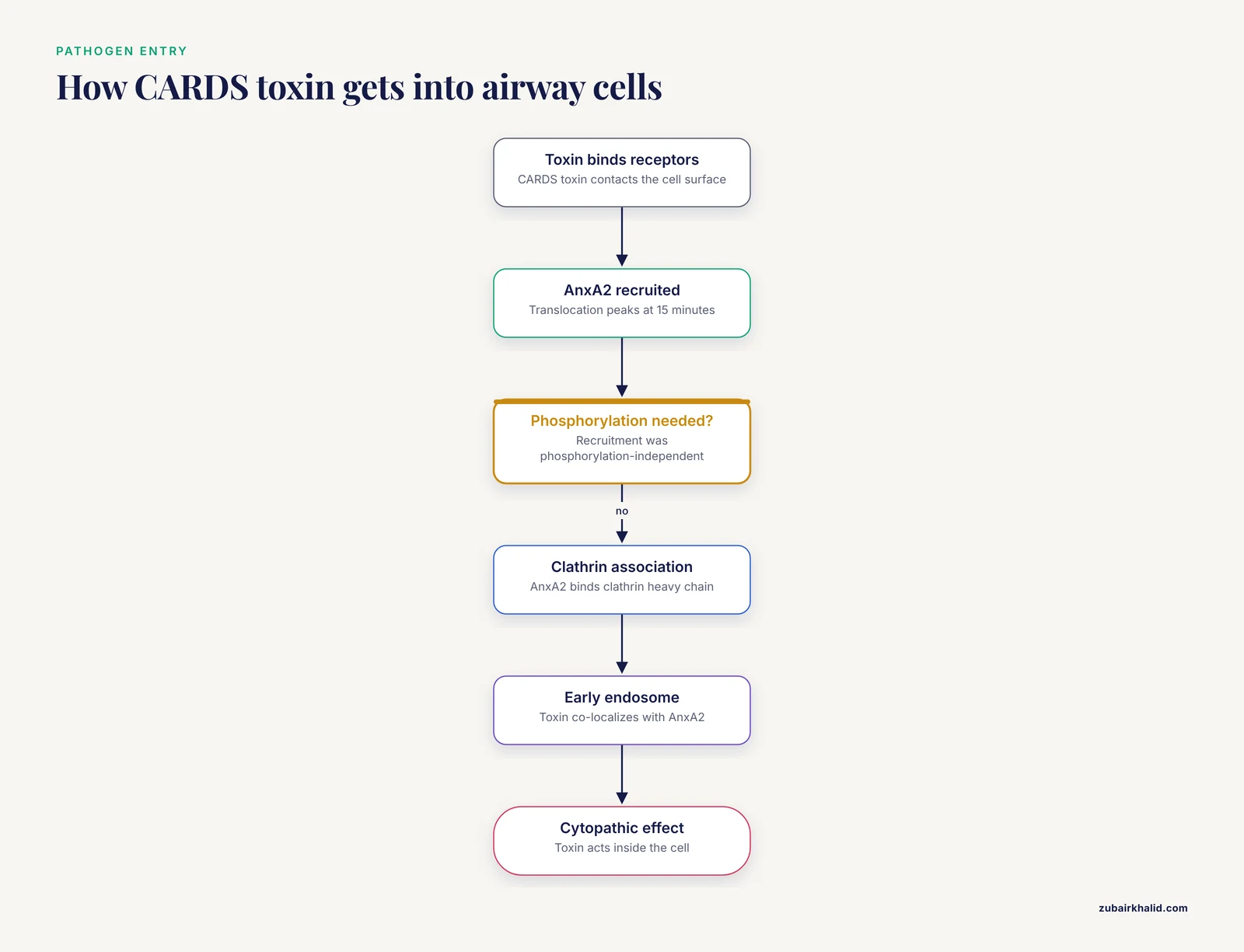
Mycoplasma pneumoniae CARDS toxin binds multiple receptors and uses host annexin A2 to enhance clathrin-mediated endocytosis [4]. AnxA2 translocates to the plasma membrane within minutes, associates with clathrin heavy chain, and co-localizes with the toxin in early endosomes. AnxA2 knockout reduces toxin internalization. This is an endocytosis example in which the pathogen does not build its own endocytic machine. It recruits a host protein that normally participates in membrane organization and endosomal traffic. The cytopathic effect depends on getting the toxin inside.
Apoptotic Cell Clearance by Efferocytosis
Efferocytosis is the phagocytic uptake of apoptotic cells. It prevents secondary necrosis and inflammation. Phagosome maturation is required for degradation of the engulfed cell, and the C. elegans study showed that SUMOylation of GDI-1 at K270 promotes phagosomal degradation through a RAB-1 and RAB-7 mechanism [12]. The pathway is an endocytic process in the broad sense, but its cargo is a whole cell and its receptor requirements differ from clathrin-mediated uptake. It belongs in the endocytosis types map because it illustrates how endocytosis can be used for tissue homeostasis, not just nutrient acquisition.
Therapeutic Delivery Through Receptor-Mediated Endocytosis
Ctx-ETAC uses cetuximab to deliver a PROTAC payload into EGFR-expressing cells by receptor-mediated endocytosis [7]. The conjugate reduced extracellular vesicle secretion and disrupted tumor-stroma crosstalk. A second example is a DNA nanoflower with AS1411 aptamers that target nucleolin. After receptor-mediated endocytosis and lysosomal escape, the nanoflower releases a DNAzyme that cleaves survivin mRNA [14]. A third example is a hyaluronic acid-polylactic acid polymersome that delivers doxorubicin to glioblastoma cells through CD44-mediated uptake [15]. These systems show why endocytosis is a central concern in drug delivery. The therapeutic must be internalized by the right cells and then escape the endosomal system in the right compartment.
How Endocytosis and Exocytosis Are Measured
Endocytosis and exocytosis are measured with tracers, surface labels, and secretion assays. The choice depends on the cargo, the desired time resolution, and whether the question is about binding, internalization, or downstream fate.
For endocytosis, a common design is to label a ligand with a fluorophore or biotin and measure cell-associated signal after washing. Flow cytometry gives a population readout. Confocal microscopy gives spatial information but needs controls for surface-bound versus internalized cargo. Acid washing or a pH-sensitive label can distinguish probe on the surface from probe inside the cell. In the AMPA receptor work, pHluorin-tagged receptors and rapid pH changes allowed individual endocytic events to be visualized around postsynaptic-like membrane [5]. In the CARDS toxin study, flow cytometry, biotinylated membrane protein analysis, co-immunoprecipitation, endosomal fractionation, and immunofluorescence were combined to connect surface binding to clathrin association and early endosome co-localization [4].
Inhibitor studies are common but require caution. Chlorpromazine, monodansylcadaverine, and sucrose were used to inhibit clathrin-mediated endocytosis in human neutrophils [1]. These reagents are not perfectly specific, so a single inhibitor result should not define a pathway. Genetic tools such as CRISPR/Cas9 knockout of AnxA2 can strengthen causal claims [4]. Dominant-negative dynamin, AP2 knockdown, and cargo-specific receptor mutants are also standard. Temperature is a practical variable because endocytosis is strongly reduced at 4°C, while binding can still occur. A 4°C control helps separate binding from internalization.
For exocytosis, secretion assays measure cargo released into the medium. ELISA and chemiluminescence were used to measure granule components released from neutrophils [1]. Surface staining can detect proteins delivered to the plasma membrane after vesicle fusion. pHluorin tags can report fusion events as fluorescence flashes when the vesicle lumen is exposed to the neutral extracellular pH. Calcium imaging and patch clamp can report the trigger for regulated exocytosis. The best experiments pair a secretion readout with an endocytosis readout because the two pathways share membrane and machinery.
For receptor traffic, co-immunoprecipitation and proximity-dependent biotin identification can identify adaptors and interactors. The ROBO1 study used proximity-dependent biotin identification to find proteins whose association with the ROBO1 cytoplasmic domain changed after NSLIT2 binding [6]. That approach is useful when a receptor has a constitutive endocytic route and a ligand-triggered route that differ in adaptor use.
Clinical and Signaling Relevance
Endocytosis and exocytosis matter in infection, immunity, kidney disease, cancer, and neuroscience. The mechanisms are also becoming therapeutic targets.
Infection is one domain. CARDS toxin uses annexin A2 and clathrin-mediated endocytosis to enter airway cells [4]. Candida albicans is recognized by host receptors that trigger phagocytosis, reactive oxygen species, and inflammatory cytokines [10]. A pathogen that blocks or exploits endocytosis can change the course of infection. A host that fails to phagocytose apoptotic cells or pathogens can lose tissue homeostasis.
Kidney protein handling is another domain. Albumin overload reduced CFTR and V-ATPase expression and impaired albumin endocytosis and degradation in proximal tubule cells [3]. This is a reminder that endocytosis is not only a receptor event. The endosomal acidification machinery is part of the functional pathway. When the degradative arm fails, the cargo can accumulate or signal in abnormal compartments.
Immune signaling uses both arms. CD47-SIRPα blockade enhances Fcγ receptor-mediated antibody-dependent cellular phagocytosis by macrophages [11]. Efferocytosis requires phagosome maturation, and SUMOylation of GDI-1 regulates that maturation [12]. Exocytosis releases granule contents and cytokines. The neutrophil priming study showed that endocytosis is required for exocytosis and priming of respiratory burst activity [1]. That coupling means that an assay of secretion alone can miss a defect in membrane retrieval.
Cancer therapy increasingly depends on receptor-mediated endocytosis. Antibody-drug conjugates, PROTAC conjugates, aptamer-targeted nanoparticles, and polymersomes all require uptake into the target cell [7][14][15]. The endosomal route determines whether the payload reaches the cytosol, the lysosome, or another compartment. A delivery system that binds a receptor but fails to internalize is not a targeted therapy in practice.
Neuroscience uses endocytosis to shape synaptic strength. AMPA receptor endocytosis during long-term depression is clathrin- and dynamin-dependent, while constitutive AMPA receptor endocytosis in the same preparation was not [5]. The difference helps explain why plasticity is input-specific. A neuron can change the surface expression of receptors at particular synapses without dismantling all receptor traffic.
Receptor signaling is also regulated by endocytosis. XCR1 internalizes constitutively through an AP2-dependent, β-arrestin-independent route [8]. ROBO1 endocytosis is required for NSLIT2-induced inhibition of cell spreading [6]. These examples show that endocytosis is part of signal processing. It can desensitize a receptor, deliver a receptor to a signaling endosome, or terminate a response by degradation.
Some signaling outcomes do not require endocytosis. EGF-functionalized nanocurved materials induced apoptosis through a PACSIN2-mediated, endocytosis-independent pathway [16]. That result is a useful control against the assumption that every receptor signal depends on uptake. The correct question is which receptor, which cell, and which output.
Common Mistakes and Limitations
Several mistakes show up repeatedly in student explanations and in primary literature discussions.
The first mistake is treating endocytosis as a synonym for clathrin-mediated endocytosis. Clathrin-coated pits are important, but caveolae, macropinocytosis, phagocytosis, and other routes exist. AMPA receptor constitutive endocytosis was not clathrin- and dynamin-dependent, while the triggered pathway was [5]. A single pathway label cannot cover all endocytosis.
The second mistake is assuming that receptor-mediated endocytosis always uses β-arrestin. XCR1 internalizes through an AP2-dependent but β-arrestin-independent pathway [8]. β-arrestin is common in G protein-coupled receptor traffic, but it is not a universal requirement.
The third mistake is confusing phagocytosis with pinocytosis. Phagocytosis takes in large particles through receptor-driven actin remodeling. Macropinocytosis takes in bulk fluid through ruffling. Efferocytosis is a specialized phagocytic clearance of apoptotic cells [12]. The cargo and the receptor logic are different, even though all three involve membrane internalization.
The fourth mistake is assuming that endocytosis always leads to degradation. Recycling is a major route. The transferrin receptor returns to the surface, and many signaling receptors are sorted between recycling and degradation. A co-localization image with an early endosome marker does not prove that the cargo will be degraded.
The fifth mistake is forgetting that exocytosis adds membrane. A cell that releases a large amount of granule content must retrieve membrane and proteins by endocytosis. The neutrophil study showed that blocking endocytosis inhibited granule exocytosis and priming of superoxide release [1]. Membrane balance is part of the physiology.
The sixth mistake is overreading inhibitor experiments. Chlorpromazine, monodansylcadaverine, and sucrose are useful tools, but they can affect multiple pathways [1]. Genetic or cargo-specific perturbations are needed to support a strong mechanistic claim. A single inhibitor result should be described as evidence for involvement, not as proof of a unique pathway.
The seventh mistake is mixing up binding and internalization. A ligand can bind a receptor without entering the cell. Flow cytometry after an acid wash, pH-sensitive labels, and temperature controls help separate the two. The CARDS toxin work combined surface biotinylation, co-immunoprecipitation, and endosomal fractionation to connect binding to internalization [4]. That level of evidence is stronger than a single uptake assay.
There are also limitations to the models. Cultured cell lines can differ from primary cells in receptor expression, membrane lipid composition, and endocytic capacity. Overexpressed fluorescent cargo can saturate or mislocalize. Inhibitors can have off-target effects. A pathway that is dominant in one cell type may be minor in another. Applying these mechanisms to a patient still requires a licensed physician or veterinarian who can interpret the whole case.
Quick Review
- Endocytosis definition: uptake of extracellular cargo by plasma membrane invagination and vesicle formation.
- Exocytosis definition: release of intracellular cargo by fusion of vesicles with the plasma membrane.
- Endocytosis types include clathrin-mediated endocytosis, caveolae, macropinocytosis, phagocytosis, and efferocytosis.
- Receptor-mediated endocytosis is a broad term, not a synonym for clathrin-mediated uptake.
- Exocytosis can be constitutive or regulated, and regulated exocytosis often uses calcium and SNARE-mediated fusion.
- The two pathways are coupled because exocytosis adds membrane and endocytosis retrieves it.
- Endocytosis can regulate signaling by removing receptors, recycling them, or delivering them to signaling endosomes.
Related Guides
- tRNA Diagram: Structure, Function, and Role in Protein Synthesis: tRNA diagram explains protein synthesis behind secretory cargo production
- Transcription Diagram: Steps, Eukaryotes, and Prokaryotes: Transcription diagram gives background for mRNA in therapeutic examples
Frequently Asked Questions
What is the difference between endocytosis and exocytosis?
Endocytosis brings material into the cell by membrane invagination, while exocytosis releases material by vesicle fusion with the plasma membrane. Endocytosis removes membrane from the surface, and exocytosis adds membrane to the surface. The two processes are coupled to keep membrane traffic balanced.
Is receptor-mediated endocytosis the same as clathrin-mediated endocytosis?
No. Receptor-mediated endocytosis describes uptake that depends on a surface receptor, and clathrin-mediated endocytosis describes one set of coat and fission machinery. Many receptor-mediated pathways use clathrin, but some use caveolae, lipid rafts, or other adaptors. The terms overlap without being identical.
What are the main types of endocytosis?
The main types are clathrin-mediated endocytosis, caveolae-mediated uptake, macropinocytosis, phagocytosis, and efferocytosis. Phagocytosis and efferocytosis take in large particles or cells, while clathrin-mediated uptake and caveolae take in smaller cargo. A single cell can use more than one type at the same time.
Does endocytosis require ATP?
Yes. Endocytosis requires energy for membrane remodeling, actin dynamics, vesicle scission, and transport. Dynamin uses GTP, and chaperones and motors use ATP. Binding to a receptor can occur without internalization, so uptake assays need controls that separate binding from energy-dependent entry.
What is an example of endocytosis?
Transferrin uptake through the transferrin receptor is a standard example of receptor-mediated endocytosis. The receptor-ligand complex enters through clathrin-coated pits, iron is released in the acidic endosome, and the receptor recycles to the surface. Albumin reabsorption in proximal tubule cells and AMPA receptor internalization in neurons are other examples.
How are endocytosis and exocytosis linked?
Endocytosis and exocytosis are linked by membrane balance and by shared trafficking machinery. Exocytosis adds membrane and delivers proteins to the surface, and endocytosis retrieves membrane and recycles proteins. In neutrophils, blocking clathrin-mediated endocytosis prevented priming and granule exocytosis, which shows the two arms can regulate each other.
Sources
- Endocytosis is required for exocytosis and priming of respiratory burst activity in human neutrophils.
- Entropy-Driven Initiation and Cytoskeletal Viscoelasticity in Endocytosis: An Onsager Variational Framework.
- Albumin overload reduces CFTR and V-ATPase expression and impairs albumin endocytosis and degradation in a renal proximal tubule cell line.
- Mycoplasma pneumoniae CARDS toxin hijacks annexin A2 to enhance clathrin-mediated endocytosis and induce a cytopathic effect.
- Detection and characterization of individual endocytosis of AMPA-type glutamate receptor around postsynaptic membrane.
- APC regulates NSLIT2-induced internalization and signaling of the chemorepellent receptor, ROBO1.
- Receptor-Guided Targeted Protein Degradation Collapses Extracellular Vesicle-Driven Tumor-Stroma Crosstalk.
- Mechanisms of XCR1 Membrane Trafficking and Constitutive Endocytosis.
- Cell Membrane Biophysics as a Therapeutic Interface for Nanomedicine: From Disease-Associated Remodeling to Translational Qualification.
- Host receptor-mediated responses to Candida albicans: An update.
- Direct Tumor Suppression by Anti-Basigin Antibody and Enhancement of Macrophage-Mediated Cancer Cell Phagocytosis under SIRPα Blockade.
- GEI-17-mediated SUMOylation of GDP dissociation inhibitor GDI-1 regulates apoptotic cell clearance.
- FOXA2-mediated GDF15 secretion drives metastasis and CD8+ T cell exhaustion in clear cell renal cell carcinoma.
- APE1/ATP dual-input AND-gated DNA nanoflowers as a colloidal nanoplatform for logic-controlled cancer cell imaging and DNAzyme gene silencing.
- Hyaluronic acid-b-polylactic acid polymersomes facilitate CD44-mediated delivery of doxorubicin to glioblastoma in vitro.
- Chimeric Nanocurved Materials Reprogram Receptor Signaling to Induce Apoptosis via a Curvature-Sensing Protein.