Cryptococcus neoformans: Biology, Disease, Lab ID
By Dr. Zubair Khalid, DVM, MS, PhD ·

Cryptococcus neoformans is an encapsulated, basidiomycetous yeast that causes cryptococcosis, a fungal disease that ranges from asymptomatic pulmonary colonization to life-threatening meningitis. Cryptococcal meningitis (CM), an infection of the meninges, the membranes that surround the brain and spinal cord, is the second leading cause of infectious morbidity and mortality in HIV-infected patients worldwide [1].
The organism is found in soil, decaying wood, and bird droppings, especially pigeon excreta. Infection begins when a person inhales aerosolized yeast cells or basidiospores, which are the sexual spores produced by basidiomycete fungi. In the lung, the fungus faces alveolar macrophages, complement, and the physical constraints of the pulmonary airspace. It can remain dormant for years or disseminate through the bloodstream to the central nervous system (CNS). Understanding the biology of C. neoformans, the disease it causes, and the laboratory methods used to identify it is essential for timely diagnosis and treatment. This article covers the organism's biology, clinical disease, laboratory identification, treatment, and the distinctions between C. neoformans and C. gattii.
Biology of Cryptococcus neoformans
Taxonomy and the C. neoformans species complex
C. neoformans belongs to the phylum Basidiomycota, order Tremellales, family Tremellaceae. The genus Cryptococcus contains many species, but only C. neoformans and C. gattii are significant human pathogens. C. neoformans is divided into two varieties: var. grubii (serotype A, molecular types VNI and VNII) and var. neoformans (serotype D, molecular type VNIV). A serotype is a classification based on antigenic differences in the capsule. Hybrids between the two varieties are classified as molecular type VNIII. C. gattii comprises molecular types VGI through VGIV and serotypes B and C.
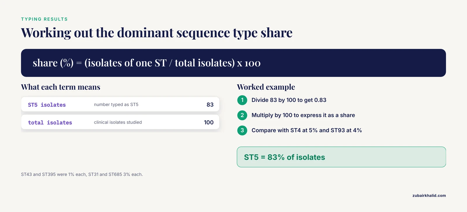
Multilocus sequence typing (MLST) is the reference method for genotyping C. neoformans isolates. MLST compares DNA sequences at multiple conserved loci to assign a sequence type (ST). In a study of 100 clinical isolates from AIDS patients with cryptococcal meningitis in southern China, seven sequence types were identified. ST5 was dominant at 83%, followed by ST4 (5%), ST93 (4%), ST31 (3%), ST685 (3%), and ST43 and ST395 (1% each) [2]. In a separate case report from Japan, an immunocompetent patient with CM was infected with C. neoformans var. grubii, sequence type 5, molecular type VNI [3]. The dominance of ST5 in Asia and its presence in immunocompetent hosts suggest that this lineage has a broad host range.
The polysaccharide capsule
The capsule is the defining virulence structure of C. neoformans. It is composed primarily of two high-molecular-weight polysaccharides: glucuronoxylomannan (GXM), which makes up about 90% of the capsule mass, and glucuronoxylomannogalactan (GXMGal). The capsule is anchored to the outer cell wall and forms a thick, gelatinous layer that can exceed the diameter of the cell body itself.
The capsule has multiple immune-evasive functions. It hinders recognition by host phagocytes, inhibits complement deposition, reduces the production of pro-inflammatory cytokines, and blocks the maturation of dendritic cells. These effects allow the fungus to survive and proliferate in the host [4]. The capsule also protects the yeast against desiccation and environmental stressors outside the host.
Capsule size is highly dynamic and regulated by environmental cues. Under carbon starvation, C. neoformans remodels its capsule and cell wall in ways that increase capsule porosity and density without changing the overall capsule diameter [5]. This remodeling is mediated by carbohydrate-active enzymes. Mutants lacking Cas1 and Cas3, enzymes that mediate O-acetylation of capsule polysaccharide, chitin synthases Chs1 through Chs8, hyaluronic acid synthase Cps1, and the β-glucan-specific glycosidase Kre64 all show altered capsule and cell wall responses to carbon limitation [5]. These findings indicate that the capsule is not a static structure but a metabolically active compartment that helps the fungus survive nutrient-poor conditions in the host.
The cell wall
The cell wall of C. neoformans is a layered structure composed of α-1,3-glucan, β-1,3-glucan, β-1,6-glucan, chitin, chitosan, and mannoproteins. The wall provides structural rigidity and integrity under diverse environmental conditions [4].
Chitin is an essential component of the cell wall. Chitin deacetylase genes encode enzymes that deacetylate chitin, converting it to chitosan. Chitosan is critical for cell wall integrity and is a major virulence determinant. Mutant strains deficient in chitosan are completely avirulent in a mouse model of pulmonary cryptococcosis [4]. Because chitosan is absent in the human host, the chitin deacetylase pathway is an attractive target for antifungal drug development [4].
The gene FKS1 encodes β-1,3-glucan synthase, the target of echinocandin drugs. Echinocandins are a class of antifungal drugs that inhibit β-1,3-glucan synthase. C. neoformans is intrinsically resistant to echinocandins, and FKS1 has long been considered essential. However, a genome-wide gene inactivation study using a method called CRISPR-insert showed that FKS1 is not essential. Targeted deletion of FKS1 produced mutants that completely lack β-1,3-glucan, and no compensatory synthase took over its function [6]. This finding overturns a long-standing assumption and has implications for understanding echinocandin resistance and for identifying new drug targets. The same study predicted 1,297 essential genes in C. neoformans and identified synthetic lethal dependencies of echinocandin resistance determinants, including the calcineurin and Cdc50-Apt1 pathways [6].
Titan cells
In the host lung, C. neoformans undergoes an unusual morphological transition from haploid yeast to large, highly polyploid cells called Titan cells [7]. Titan cells are 10 to 100 times the volume of normal yeast cells and have a thickened capsule and a highly cross-linked cell wall. They were first described in infected mouse lungs and were difficult to study because no reproducible in vitro induction system existed.
A reproducible in vitro system was developed using environmental stimuli consistent with the host lung. In vitro Titan cells exhibit all the properties of in vivo-generated Titan cells, including altered capsule, cell wall, size, high mother cell ploidy, and aneuploid progeny [7]. Bacterial peptidoglycan, which can be present in serum, was identified as a trigger for the shift in cell size and ploidy. Bronchial lavage fluid and E. coli co-culture also induce Titan cell formation [7].
Titan cells influence pathogenesis in several ways. They are more resistant to antifungal drugs, they alter the exposure of pathogen-associated molecular patterns (PAMPs) to host immune receptors, and their large size prevents phagocytosis by alveolar macrophages [7]. PAMPs are molecular signatures of microbes that are recognized by host immune receptors. Titan cells also produce aneuploid progeny, which may increase genetic diversity and adaptive potential within the host.
Virulence factors beyond the capsule
In addition to the capsule, C. neoformans produces several other virulence factors:
- Melanin: a dark pigment synthesized from catecholamines by the enzyme laccase. Melanin is deposited in the cell wall and protects the fungus against oxidative stress, reactive nitrogen species, and antifungal peptides.
- Urease: an enzyme that hydrolyzes urea to ammonia and carbon dioxide. Ammonia alkalinizes the local environment and damages host tissues. Urease also promotes dissemination from the lung to the brain.
- Phospholipase B: an enzyme that disrupts host cell membranes and facilitates tissue invasion.
- Extracellular enzymes: including proteases and nucleases that degrade host components.
These factors are used as markers of virulence in experimental studies. Riboflavin, also known as vitamin B2, has been shown to inhibit C. neoformans growth and to reduce capsule, melanin, and urease production in vitro, which makes it a candidate for antifungal therapy [8].
Clinical Disease
Epidemiology and risk factors
Cryptococcal meningitis remains most common in HIV-infected individuals [9]. In Sub-Saharan Africa, CM is a major contributor to mortality and morbidity in people living with HIV/AIDS, largely due to delays in diagnosis, limited access to antifungal treatment, and a lack of healthcare resources [10].
The population at risk has expanded to include patients with non-HIV immunosuppression. This group includes patients receiving glucocorticoid treatment, organ transplant recipients, and patients with hematological cancer [9]. In solid organ transplant recipients, cryptococcosis is the third most common invasive fungal infection [11]. In a series of kidney transplant patients, the median time from transplant to infection was 6 months, with a range of 3 to 9 months [11]. Cases have also been reported in patients treated with cladribine for hairy cell leukemia [9], in a kidney transplant recipient who recovered from COVID-19 [12], and in a patient with chronic renal failure on immunosuppressants who developed simultaneous CM and tuberculosis meningitis [13].
Pulmonary cryptococcosis
The lung is the portal of entry. Infection can be asymptomatic or present as pneumonia with cough, fever, chest pain, and dyspnea. Chest CT often reveals pulmonary nodules, which can be mistaken for malignancy. In a study of 90 patients with CM, 44.4% had pulmonary nodules on chest CT [14]. Patients with pulmonary nodules had higher CSF/serum albumin ratios, higher rates of CSF protein above 1000 mg/L, and CSF glucose below 2.5 mmol/L. They also had worse overall treatment response and more unfavorable clinical outcomes [14]. Multivariate analysis showed that immunocompromise and CSF glucose below 2.5 mmol/L were independent predictors of poor outcome in patients with pulmonary nodules, while CSF glucose below 2.5 mmol/L also predicted poor outcome in patients without nodules [14].
Cryptococcal meningitis
CM is the most severe manifestation of cryptococcosis. It usually presents as a subacute or chronic meningoencephalitis, but can occasionally be fulminant [1]. Common symptoms include headache, fever, and depressed level of consciousness [1]. In a study of 100 AIDS patients with CM in southern China, headache was the most common symptom, occurring in 85% of patients [2]. Other symptoms include nausea, vomiting, neck stiffness, photophobia, and focal neurological deficits.
The infection affects both the subarachnoid space and the brain parenchyma. A hallmark of CM in HIV patients is a paucity of inflammation in the CSF despite a large fungal burden [1]. CSF analysis typically shows a raised opening pressure, normal or mildly elevated white blood cell count, elevated protein, and low glucose. In immunocompetent patients, the inflammatory response is stronger and the presentation can be misleading. A 35-year-old immunocompetent woman with acute meningoencephalitis had CSF cytological and biochemical abnormalities and a positive India ink stain. Despite treatment with intravenous fluconazole, she died after 10 days [15].
CM can also cause severe complications. Massive cerebral edema resulting in brain death has been reported as a complication of C. neoformans meningitis [1]. In that case, the diagnosis was based on clinical neurological examinations and supported by the absence of cerebral blood flow on brain angiography [1].
Atypical presentations and diagnostic delay
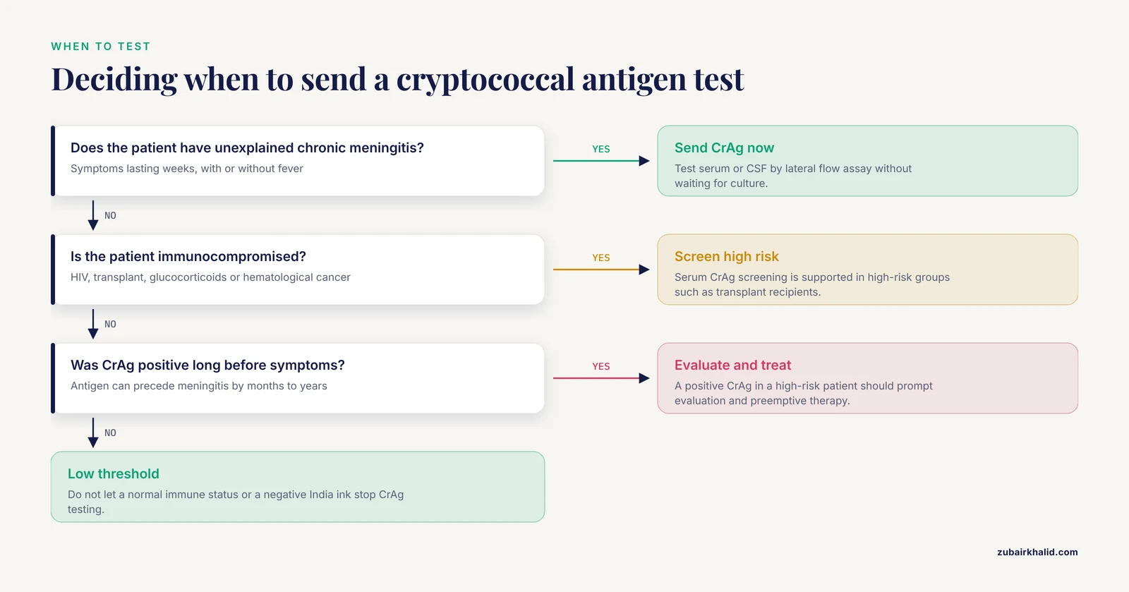
CM in non-HIV immunocompromised patients often has an atypical presentation that delays diagnosis. A fatal case of chronic CM progressing over 4 years was reported in a patient treated with cladribine for hairy cell leukemia [9]. Retrospective analysis showed that the cryptococcal antigen (CrAg) test was already positive 3 years before the CM diagnosis, when the patient first presented with pleocytosis [9]. The case demonstrates important pitfalls in HIV-negative patients, including atypical disease presentation and diagnostic challenges that resulted in a 3-year delay [9].
Immune reconstitution inflammatory syndrome
Immune reconstitution inflammatory syndrome (IRIS) is a paradoxical worsening of symptoms that occurs when the immune system recovers and mounts an inflammatory response against the fungus. It is well described in HIV patients starting antiretroviral therapy and can also occur in immunocompetent hosts during treatment. A young immunocompetent adult with CM developed neurological deterioration due to an IRIS-like phenomenon that required prolonged use of steroids [16]. He also suffered serious adverse effects from liposomal amphotericin B and flucytosine. He recovered after 16 months of treatment [16]. The case highlights the possible difficulties in managing CM in immunocompetent hosts and the need for new trial data and guidelines for this population [16].
Laboratory Identification
Specimen collection and transport
The diagnosis of cryptococcal meningitis relies on examination of cerebrospinal fluid (CSF). Lumbar puncture should be performed in all patients with suspected CM. CSF should be sent for India ink stain, CrAg testing, culture, and routine chemistry and cell count. Blood cultures and serum CrAg testing are also useful. In kidney transplant recipients, CrAg was detected in plasma up to 4 weeks before the diagnosis of meningitis, which supports the use of serum CrAg screening in high-risk populations [11].
India ink stain
The India ink preparation is the classic rapid diagnostic test for CM. A drop of CSF is mixed with a drop of India ink on a glass slide, covered with a coverslip, and examined under a light microscope at 400x magnification. The ink particles cannot penetrate the polysaccharide capsule, so the yeast cells appear as round, budding cells surrounded by a clear halo against a dark background. India ink revealed budding capsulated yeast cells in an HIV-positive patient with CM and renal involvement [17]. The test is rapid and inexpensive, but its sensitivity depends on the fungal burden and the skill of the microscopist. In early infection or in patients with few organisms, the sensitivity is low. A negative India ink does not exclude CM.
Cryptococcal antigen testing
The cryptococcal antigen (CrAg) test detects GXM polysaccharide in serum, plasma, or CSF. The lateral flow assay (LFA) is a point-of-care test that gives results within 10 minutes and requires no specialized equipment [18]. In a study of 37 CSF samples from immunocompromised patients with suspected meningitis, 2 samples (5.4%) were strongly positive by LFA, and the same 2 samples grew C. neoformans in culture after 3 to 5 days of incubation [18]. The sensitivity, specificity, positive predictive value, and negative predictive value of the LFA were all 100% in that study [18]. The LFA is particularly valuable in remote and resource-limited settings because it is simple, rapid, and stable at room temperature [18].
Culture
CSF, blood, and respiratory specimens can be cultured for C. neoformans. The fungus grows on Sabouraud Dextrose Agar (SDA) and on Bird Seed Agar, also called niger seed agar or Staib agar [18]. On SDA, colonies appear creamy, mucoid, and yeast-like within 3 to 5 days. On Bird Seed Agar, C. neoformans produces brown to black colonies because the fungus oxidizes caffeic acid in the medium to melanin. This characteristic helps distinguish C. neoformans from other encapsulated yeasts. C. neoformans grows well at 37°C, a trait that most environmental Cryptococcus species lack, although growth at 37°C alone is not enough to identify it.
Molecular methods
Multiplex PCR assays can identify Cryptococcus species directly from CSF and can detect unanticipated pathogens in patients with mixed or atypical presentations [3]. In one case, a multiplex PCR assay of CSF identified Cryptococcus species in an immunocompetent patient who was initially treated for tuberculous meningitis and varicella-zoster virus vasculopathy [3]. The multiplex PCR assay, which can identify multiple pathogens simultaneously, enabled swift treatment and a favorable clinical outcome [3]. MLST is used for epidemiological typing. The isolate in that case was identified as C. neoformans var. grubii, sequence type 5, molecular type VNI [3].
MALDI-TOF mass spectrometry
Matrix-assisted laser desorption/ionization time-of-flight mass spectrometry (MALDI-TOF MS) is a rapid and accurate method for identifying yeast isolates from culture. In a study of 100 clinical isolates from AIDS patients with CM, all isolates were identified by MALDI-TOF MS and genotyped by MLST [2]. Although simplified microbial identification techniques such as mass spectrometry have been developed, molecular biological assays remain essential for the accurate identification of infectious strains [3].
Antifungal susceptibility testing
Antifungal susceptibility testing is performed by broth microdilution following CLSI guidelines. In a study of 100 C. neoformans isolates from southern China, susceptibility rates were 98.9% for 5-flucytosine, 71.9% for amphotericin B, 82.3% for fluconazole, 59.4% for itraconazole, and 86.8% for voriconazole [2]. These data show that resistance to amphotericin B and azoles exists and that susceptibility testing is clinically valuable, especially in patients who fail to respond to standard therapy.
Treatment
Induction, consolidation, and maintenance
The standard treatment for CM has three phases. Induction therapy uses amphotericin B (liposomal or deoxycholate) plus flucytosine for at least 2 weeks. Consolidation therapy uses fluconazole for 8 weeks. Maintenance therapy with fluconazole continues for 6 to 12 months or until immune recovery. In a series of kidney transplant recipients, induction with liposomal amphotericin B and flucytosine lasted a median of 10 days, with a range of 7 to 14 days, followed by fluconazole maintenance [11]. Acute kidney injury from liposomal amphotericin B occurred in one of three patients, and all patients had tacrolimus overdose after starting fluconazole due to drug-drug interactions between fluconazole and calcineurin inhibitors [11]. Close monitoring of renal function and drug levels is essential during treatment. In an HIV-positive patient with CM and renal involvement, fluconazole maintenance was discontinued when the CD4+ T cell count rose above 100/mm3 after 12 months of therapy [17].
Drug tolerance and resistance
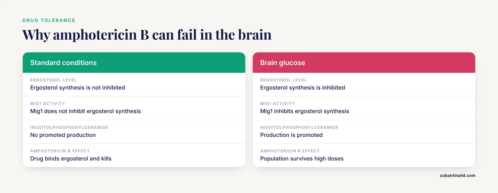
Amphotericin B is the most potent fungicidal drug for CM, but its efficacy can be limited by fungal tolerance. Antibiotic tolerance is the ability of a susceptible population to survive high doses of cidal drugs. Brain glucose induces tolerance of C. neoformans to amphotericin B in mouse brain tissue and in patient cerebrospinal fluid [19]. This tolerance is mediated by the fungal glucose repression activator Mig1, which inhibits the synthesis of ergosterol, the main sterol in fungal cell membranes and the target of amphotericin B. Mig1 also promotes the production of inositolphosphorylceramide, a fungal sphingolipid that competes with amphotericin B for ergosterol [19]. Combining amphotericin B with aureobasidin A, an inhibitor of fungal-specific inositolphosphorylceramide synthase, showed better efficacy against cryptococcal meningitis in mice than clinically recommended therapies [19]. This combination approach is a promising strategy for overcoming drug tolerance.
Emerging therapies
Riboflavin (vitamin B2) has antifungal activity against C. neoformans. It inhibits growth and biofilm formation, damages the cell membrane, compromises cell wall integrity, and promotes accumulation of intracellular reactive oxygen species [8]. Riboflavin also reduces virulence by decreasing capsule, melanin, and urease production [8]. In mouse models of pulmonary cryptococcosis and meningitis, riboflavin treatment significantly reduced fungal burden in multiple organs, reduced lung and brain damage, and decreased plasma levels of interferon-gamma, tumor necrosis factor-alpha, and interleukin-4 in the early stage of infection [8]. These findings suggest that riboflavin could be developed as an affordable adjunctive therapy, particularly in resource-limited settings.
Cryptococcus neoformans vs Cryptococcus gattii
The two pathogenic Cryptococcus species differ in ecology, epidemiology, and clinical presentation. The table below summarizes the key differences.
| Feature | Cryptococcus neoformans | Cryptococcus gattii |
|---|---|---|
| , - | , - | , - |
| Ecology | Worldwide, associated with bird droppings, especially pigeon excreta | Primarily tropical and subtropical, associated with eucalyptus and other hardwood trees |
| Molecular types | VNI, VNII (var. grubii), VNIII, VNIV (var. neoformans) | VGI, VGII, VGIII, VGIV |
| Serotypes | A, D, and AD hybrids | B and C |
| Typical host | Immunocompromised (HIV, transplant, hematologic malignancy) | Immunocompetent individuals, though immunocompromised hosts are also affected |
| Typical clinical form | Meningitis, pulmonary disease | Pulmonary cryptococcomas, meningitis, skin and bone lesions |
| Geographic association | Global | Pacific Northwest (VGII), Australia, South America, Asia, Africa |
| Treatment | Amphotericin B plus flucytosine induction, fluconazole consolidation | Similar, but prolonged therapy often needed, especially for cryptococcomas |
The table reflects standard textbook knowledge. The practical distinction matters because C. gattii infection should be suspected in immunocompetent patients with pulmonary cryptococcomas and a history of travel to endemic regions. Species identification can be performed with culture on canavanine-glycine-bromothymol blue (CGB) agar, where C. gattii turns the medium blue, or by molecular methods.
Quick Review
- Cryptococcus neoformans is an encapsulated basidiomycetous yeast that causes pulmonary cryptococcosis and cryptococcal meningitis.
- The polysaccharide capsule is the primary virulence factor. It prevents immune recognition and is detected by India ink stain and CrAg tests.
- Titan cells are large polyploid forms that form in the lung and increase drug resistance and immune evasion.
- Chitosan, produced by chitin deacetylases such as Cda1, is essential for cell wall integrity and virulence.
- Laboratory diagnosis combines India ink, CrAg LFA, culture on SDA and Bird Seed Agar, and molecular methods such as multiplex PCR and MLST.
- Standard treatment is amphotericin B plus flucytosine induction, followed by fluconazole consolidation and maintenance.
- Brain glucose induces amphotericin B tolerance through the Mig1 pathway. Aureobasidin A can reverse this tolerance in experimental models.
Common Mistakes and Limitations
- India ink is not a screening test. Its sensitivity depends on fungal burden and operator skill. A negative India ink does not exclude CM. The CrAg LFA is more sensitive and should be performed on all suspected cases.
- Culture can take 3 to 5 days. Do not delay treatment while waiting for culture results. The CrAg LFA gives results in 10 minutes and should be used for rapid diagnosis [18].
- A normal CSF cell count does not exclude CM. In HIV patients, the CSF often shows a paucity of inflammation despite a high fungal burden [1].
- Cryptococcal antigen can be positive months to years before clinical meningitis develops. A positive CrAg in a high-risk patient should prompt evaluation and preemptive therapy.
- Fluconazole interacts with calcineurin inhibitors. In kidney transplant recipients, fluconazole maintenance caused tacrolimus overdose in all three patients in one series [11]. Drug monitoring is essential.
- Immunocompetent patients can die from CM. Do not assume a normal immune status predicts a mild course.
- Amphotericin B tolerance is induced by brain glucose. Standard susceptibility testing may show that the isolate is susceptible, yet the drug can fail in the CNS because of this metabolic effect [19].
- The diagnosis can be delayed in non-HIV patients because the presentation is atypical. Clinicians should maintain a low threshold for CrAg testing in any patient with unexplained chronic meningitis [9].
- Individual patient management decisions require clinical correlation and consultation with an infectious disease specialist. Laboratory results must always be interpreted in the context of the full clinical picture.
Related Guides
- Inoculum Size and Culture Age: Reviews culture age and inoculum effects on microbiology results
Frequently Asked Questions
What is Cryptococcus neoformans?
Cryptococcus neoformans is an encapsulated yeast that causes cryptococcosis, a fungal infection that starts in the lungs and can spread to the brain to cause meningitis. It is a leading cause of fungal meningitis worldwide and is especially dangerous in people with weakened immune systems [1].
How is Cryptococcus neoformans meningitis diagnosed?
The diagnosis is made by examining cerebrospinal fluid with India ink stain, detecting cryptococcal antigen with a lateral flow assay, and growing the fungus in culture on Sabouraud Dextrose Agar or Bird Seed Agar [18]. Molecular methods such as multiplex PCR can also identify the pathogen directly from CSF [3].
What does Cryptococcus neoformans look like on India ink stain?
On India ink stain, C. neoformans appears as round, budding yeast cells surrounded by a clear halo. The halo is the polysaccharide capsule, which excludes the ink particles.
How is Cryptococcus neoformans treated?
The standard treatment is induction with amphotericin B plus flucytosine, followed by consolidation and maintenance with fluconazole [11]. Newer approaches include combining amphotericin B with aureobasidin A to overcome brain glucose-induced drug tolerance [19].
What is the difference between Cryptococcus neoformans and Cryptococcus gattii?
C. neoformans is found worldwide in bird droppings and mainly infects immunocompromised people. C. gattii is found in tropical and subtropical trees and more often infects immunocompetent people. The two species are distinguished by molecular typing and by differences in ecology and clinical presentation.
Can immunocompetent people get cryptococcal meningitis?
Yes. Cryptococcal meningitis is increasingly reported in immunocompetent adults [16], [3], [15]. The presentation can be misleading and the prognosis is severe, so a low threshold for CrAg testing is warranted.
Sources
- Massive cerebral edema resulting in brain death as a complication of Cryptococcus neoformans meningitis
- Sequence Types of Cryptococcus neoformans and Their Associations with Clinical Characteristics and Outcomes of AIDS Patients with Cryptococcal Meningitis in Southern China.
- Early diagnosis of Cryptococcus neoformans var. grubii meningitis using multiplex PCR assay in an immunocompetent patient.
- Cryptococcus neoformans Cda1 and Its Chitin Deacetylase Activity Are Required for Fungal Pathogenesis
- Carbon starvation induces coincident capsule and cell wall remodeling in Cryptococcus neoformans.
- Therapeutic vulnerabilities of the drug-resistant fungal meningitis pathogen Cryptococcus neoformans
- The Cryptococcus neoformans Titan cell is an inducible and regulated morphotype underlying pathogenesis
- Riboflavin inhibits growth and reduces virulence of Cryptococcus neoformans in vitro by membrane disruption and excessive accumulation of reactive oxygen species and exhibits efficacy against pulmonary cryptococcosis and meningitis
- A fatal case of chronic Cryptococcus neoformans meningitis progressing over a 4-year period following cladribine therapy
- Systematic Review of Cryptococcus neoformans Seroprevalence, Antifungal Susceptibility, and Pathogenesis in Patients With HIV/AIDS on Combination Antiretroviral Therapy in Abuja, Nigeria
- Cryptococcus neoformans meningitis in kidney transplant recipients: A diagnostic and therapeutic challenge
- A case report of Cryptococcus neoformans meningitis after recovery from COVID-19 infection in a kidney transplant recipient patient
- Meningitis Simultaneuously Due to Cryptococcus neoformans and Mycobacterium tuberculosis in a Inmunosupressed Patient
- Comparison of Clinical Features and Prognostic Factors of Cryptococcal Meningitis Caused by Cryptococcus neoformans in Patients With and Without Pulmonary Nodules
- Cryptococcus neoformans meningitis in an immunocompetent adult: A case report
- Neurological worsening during treatment of an immunocompetent adult with Cryptococcus neoformans meningitis
- Cryptococcus neoformans Meningitis With Renal Involvement in an HIV-Positive Patient: A Case Report
- Early Detection Of Cryptococcus neoformans in Suspected Cases of Fungal Meningitis Using Point of Care Laboratory Testing.
- Brain glucose induces tolerance of Cryptococcus neoformans to amphotericin B during meningitis