Archaea vs Bacteria: Key Differences
By Dr. Zubair Khalid, DVM, MS, PhD ·
Archaea and bacteria are two distinct domains of single-celled prokaryotic microorganisms that lack a membrane-bound nucleus but differ fundamentally in cell wall chemistry, membrane lipid architecture, and the molecular machinery that reads their genes. The distinction matters in clinical and environmental microbiology because standard bacterial stains, culture media, antibiotics, and molecular assays do not always perform as expected when archaea are present.
For most of the history of microbiology, prokaryotes were treated as a single group. The realization that archaea are a separate domain came from comparisons of 16S ribosomal RNA gene sequences, and it has since reshaped how we classify, identify, and study microorganisms. In the laboratory, the differences between archaea and bacteria determine which antibiotics work, which stains are informative, which culture conditions are needed, and which molecular markers can be trusted.
What Are Archaea and Bacteria?
The three-domain system and its history
All cellular life falls into one of three domains: Bacteria, Archaea, and Eukarya. This classification rests primarily on phylogenetic analysis of the 16S ribosomal RNA (rRNA) gene, which is present in every prokaryote, is functionally conserved, and contains enough variable regions to distinguish closely related organisms. The All-Species Living Tree project has assembled a 16S rRNA-based reference tree from all sequenced type strains, providing a common framework for naming and comparing bacteria and archaea [1].
Early applications of 16S rRNA sequencing to veterinary pathogens demonstrated the power of the approach. The intestinal spirochetes of pigs, then classified in the genus Serpulina, were difficult to separate by biochemical tests alone. Sequence analysis of the 16S rRNA gene resolved them into distinct species and clarified their phylogenetic relationships [2][3]. These studies were among the first to show that 16S rRNA phylogeny could outperform biochemical classification for fastidious anaerobes [3].
Archaea were initially discovered in extreme environments, and for decades they were regarded as oddities. The development of culture-independent methods, particularly 16S rRNA gene amplicon sequencing and shotgun metagenomics, showed that archaea are abundant in soil, seawater, sediments, and the human body [4][5]. The term prokaryote is still used to describe both bacteria and archaea, but it describes the absence of a nucleus, not a close evolutionary relationship.
Why the distinction matters in the laboratory
Most clinical and environmental laboratories were designed around bacterial cell biology. Standard media, Gram staining, biochemical panels, and antibiotic susceptibility tests were optimized for bacteria. Archaea often fail in these workflows. They may not grow on standard media, they do not have peptidoglycan to be detected by the Gram stain, and they are not inhibited by cell wall antibiotics [6][7]. A laboratory that cannot distinguish archaea from bacteria will misidentify organisms and miss entire branches of a microbial community.
The distinction also matters for education. Archaea are rarely included in high school and undergraduate microbiology curricula, which leaves students with the impression that prokaryotes are all bacteria. A modified Kirby-Bauer disk diffusion test that pairs the bacterium Escherichia coli with the archaeon Haloferax volcanii gives students a direct, hands-on demonstration of the differences between the two domains [6].
Key Differences Between Archaea and Bacteria
Cell wall composition
The most reliable biochemical difference between the two domains is the cell wall.
Bacteria have a cell wall made of peptidoglycan, also called murein. Peptidoglycan is a polymer of alternating N-acetylglucosamine and N-acetylmuramic acid residues linked by β-1,4 glycosidic bonds, with short peptide cross-links that contain D-amino acids [8][7]. This mesh-like sacculus surrounds the cytoplasmic membrane and gives the cell its shape and osmotic protection. Because human cells have no peptidoglycan, the bacterial cell wall is one of the best targets for antibacterial therapy [9].
Archaea never make peptidoglycan [7]. Some archaea, particularly methanogens in the orders Methanobacteriales and Methanopyrales, make a structural analogue called pseudomurein, also called pseudopeptidoglycan [8][7]. Pseudomurein differs from murein in three ways. First, it contains the archaeal sugar N-acetyltalosaminuronic acid instead of N-acetylmuramic acid. Second, the glycosidic bonds are β-1,3 rather than β-1,4. Third, the peptide cross-links contain only L-amino acids, never D-amino acids [7].
The enzymes that build pseudomurein, called pMurE peptide ligases, are structurally homologous to the bacterial MurE and MurF murein peptide ligases. Crystal structures of pMurE from Methanothermus fervidus and Methanothermobacter thermautotrophicus show the same three-domain architecture as the bacterial enzymes, with conserved ATP-binding pockets [7]. This homology indicates that the two pathways share an ancient common ancestor, even though the final products are chemically distinct.
Other archaea have no pseudomurein at all. Many have a paracrystalline protein surface layer, or S-layer, as the outermost cell envelope. Some produce other polysaccharides. Archaea also produce functional amyloids, which are cross-β-sheet protein fibers that stabilize cell envelopes and biofilms. The archaeon Methanosaeta produces an amyloid sheath called MspA that forms its cell wall, and this amyloid can be detected within intact cells using infrared nanospectroscopy (AFM-IR) [10].
The practical consequence is that any antibiotic that blocks peptidoglycan synthesis, such as penicillin or vancomycin, is inactive against archaea [6]. Conversely, the bacterial cell wall remains an excellent target for antibacterial drugs precisely because it is absent from both archaea and human cells [9].
Cell membrane lipids
Bacteria and eukaryotes build their membranes from fatty acids linked to glycerol by ester bonds. Archaea instead link isoprenoid chains to glycerol by ether bonds. Archaeal membranes can also form a monolayer, in which a single long isoprenoid tetraether spans the entire membrane instead of two separate leaflets. This ether-linked architecture is more resistant to heat, acid, and salt, which helps explain why archaea dominate in extreme environments. The chemical difference is absolute: no bacterium makes ether-linked isoprenoid membranes, and no archaeon makes ester-linked fatty acid membranes.
Size and morphology
Archaea and bacteria overlap in size, and no single size cutoff separates them. Most cultured species in both domains are rods, cocci, spirals, or pleomorphic cells roughly 0.5 to 5 micrometers in the longest dimension. Cell size is nonetheless an ecologically meaningful trait. In a comparative analysis of bacterial and archaeal trait dimensions, cell size was identified as one of three main axes that influence diffusive nutrient uptake and the relative cost of building a cell wall [11]. Smaller cells take up nutrients more efficiently per unit volume but pay a higher relative wall cost, while larger cells have lower diffusive uptake efficiency [11]. Morphology is not diagnostic either: archaea can be rods, cocci, flattened squares, or filaments, and bacteria can be nearly as varied.
Genome organization and gene expression
Both archaea and bacteria usually have a single circular chromosome, but the way they express genes is different.
Bacteria use a single RNA polymerase whose promoter recognition depends on sigma (σ) factors. Archaea use a more complex transcription machinery that includes TATA-box binding protein (TBP) and TFIIB-related factors, which are close homologs of the eukaryotic transcription apparatus [12]. This is one of the strongest molecular arguments that archaea and eukaryotes share a more recent common ancestor than archaea and bacteria do.
Genome size varies widely in both domains and is associated with versatility. In the trait analysis by Westoby and colleagues, genome size correlated with signal transduction capacity, meaning organisms with larger genomes can sense and respond to more environmental signals [11]. Archaeal genomes tend to be smaller on average than bacterial genomes, but the ranges overlap broadly.
Repetitive extragenic palindromic (REP) sequences, first discovered in Escherichia coli, are also present in archaea. A comprehensive analysis using the web tool RepRanger identified more than 4,000 REPs in the E. coli MG1655 genome and found that about 52% of its 157 small noncoding RNAs contain REPs [13]. These elements appear to contribute to adaptation by rewiring metabolic pathways, and similar elements exist in archaeal genomes, though their functions are less well characterized [13].
Reproduction
Both archaea and bacteria reproduce primarily by binary fission, in which the cell replicates its chromosome and divides into two genetically identical daughter cells. Some archaea also reproduce by budding or fragmentation. Neither domain has mitosis or meiosis.
The evolutionary origin of cell division is addressed in a recent hypothesis by Yoshimura and Seki, who propose that bacteria and archaea emerged from a non-free-living pool of single operon-type genomes at an ancient submarine alkaline vent [12]. In their model, the differentiation into Bacteria and Archaea was driven by which transcription system, σ or TBP, stabilized the fused DNA operons. Both lineages independently acquired DNA replication and repair, then recruited lipids to form coacervates and eventually bilayer membranes, leading to proto-cell division [12]. This is a hypothesis, not settled fact, but it illustrates that the division machinery in the two domains, while functionally similar, evolved along parallel paths.
Horizontal gene transfer is common in both domains and can cross the domain boundary. Haloarchaea, for example, acquired Rieske/cytochrome b complexes, which are proton-pumping quinol oxidases, from bacteria via interdomain lateral gene transfer. The phylogeny of these complexes otherwise follows 16S rRNA phylogeny closely, indicating that chemiosmotic coupling was already present in the last universal common ancestor of archaea and bacteria [14].
Metabolism and bioenergetics
Many metabolic pathways are shared between archaea and bacteria, including glycolysis, the citric acid cycle, and oxidative phosphorylation. The Rieske/cytochrome b complex is present in most bacteria and most archaea, and its phylogeny mirrors 16S rRNA phylogeny [14].
The most famous archaeal metabolism is methanogenesis, the production of methane from carbon dioxide, hydrogen, acetate, or methyl compounds. Methanogens are strictly anaerobic archaea and are the only organisms that produce methane as a metabolic end product. They are abundant in the gut of ruminants and in anaerobic sediments. The methanogen cell wall, when present, is pseudomurein [7].
Bacteria do not perform methanogenesis. Instead, they dominate in processes such as aerobic respiration, fermentation, denitrification, and sulfate reduction. Some bacteria, such as the spirochetes in the genus Serpulina, are fastidious anaerobes that require specialized media and are best identified by 16S rRNA sequencing [2][3].
Archaea vs Bacteria vs Eukarya: The Three Domains
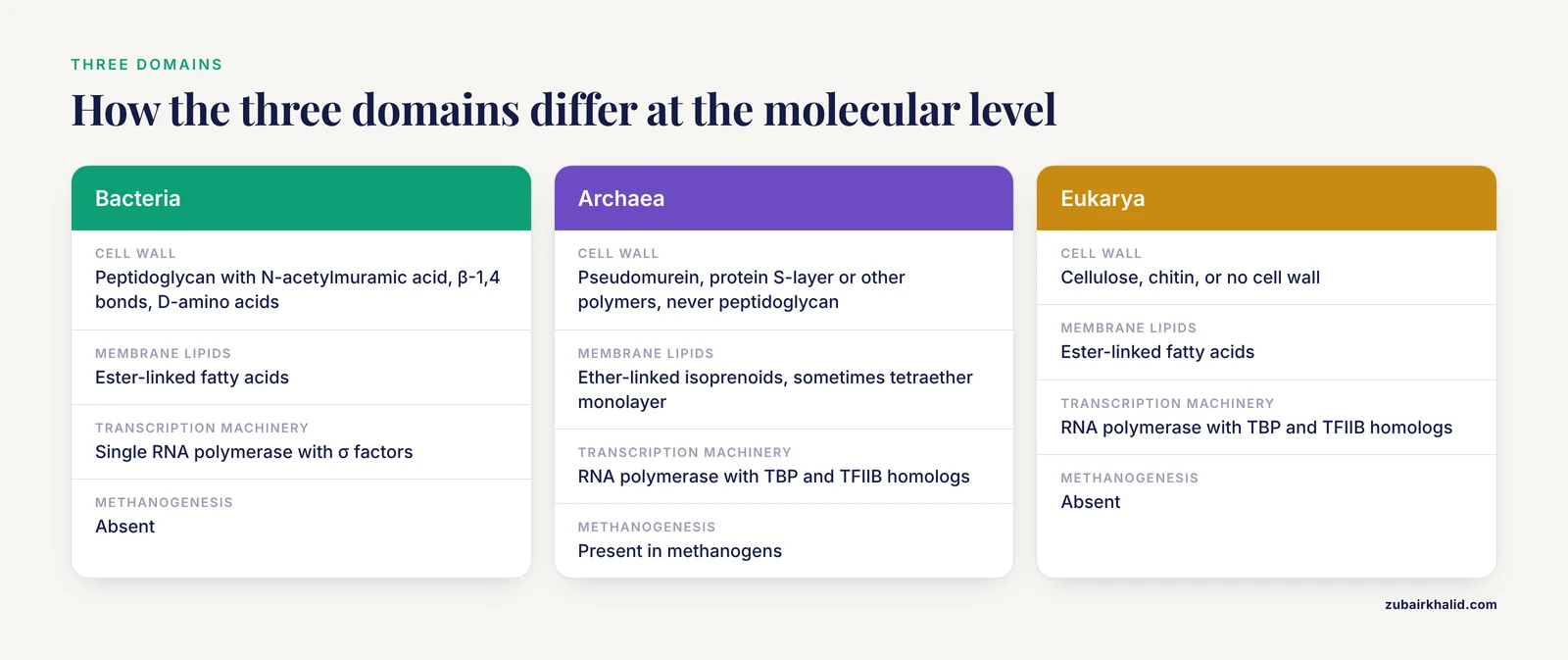
A three-way comparison is useful because archaea often look like bacteria under the microscope but behave like eukaryotes at the molecular level.
- Cell type: Bacteria and archaea are prokaryotes, meaning they lack a nucleus and membrane-bound organelles. Eukarya have a true nucleus, mitochondria, and an endomembrane system.
- Cell wall: Bacteria have peptidoglycan. Archaea have pseudomurein, protein S-layers, or other polymers, but never peptidoglycan [8][7]. Eukarya have cellulose, chitin, or no cell wall.
- Membrane lipids: Bacteria and eukarya use ester-linked fatty acids. Archaea use ether-linked isoprenoids.
- Transcription: Bacteria use a single RNA polymerase with σ factors. Archaea use RNA polymerase with TBP and TFIIB homologs, similar to eukaryotes [12].
- Ribosomes: All three domains have ribosomes, but their sizes and antibiotic sensitivities differ. The archaeal ribosome is more similar to the eukaryotic ribosome in structure, yet it is sensitive to some antibacterial drugs that target the bacterial ribosome, which makes antibiotic susceptibility testing a useful teaching tool [6].
A Venn diagram of the two prokaryotic domains would place shared features, such as prokaryotic cell organization, 16S rRNA genes, and binary fission, in the overlap. Peptidoglycan, ester-linked lipids, and σ-factor transcription would sit on the bacterial side. Pseudomurein, ether-linked lipids, and TBP-based transcription would sit on the archaeal side. The practical takeaway is that archaea are not "weird bacteria." They are a third form of life with a mosaic of bacterial, eukaryotic, and unique features.
How Archaea and Bacteria Are Identified in the Laboratory
16S rRNA gene sequencing and phylogenetic analysis
The 16S rRNA gene is the standard marker for identifying and classifying prokaryotes. It is universal, functionally conserved, and long enough to provide phylogenetic signal. The All-Species Living Tree project has used 16S rRNA sequences to build a reference tree of all sequenced type strains [1]. Studies of Serpulina species in pigs showed that 16S rRNA phylogeny could distinguish species that were nearly identical by biochemical typing [2][3].
Traditional short amplicons, typically 250 base pairs of the 16S rRNA gene, often lack enough information to resolve closely related species. A cost-effective alternative is long-read amplicon sequencing of the entire 16S-ITS-23S rRNA operon using PacBio technology. This method reduces read error to about 0.18% and produces near full-length operon sequences that allow substantially more confident phylogenies [15]. It also captures the internal transcribed spacer, which is useful for species-level discrimination.
A simpler bioinformatic approach is BLASTN, which compares a query 16S rRNA sequence against a reference database to classify an uncultured organism to the species level [16]. This is the method most routine laboratories use for identification.
One important caveat: a single cell can carry multiple copies of the 16S rRNA gene that differ slightly from each other. This intragenomic heterogeneity can cause overestimation of prokaryotic diversity when amplicon sequencing is used, because each variant is counted as a separate operational taxonomic unit [17]. Archaea are not exempt from this problem, and the effect can be large enough to distort community comparisons.
Culture-based approaches and antibiotic susceptibility
Most archaea are difficult to culture because they require extreme conditions. A notable exception is Haloferax volcanii, a halophilic archaeon that grows aerobically at ambient temperature in high-salt media. The high salt concentration prevents contamination by bacteria and fungi, making it safe and inexpensive for teaching laboratories [6].
The Kirby-Bauer disk diffusion test, normally used for bacteria, can be adapted to compare archaea and bacteria. In one published exercise, H. volcanii and Escherichia coli were tested against antibiotics that target the peptidoglycan cell wall or the ribosome. Bacteria were inhibited by cell wall antibiotics, while the archaeon was not, which directly demonstrates the absence of peptidoglycan in archaea [6]. Ribosome-targeting antibiotics produced a more complex pattern, reflecting the structural similarity between archaeal and eukaryotic ribosomes [6]. Two versions of the exercise exist: one for undergraduate laboratories and a simplified version limited to H. volcanii for high school classrooms [6].
DNA extraction and metagenomics
DNA extraction is the first step in any molecular workflow, and the choice of protocol strongly affects whether archaea are detected at all. In a reference soil study, different extraction kits and buffers produced wide differences in total DNA yield, and the relative recovery of bacterial, archaeal, and fungal fingerprints changed with the protocol [18]. The Macherey-Nagel SoilExtract II kit with buffer SL1 and enhancer solution gave the highest yields, but the enhancer also increased co-extracted contaminants [18]. Repeated extractions increased total DNA but changed the apparent diversity of archaea and fungi by the fifth extraction [18].
Shotgun metagenomics detects archaea more reliably than 16S amplicon sequencing alone. In a study of water hyacinth vermicompost, 16S rRNA amplicon data were dominated by bacteria, with more than 97% of reads assigned to bacterial phyla, while shotgun metagenomics revealed archaea and fungi that were underrepresented in the amplicon dataset [5]. For any clinical or environmental sample, a negative archaeal result by amplicon sequencing should be interpreted with caution.
Examples of Archaea and Bacteria
Representative bacteria
- Escherichia coli: a Gram-negative rod in the family Enterobacteriaceae, used as a model organism in the Kirby-Bauer antibiotic susceptibility test [6].
- Staphylococcus and Streptococcus: Gram-positive cocci that dominate the gut of cesarean-section-delivered infants in the first days of life [4].
- Bifidobacterium: a genus of Gram-positive anaerobes that dominate the gut of vaginally delivered infants [4].
- Serpulina species: fastidious anaerobic spirochetes that cause intestinal disease in pigs and were among the first veterinary pathogens classified by 16S rRNA sequencing [2][3].
- Pseudomonas: a genus of environmental Gram-negative rods that produce functional amyloid fibers called Fap [10].
Representative archaea
- Haloferax volcanii: a halophilic archaeon that grows in high-salt media and is used as a model organism in teaching laboratories [6].
- Methanosaeta: a methanogenic archaeon that produces the MspA amyloid sheath and is abundant in anaerobic digesters [10].
- Methanothermus fervidus and Methanothermobacter thermautotrophicus: hyperthermophilic methanogens whose pseudomurein peptide ligases (pMurE) have been studied by X-ray crystallography [7].
- Methanobrevibacter: a genus of methanogens that colonizes the human gut [4].
Summary Table: Archaea vs Bacteria
| Feature | Bacteria | Archaea |
|---|---|---|
| , - | , - | , - |
| Domain | Bacteria | Archaea |
| Cell type | Prokaryotic | Prokaryotic |
| Nucleus | Absent | Absent |
| Cell wall | Peptidoglycan (murein) with N-acetylmuramic acid, β-1,4 bonds, D-amino acids [8][7] | No peptidoglycan. Pseudomurein with N-acetyltalosaminuronic acid, β-1,3 bonds, L-amino acids, or protein S-layer [8][7] |
| Membrane lipids | Ester-linked fatty acids | Ether-linked isoprenoids, sometimes tetraether monolayer |
| Transcription machinery | Single RNA polymerase with σ factors [12] | RNA polymerase with TBP and TFIIB homologs [12] |
| Methanogenesis | Absent | Present in methanogens |
| 16S rRNA gene | Present, multiple copies with possible heterogeneity [17] | Present, multiple copies with possible heterogeneity [17] |
| Antibiotic susceptibility | Inhibited by cell wall antibiotics such as penicillin and vancomycin [6] | Not inhibited by cell wall antibiotics [6] |
| Habitat | Ubiquitous | Ubiquitous, including extreme environments and the human gut [4] |
| Examples | E. coli, Staphylococcus, Bifidobacterium [6][4] | Haloferax volcanii, Methanosaeta, Methanobrevibacter [6][10][4] |
Clinical and Ecological Relevance
The human microbiome
Archaea are a normal part of the human gut microbiome. In a study of infants followed through the first year of life, sequences from bacteria, archaea, and microeukaryotes were all detectable in meconium, the first stool [4]. The composition of the gut community changed over time, and the mode of delivery had a measurable effect. Cesarean-section-delivered infants experienced a delay in colonization and succession across all three domains of life, with increased relative abundances of Streptococcus and Staphylococcus and decreased Bifidobacterium in the first days of life [4]. Archaea such as Methanobrevibacter colonized the gut alongside bacteria, which shows that archaea are not restricted to extreme environments [4].
Environmental and agricultural systems
Archaea and bacteria coexist in virtually every ecosystem, and their relative abundance depends on the substrate and conditions. In vermicomposting systems fed with water hyacinth biomass, bacterial communities dominated, with Proteobacteria, Firmicutes, Actinobacteria, and Bacteroidota as the core phyla [5]. Substrate pretreatment shaped the community: composted biomass was enriched in lignin degradation and carbon cycling pathways, dry biomass showed methanogenesis and fermentation signatures, and burnt biomass was associated with nitrogen fixation and sulfur metabolism [5]. Methanogenic archaea such as Methanosaeta are key players in anaerobic digestion, where they produce methane and stabilize the system [10].
Common Mistakes and Limitations
- Calling archaea "extremophilic bacteria." Many archaea are mesophiles that live in soil, seawater, and the human gut [4]. Extremophiles exist in both domains.
- Assuming Gram stain results have the same meaning across domains. The Gram stain detects peptidoglycan thickness in bacteria. Archaea lack peptidoglycan, so a Gram result does not have the same structural meaning [7].
- Treating 16S rRNA amplicon data as a complete census. Intragenomic heterogeneity inflates diversity estimates [17], and 16S primers can underrepresent archaea [5].
- Assuming antibiotics that work on bacteria will work on archaea. Cell wall synthesis inhibitors are inactive against archaea [6].
- Assuming archaea are always hard to culture. Halophiles like H. volcanii grow easily in high-salt media at room temperature [6].
- Overinterpreting evolutionary hypotheses. The crystallization model of archaeal and bacterial origins is a proposal, not an established fact [12].
- Ignoring DNA extraction bias. The choice of extraction kit can determine whether archaea are detected in a sample [18].
- Individual clinical isolates should be interpreted by a qualified clinical microbiologist or infectious disease physician, especially when unusual organisms are encountered.
Quick Review
- Archaea and bacteria are both prokaryotes, but they are separate domains of life.
- Bacteria have peptidoglycan cell walls; archaea have pseudomurein, S-layers, or other polymers, never peptidoglycan [8][7].
- Archaeal membranes use ether-linked isoprenoid lipids, while bacterial membranes use ester-linked fatty acids.
- Archaeal transcription uses TBP and TFIIB homologs, making it more similar to eukaryotes than to bacteria [12].
- Cell wall antibiotics such as penicillin do not inhibit archaea [6].
- The 16S rRNA gene is the standard identification marker, but intragenomic heterogeneity and primer bias can distort results [15][17].
- Archaea are common in the human gut and environment, not just in extreme habitats [4].
How to Choose 16S rRNA Primers That Detect Archaea
Most "universal" 16S primers were designed from bacterial reference sequences and carry mismatches to archaeal templates, so archaea are often underrepresented in amplicon data [5]. To capture both domains, use primers that anneal to conserved regions shared by bacteria and archaea. The V4 pair 515F (GTGYCAGCMGCCGCGGTAA) and 806R (GGACTACNVGGGTWTCTAAT) amplifies both domains with an expected product of about 253 bp. The V3-V4 pair 341F (CCTACGGGNGGCWGCAG) and 805R (GACTACHVGGGTATCTAATCC) gives about 460 bp.
Worked example: amplify mixed bacterial-archaeal DNA with 515F/806R. Use 35 cycles of 95°C 30 s, 55°C 45 s, 72°C 45 s. Run 5 µL on a 1.5% agarose gel. A single band near 250 bp indicates successful amplification. No band suggests inhibition or primer-template mismatch; dilute the DNA or switch to archaea-specific primers such as Arch349F/Arch806R. A smear indicates degraded DNA or excessive cycles.
Worked Example: Interpreting a Mixed Archaea-Bacteria Sample
A soil sample is extracted for total DNA, and the 16S rRNA gene is amplified with universal primers. Reads are clustered into operational taxonomic units (OTUs) at 97% identity. The output shows, for example, 97% bacterial OTUs and only 3% archaeal OTUs.
Before concluding the community is bacteria-dominated, run four quality checks. First, confirm the extraction kit recovers archaeal DNA; some protocols bias against archaea [18]. Second, check primer coverage against known archaeal 16S sequences; mismatches suppress archaeal amplification. Third, examine OTU clustering for inflated diversity caused by intragenomic 16S heterogeneity [17]. Fourth, include a negative extraction control; contaminating DNA can distort low-abundance OTUs.
If all checks pass, low archaeal reads may still reflect primer bias rather than true abundance. Shotgun metagenomics is the confirmatory step, as it detects archaea that amplicon sequencing underrepresents [5]. The most common mistake is reporting relative abundance as a complete census, or concluding archaea are absent when they are merely underrepresented.
How to Read a Phylogenetic Tree of Archaea and Bacteria
A 16S rRNA tree is a hypothesis of evolutionary relationships. Terminal labels name the organisms, usually species names with strain or accession identifiers. In the All-Species Living Tree, every tip is a sequenced type strain [1].
Branch lengths show the amount of sequence change. Horizontal distance from a node to a tip represents substitutions per site; longer branches mean more divergence. Vertical lines are only layout and carry no meaning.
Numbers at internal nodes are support values. Bootstrap percentages above 70 or posterior probabilities above 0.95 indicate a robust grouping. Low support means that part of the tree is uncertain.
The root determines direction. A tree with bacteria on one side and archaea on the other does not mean bacteria are ancestral or that archaea evolved from bacteria. Both domains descend from a common ancestor. A common misinterpretation is reading tip order as an evolutionary ladder; the tips are contemporary organisms, not ancestors.
Related Guides
- Microbiology vs. Cell Biology: Key Differences: Explains the disciplinary context behind bacterial and archaeal laboratory work
- Gene Promoter vs Enhancer: Key Differences in Gene Regulation: Clarifies promoter versus enhancer roles in gene regulation discussed here
- DNA and RNA Differ: Key Structural and Functional Differences: Useful for understanding nucleic acid differences underlying archaeal and bacterial identification
- Small RNA vs microRNA: Key Differences Explained: Explains small RNA classes, relevant to REP-associated noncoding RNAs in bacteria
- Helicase vs Polymerase: Key Differences in DNA Replication: Covers DNA replication enzymes, relevant to genome duplication in prokaryotes
- Primase vs Polymerase: Key Differences in DNA Replication: Another DNA replication enzyme comparison useful for understanding prokaryotic genome copying
Frequently Asked Questions
Are archaea bacteria?
No. Archaea are a separate domain of life. They look like bacteria under the microscope and are prokaryotes, but their cell walls, membrane lipids, and transcription machinery are fundamentally different [12][7].
How can you tell archaea and bacteria apart in the laboratory?
The most reliable methods are 16S rRNA gene sequencing and, for some species, antibiotic susceptibility testing. Archaea are resistant to cell wall synthesis inhibitors such as penicillin, while most bacteria are not [6]. DNA extraction and sequencing protocols must be chosen carefully, because some methods underrepresent archaea [18].
Do antibiotics work against archaea?
Some do and some do not. Antibiotics that target peptidoglycan synthesis, such as penicillin and vancomycin, are inactive against archaea because archaea lack peptidoglycan [6]. Antibiotics that target the ribosome can inhibit archaea, depending on the drug and the organism [6].
Are all archaea extremophiles?
No. Archaea were first discovered in extreme environments, but they are now known to be widespread in soil, seawater, and the human gut [4]. Many archaea are mesophiles that grow best at moderate temperatures.
Which is more closely related to eukaryotes, archaea or bacteria?
Archaea are more closely related to eukaryotes in terms of information processing, including transcription and translation. Archaeal transcription uses TBP and TFIIB homologs that are close relatives of the eukaryotic factors [12]. In terms of metabolic genes, however, archaea share many pathways with bacteria [14].
Can archaea cause disease?
No archaeon is a confirmed primary human pathogen. Archaea such as Methanobrevibacter colonize the human gut and may be associated with certain conditions, but they are not known to cause infectious disease [4].
Sources
- The All-Species Living Tree project: a 16S rRNA-based phylogenetic tree of all sequenced type strains.
- The phylogeny of intestinal porcine spirochetes (Serpulina species) based on sequence analysis of the 16S rRNA gene
- Phylogeny of Serpulina based on sequence analyses of the 16S rRNA gene and comparison with a scheme involving biochemical classification.
- Colonization and Succession within the Human Gut Microbiome by Archaea, Bacteria, and Microeukaryotes during the First Year of Life
- Substrate-Driven Microbiome Assembly in Water Hyacinth Vermicompost: Combined 16S rRNA and Shotgun Metagenomics for Sustainable Agriculture.
- A Twist to the Kirby-Bauer Disk Diffusion Susceptibility Test: an Accessible Laboratory Experiment Comparing Haloferax volcanii and Escherichia coli Antibiotic Susceptibility to Highlight the Unique Cell Biology of Archaea.
- Structural characterisation of methanogen pseudomurein cell wall peptide ligases homologous to bacterial MurE/F murein peptide ligases.
- Reevaluation and novel insights into amino sugar and neutral sugar necromass biomarkers in archaea, bacteria, fungi, and plants.
- The Bacterial Cell Wall and Membrane-A Treasure Chest for Antibiotic Targets.
- In situ Sub-Cellular Identification of Functional Amyloids in Bacteria and Archaea by Infrared Nanospectroscopy.
- Trait dimensions in bacteria and archaea compared to vascular plants.
- The Possible Crystallization Process in the Origin of Bacteria, Archaea, Viruses, and Mobile Elements.
- Comprehensive analysis of repetitive extragenic palindrome sequences identified in bacteria and archaea using a new web-based tool, RepRanger.
- Phylogeny of Rieske/cytb Complexes with a Special Focus on the Haloarchaeal Enzymes
- Confident phylogenetic identification of uncultured prokaryotes through long read amplicon sequencing of the 16S-ITS-23S rRNA operon
- BLASTN Based Methodology to Classify Benzene Degrading Uncultured Bacteria into Bacterial Species using 16S rRNA Gene
- Intragenomic Heterogeneity of 16S rRNA Genes Causes Overestimation of Prokaryotic Diversity
- Effect of DNA extraction procedure, repeated extraction and ethidium monoazide (EMA)/propidium monoazide (PMA) treatment on overall DNA yield and impact on microbial fingerprints for bacteria, fungi and archaea in a reference soil.