Mitotic vs Meiotic: Key Differences in Cell Division
By Dr. Zubair Khalid, DVM, MS, PhD ·
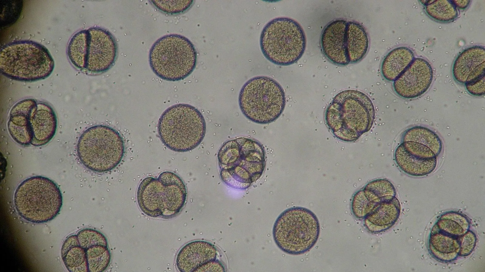
Mitosis is one round of nuclear division that produces two diploid daughter cells genetically identical to the parent, while meiosis is two consecutive rounds of nuclear division that produce four haploid gametes that are genetically unique. That single sentence is the whole answer in miniature. The rule of thumb for choosing between them: if the goal is to copy a cell or grow a body, the cell uses mitosis, and if the goal is to make sperm or eggs for sexual reproduction, the cell uses meiosis.
Eukaryotic life depends on both programs. A fertilized egg must divide billions of times to build an organism, and every one of those divisions is mitotic. A sexually reproducing adult must produce gametes with half the normal chromosome number, and every one of those divisions is meiotic. The two processes share a core machinery of spindle, kinetochore, cohesin and cyclin-dependent kinase control, yet they differ in purpose, division count, ploidy, daughter cell number and genetic outcome. This guide walks through each stage, explains where variation comes from, and gives a side-by-side table that answers the practical question of mitosis versus meiosis in one glance.
What Mitosis Is: A Short Definition
Mitosis is a single nuclear division that separates duplicated chromosomes into two daughter nuclei, each with the same chromosome number and the same genetic content as the parent cell. The full cell cycle around it includes interphase (G1, S and G2), during which DNA is replicated in S phase, followed by the mitotic phase (karyokinesis plus cytokinesis). A diploid human somatic cell with 46 chromosomes enters mitosis with 46 duplicated chromosomes, and each daughter cell exits with 46 unduplicated chromosomes.
The official glossary definition from the National Human Genome Research Institute states that mitosis is "a type of cell division that results in two daughter cells each having the same number and kind of chromosomes as the parent nucleus, typical of ordinary tissue growth" [1]. That phrasing captures the two defining features: equal chromosome number and equal kind of chromosomes. Nothing is shuffled, nothing is halved, and nothing is lost.
Mitosis is the division program of somatic cells. Skin, gut lining, bone marrow, liver and every other renewing tissue uses it. In plants, cambium stem cells in wood-forming tissues divide mitotically along their longest axis to produce xylem and phloem in a strictly bidirectional pattern [2]. In the malaria parasite Plasmodium berghei, rapid mitoses generate male gametes inside the mosquito host before meiosis takes over in the zygote [3]. The same core logic applies across these very different organisms.
What Meiosis Is: A Short Definition
Meiosis is a specialized cell division that produces haploid gametes through two consecutive nuclear divisions after a single round of DNA replication. A diploid germ cell with 46 chromosomes produces four haploid cells with 23 chromosomes each, and those four cells are genetically distinct from each other and from the parent. Meiosis is the only division program in sexually reproducing eukaryotes that reduces chromosome number and reshuffles genetic information.
The meiotic program is not simply mitosis run twice. It is a distinct cell cycle with meiosis-specific cohesin complexes (SMC1β, REC8, RAD21L, STAG3 in mammals) that build a chromosome axis, organize chromatin loops and provide the framework for homolog recognition, synapsis and crossover control [4]. It has its own spindle behavior, its own checkpoint logic and its own coupling to cellular remodeling. Loss of a single meiotic regulator such as the prefoldin subunit Gim3 in budding yeast causes severe chromosome missegregation and gamete inviability, while the same deletion barely affects mitosis, which shows how differently the two programs are wired [5].
Meiosis happens in the germline. In mammals, it happens in the testis and ovary. In plants, it happens in the anther and ovule. In the malaria parasite, it happens in the zygote after fertilization in the mosquito midgut [3]. In the ciliate Tetrahymena thermophila, it happens in the germline micronucleus during sexual development [6].
The Stages of Mitosis
Mitosis has four classical stages: prophase, metaphase, anaphase and telophase. Some textbooks add prometaphase between prophase and metaphase, and most add cytokinesis as the final physical split. The four-stage version is the standard for comparison with meiosis.
Prophase
Chromatin condenses into visible chromosomes, each consisting of two sister chromatids joined at a centromere. The centrosomes duplicate and begin to move apart, and the mitotic spindle starts to form from microtubules. The nuclear envelope begins to break down. In plant cells, a preprophase band of microtubules often marks the future division plane, though in some stem cell types such as Arabidopsis cambium, division plane orientation is established independently of the preprophase band and depends on the cortical division zone instead [2].
Metaphase
Chromosomes align along the metaphase plate at the cell equator. Each chromosome attaches to spindle microtubules from both poles through its kinetochore, and the spindle assembly checkpoint verifies that every kinetochore is properly attached before anaphase begins. In fission yeast, the timing of anaphase is dictated partly by molecular noise from small-number stochastic effects rather than by feedback loops alone, which is a reminder that even a tightly controlled checkpoint has physical limits [7].
Anaphase
Cohesin rings holding sister chromatids together are cleaved, and the two sister chromatids of each chromosome separate and move to opposite poles. Sister chromatids are identical copies, so each pole receives the same genetic information. Anaphase is fast, typically lasting only a few minutes in a mammalian somatic cell.
Telophase
Chromosomes decondense, the nuclear envelope reforms around each set, and the spindle disassembles. Cytokinesis then physically separates the cell into two daughters. In animal cells, an actin-myosin contractile ring pinches the cell in two. In plant cells, a cell plate forms and matures into a new cell wall.
The outcome: two diploid daughter cells, each genetically identical to the parent cell and to each other.
The Stages of Meiosis
Meiosis has two rounds of division, meiosis I and meiosis II, each with prophase, metaphase, anaphase and telophase. The stages of meiosis I are the ones that make meiosis unique, because meiosis I separates homologous chromosomes, while meiosis II separates sister chromatids.
Meiosis I Stages
Prophase I. This is the longest and most complex phase of meiosis. Chromosomes condense, homologous chromosomes pair along their length, and the synaptonemal complex forms between them. The synaptonemal complex is a protein scaffold essential for pairing, recombination and segregation of homologs, and its assembly is hierarchical. In Brassica napus, the protein ZSL bridges the transverse filament protein ZYP1 with the central element proteins SCEP1/2, and loss of ZSL abolishes continuous central region assembly, causing synapsis failure and chromosome missegregation [8]. In Drosophila, the RNA-binding protein Ataxin-2 positively regulates synaptonemal complex components, and its depletion reduces SC mRNA and protein levels, leading to failed homolog pairing [9]. In mammals, the axis protein SYCP2 recruits HORMAD2 to unsynapsed chromosome axes, and that recruitment drives ATR checkpoint signaling that monitors synapsis fidelity [10].
Within prophase I, programmed DNA double-strand breaks are introduced, and some are repaired as crossovers between homologs. Crossovers physically link homologs and are required for accurate segregation at meiosis I [11]. Prophase I is subdivided into leptotene, zygotene, pachytene, diplotene and diakinesis, and rapid prophase chromosome movements occur during early prophase I to facilitate homolog pairing [12].
Metaphase I. Homolog pairs (bivalents) align at the metaphase plate. Kinetochores of sister chromatids attach to microtubules from the same pole, so the two homologs face opposite poles. This is the opposite of mitosis, where sister kinetochores face opposite poles.
Anaphase I. Homologs separate and move to opposite poles. Sister chromatids stay together because cohesin at the centromere is protected during meiosis I. The stepwise release of cohesion across the two meiotic divisions is a defining feature of meiosis-specific cohesin [4].
Telophase I and cytokinesis. Two haploid cells form, each with duplicated chromosomes. In oocytes, this division is highly asymmetric, producing one large secondary oocyte and one small first polar body. Depletion of the regulator DEPDC7 in mouse oocytes disrupts actin cap assembly and first polar body extrusion, producing diploid oocytes that can go on to form triploid zygotes [13]. Similar defects follow loss of PARP7, which stabilizes MYH9 to maintain actin cap integrity during anaphase I [14].
Meiosis II Stages
Meiosis II looks much like mitosis, except that the starting cells are haploid. Prophase II, metaphase II, anaphase II and telophase II proceed with sister chromatids separating at anaphase II. The outcome is four haploid cells. In males, all four become sperm. In females, only one becomes a mature egg, and the other three become polar bodies that are discarded.
The two divisions together reduce chromosome number by half and produce four genetically distinct cells.
Side-by-Side Comparison: Mitosis vs Meiosis


The table below is the core reference for mitosis versus meiosis. Read it left to right for a single feature, or top to bottom for a full comparison.
| Feature | Mitosis | Meiosis |
|---|---|---|
| Purpose | Growth, repair, asexual reproduction, tissue renewal | Production of haploid gametes for sexual reproduction |
| Number of divisions | One | Two (meiosis I and meiosis II) |
| DNA replication | Once, before division | Once, before meiosis I |
| Ploidy in | Diploid (2n) in typical somatic cells | Diploid (2n) in germline cells |
| Ploidy out | Diploid (2n), same as parent | Haploid (n) |
| Daughter cells | Two | Four |
| Genetic outcome | Identical to parent and to each other | Genetically unique, with new allele combinations |
| Homolog pairing | No | Yes, in prophase I |
| Crossing over | No (or negligible) | Yes, in prophase I |
| Independent assortment | No | Yes, at metaphase I |
| What separates in anaphase | Sister chromatids | Homologs in anaphase I, sister chromatids in anaphase II |
| Cohesin type | Canonical (SMC1α, SMC3, RAD21, STAG1/2) | Meiosis-specific (SMC1β, REC8, RAD21L, STAG3) |
| Where it occurs | Somatic tissues throughout the body | Germline: testis, ovary, anther, ovule |
| Duration in humans | Typically under an hour for the nuclear division | Days to decades, especially in oocytes |
| Cytokinesis symmetry | Usually symmetric | Often asymmetric in oocytes |
The table makes the practical difference clear. Mitosis is a copying machine. Meiosis is a shuffling and halving machine. The two divisions of meiosis are not a doubled-up mitosis, and the four daughter cells of meiosis are not simply "twice as many" as the two of mitosis. The chromosome number is halved, the genetic content is reshuffled, and the regulatory logic is different at every stage.
Why the Difference Is Not Just Two Versus Four
A common shorthand says mitosis makes two cells and meiosis makes four. That shorthand misses three deeper differences that matter for understanding fertility, aneuploidy and development.
First, ploidy changes. Mitosis preserves chromosome number, so a diploid cell produces diploid daughters. Meiosis reduces chromosome number by half, so a diploid germ cell produces haploid gametes. Without that reduction, fertilization would double the chromosome number every generation.
Second, genetic content changes. Mitosis produces identical daughters because sister chromatids are identical copies. Meiosis produces unique daughters because crossing over and independent assortment create new allele combinations in every gamete.
Third, the regulatory machinery is different. Meiosis uses specialized cohesin variants that build a chromosome axis and support homolog pairing, synapsis and crossover control [4]. It uses different spindle behavior and checkpoint logic, and it is coupled to cellular remodeling that excludes age-associated protein aggregates from newly forming gametes [5]. The meiotic spindle is also more sensitive to tubulin abundance than the mitotic spindle, which is why loss of the prefoldin subunit Gim3 disrupts meiosis but leaves mitosis largely intact [5].
The transition from meiosis back to mitosis after fertilization is itself a major reprogramming event. Following fertilization, the highly asymmetric meiotic divisions of the oocyte are followed one cell cycle later by the symmetric first embryonic mitosis of the zygote, and the spindle, centrosomes and chromosomes are all remodeled to enable that switch [15]. The two programs are not interchangeable. They are separate cell cycle modes with separate molecular requirements.
Where Each Process Occurs
Mitosis occurs in somatic tissues throughout the body. Any tissue that renews itself uses mitosis: skin epithelium, intestinal crypts, bone marrow, liver, and the cambium of woody plants [2]. It also occurs in some pathogens during specific life stages, such as the rapid mitoses that generate male gametes in Plasmodium berghei [3].
Meiosis occurs only in the germline. In mammals, it occurs in the testis (spermatogenesis) and ovary (oogenesis). In flowering plants, it occurs in the anther and ovule. In the malaria parasite, it occurs in the zygote after fertilization in the mosquito midgut [3]. In Tetrahymena thermophila, it occurs in the germline micronucleus during sexual development, while the somatic macronucleus divides by a different process called amitosis [6].
The location matters because it determines the consequences of failure. A mitotic error in a renewing tissue can contribute to cancer or tissue dysfunction. A meiotic error in the germline produces aneuploid gametes, which are a leading contributor to infertility, miscarriage and congenital disorders [11].
Worked Scenarios: Choosing the Right Framework
Scenario 1: A researcher studies a human skin biopsy and wants to know how the epidermis renews. The answer is mitosis. Basal keratinocytes divide mitotically to produce two diploid daughters, one of which remains a stem cell and one of which differentiates. No homolog pairing, no crossing over, no ploidy change.
Scenario 2: A clinician evaluates a couple with recurrent miscarriage. The most likely meiotic contributor is aneuploidy from chromosome missegregation during meiosis I or meiosis II, often linked to crossover failure, synapsis defects or loss of cohesin integrity with maternal age [4]. The relevant framework is meiosis, not mitosis.
Scenario 3: A plant biologist studies wood formation in Arabidopsis and wants to know how cambium stem cells orient their divisions. The answer involves mitosis, and specifically the cortical division zone rather than the preprophase band [2]. Meiosis is not involved.
Scenario 4: A parasitologist studies transmission of malaria from humans to mosquitoes. The relevant framework includes both: rapid mitoses generate male gametes, and meiosis in the zygote seeds the next round of infection [3]. This is a case where the two programs sit side by side in the same life cycle.
Scenario 5: A developmental biologist studies the first division of a zygote. The answer is mitosis, but the transition from the highly asymmetric meiotic divisions of the oocyte to the symmetric first embryonic mitosis requires extensive subcellular remodeling of the spindle, centrosomes and chromosomes [15]. Understanding the switch requires knowing both programs.
Common Mistakes and Limitations
A few mix-ups come up again and again.
Saying meiosis is "two mitoses." It is not. Meiosis I separates homologs, which mitosis never does, and it includes pairing, synapsis and crossing over. Meiosis II resembles mitosis in its mechanics, but it starts from a haploid cell and produces haploid daughters.
Confusing sister chromatids with homologs. Sister chromatids are identical copies joined at a centromere. Homologs are the maternal and paternal versions of the same chromosome. Mitosis separates sister chromatids. Meiosis I separates homologs. Meiosis II separates sister chromatids.
Assuming crossing over is optional. Crossovers are required for accurate homolog segregation at meiosis I, not just for generating diversity [11]. Loss of crossover control causes missegregation and aneuploidy.
Treating oocyte meiosis as a scaled-up version of spermatocyte meiosis. It is not. Oocyte meiosis is highly asymmetric, produces one functional egg and up to three polar bodies, and has unique regulatory features. Depletion of DEPDC7 or PARP7 in mouse oocytes disrupts the actin cap and first polar body extrusion, which can produce diploid oocytes and triploid zygotes [13][14]. The oocyte cell cycle has many adaptations that a typical mitosis does not need [16].
Ignoring the meiotic spindle's sensitivity to tubulin levels. The meiotic spindle is more sensitive to reduced tubulin abundance than the mitotic spindle, which is one reason some mutations affect fertility without affecting somatic tissue [5].
Assuming all four meiotic products survive. In males, all four become sperm. In females, only one becomes a mature egg. The other three are polar bodies that are discarded.
Forgetting that individual cases need professional evaluation. A veterinarian, physician or clinical geneticist should assess any specific fertility, miscarriage or developmental concern, because the underlying cause may involve meiotic errors, mitotic errors or both.
Frequently Asked Questions
What is the main difference between mitosis and meiosis?
Mitosis is one division that produces two diploid cells identical to the parent, while meiosis is two divisions that produce four haploid cells that are genetically unique. The ploidy change and the genetic reshuffling in meiosis are the defining differences.
What is a short definition of mitosis?
Mitosis is a type of cell division that produces two daughter cells with the same number and kind of chromosomes as the parent cell [1]. It is the ordinary division program of somatic tissues.
What are the stages of meiosis?
Meiosis has two rounds. Meiosis I has prophase I, metaphase I, anaphase I and telophase I. Meiosis II has prophase II, metaphase II, anaphase II and telophase II. Prophase I is the longest stage and includes homolog pairing, synapsis and crossing over.
What happens in meiosis I that does not happen in mitosis?
In meiosis I, homologous chromosomes pair, form the synaptonemal complex, undergo crossing over, and then separate at anaphase I. None of those events occur in mitosis.
Why does meiosis produce genetic variation?
Crossing over in prophase I swaps segments between homologs, and independent assortment at metaphase I randomly orients each homolog pair. Together they create new allele combinations in every gamete.
Does meiosis occur in all cells?
No. Meiosis occurs only in germline cells that produce gametes, such as cells in the testis and ovary in mammals, the anther and ovule in plants, and the zygote in the malaria parasite. Somatic cells use mitosis.
Related Articles
- Cell Division
- Primary and Secondary Cell Culture: Definitions and Key Differences
- Single-Cell vs. Single-Nucleus RNA-Seq for Differential Expression: Key Differences and Best Practices
- RNA-seq Quality Control for Single-Cell Data: Key Differences and Best Practices
- Epigenetics vs Epigenesis: Key Differences Explained
- A Beginner's Guide to Cell Type Annotation in Single-Nucleus RNA-Seq: Key Differences and Best Practices
- Animal vs Plant Cell: Key Differences Compared
- Centriole Function: Roles in Cell Division
- In Vitro vs In Vivo: Key Differences Explained
Sources
- Mitosis
- The preprophase band is dispensable for robust cell division orientation in wood-forming cambium stem cells.
- A modular chromosomal passenger complex rewires chromosome segregation in Plasmodium berghei.
- Chromosome Architecture Defined by the Meiosis-Specific Cohesin in Mammalian Germ Cells.
- Prefoldin function links meiotic chromosome segregation with cellular remodeling and reveals tubulin sensitivity of the meiotic spindle.
- The nucleosome assembly protein Nap1 regulates chromatin stability and nuclear division in Tetrahymena thermophila.
- Small-number effects limit chromosome segregation synchrony.
- ZSL Orchestrates Synaptonemal Complex Assembly as a Central Region Scaffold to Ensure Synapsis Fidelity and Crossover Control in Polyploid Meiosis.
- Ataxin-2 regulates the synaptonemal complex to ensure chromosome pairing during female meiosis.
- SYCP2 recruits HORMAD2 to chromosome axes for unsynapsed chromatin silencing and synapsis surveillance in meiosis.
- Signaling Cascades in Meiotic Chromosome Dynamics.
- Rapid chromosome movements persist within nuclear protrusions in Arabidopsis thaliana meiocytes.
- DEPDC7 couples homologous chromosome segregation to asymmetric cytokinesis through recruiting ARP2/3 complex in oocyte meiosis.
- PARP7-mediated mono-ADP-ribosylation stabilizes MYH9 to ensure actin cap integrity and chromosome segregation in mouse oocytes.
- Setting up for embryogenesis: subcellular changes at the meiosis to mitosis transition.
- Peculiarities of the Mammalian Oocyte Cell Cycle.