
Pasteurella multocida in Livestock: Fowl Cholera and Bovine Respiratory Disease
Introduction
Pasteurella multocida is a Gram-negative, facultatively anaerobic, nonmotile coccobacillus belonging to the family Pasteurellaceae. This bacterium is a commensal of the upper respiratory tract and gastrointestinal mucosa in many domestic and wild animals, yet it can transition to a primary or secondary pathogen under conditions of host stress, immunosuppression, or viral co-infection [1, 2]. In livestock production systems, P. multocida is responsible for two clinically and economically devastating disease syndromes: fowl cholera in avian species and bovine respiratory disease (BRD) in cattle. Fowl cholera is a septicemic disease of poultry and waterfowl characterized by acute mortality and chronic localized infections [3]. Bovine respiratory disease, often termed shipping fever, is a multifactorial polymicrobial pneumonia in which P. multocida acts as a key bacterial agent alongside Mannheimia haemolytica and Histophilus somni [4, 5].
This review provides a detailed comparison of these two disease presentations, focusing on capsular typing, virulence factors, antimicrobial susceptibility profiles, vaccination strategies, and the role of co-infections. The discussion emphasizes the biophysical and molecular mechanisms underlying host-pathogen interactions and diagnostic approaches.
Taxonomy and Capsular Typing
Pasteurella multocida is classified into five capsular serogroups (A, B, D, E, F) based on the antigenic properties of the polysaccharide capsule, and 16 somatic lipopolysaccharide (LPS) serotypes (1 through 16) determined by heat-stable antigens [6, 7]. Capsular typing is performed using multiplex PCR targeting capsule biosynthesis genes: hyaD-hyaC for capsular type A, bcbD for type B, dcbF for type D, ecbJ for type E, and fcbD for type F [8].
The capsular serogroup correlates strongly with host predilection and disease manifestation. Capsular type A is the predominant isolate in avian fowl cholera and bovine respiratory disease [9, 10]. Capsular type B and type E are associated with hemorrhagic septicemia in cattle and water buffalo in tropical regions [11]. Capsular type D is linked to atrophic rhinitis in swine and occasional respiratory infections in cattle [12]. Capsular type F has been reported in avian cholera outbreaks in turkeys and waterfowl [13].
Fowl Cholera: Pathogenesis and Clinical Presentation
Fowl cholera is an acute or chronic septicemic disease affecting chickens, turkeys, ducks, geese, and other avian species. The disease occurs worldwide and causes significant economic losses due to high mortality, decreased egg production, and carcass condemnation [3, 14].
Pathogenesis
Transmission occurs via inhalation or ingestion of P. multocida shed from the oral, nasal, or conjunctival secretions of infected or carrier birds [15]. The bacterium colonizes the upper respiratory tract mucosa and invades the epithelium through mechanisms involving fimbriae, outer membrane proteins, and the polysaccharide capsule [16]. The capsule, particularly hyaluronic acid in type A strains, inhibits phagocytosis and complement-mediated killing [17]. Once in the bloodstream, P. multocida multiplies rapidly, causing a fulminant septicemia with endotoxic shock [18].
Key virulence factors include the following:
- Capsule: Antiphagocytic; type A capsule composed of hyaluronic acid; type D capsule composed of heparin-like glycosaminoglycans [17, 19].
- Lipopolysaccharide (LPS): Induces strong inflammatory responses via Toll-like receptor 4 (TLR4) activation; contributes to endotoxic shock [20].
- Outer membrane proteins (OMPs): OmpA, OmpH, and Omp87 mediate adhesion to host cells and serum resistance [21].
- Sialidases (NanB, NanH): Cleave sialic acid from host glycoproteins, exposing receptors for bacterial adhesion and providing a carbon source [22].
- Toxins: Dermonecrotic toxin (PMT) is produced primarily by type D strains; PMT is a potent mitogen that activates G-protein signaling pathways, leading to osteoclast activation and turbinate atrophy in swine, but its role in avian disease is less clear [23].
Clinical Signs
Acute fowl cholera presents with sudden death in well-conditioned birds, often without premonitory signs. Morbidity and mortality can reach 50% or higher in untreated flocks [3]. Clinical signs in surviving birds include fever, depression, anorexia, ruffled feathers, oral and nasal discharge, cyanosis of the comb and wattles, and diarrhea [14]. Chronic fowl cholera manifests as localized infections including swollen wattles, conjunctivitis, sinusitis, arthritis, and torticollis due to middle ear infection [24].
Pathology
Gross lesions in acute cases include petechial hemorrhages on the heart, serosal surfaces, and abdominal fat; hepatomegaly and splenomegaly with multifocal necrosis; and fibrinous pericarditis and peritonitis [25]. Chronic cases show caseous exudate in the wattles, joints, and sinuses.
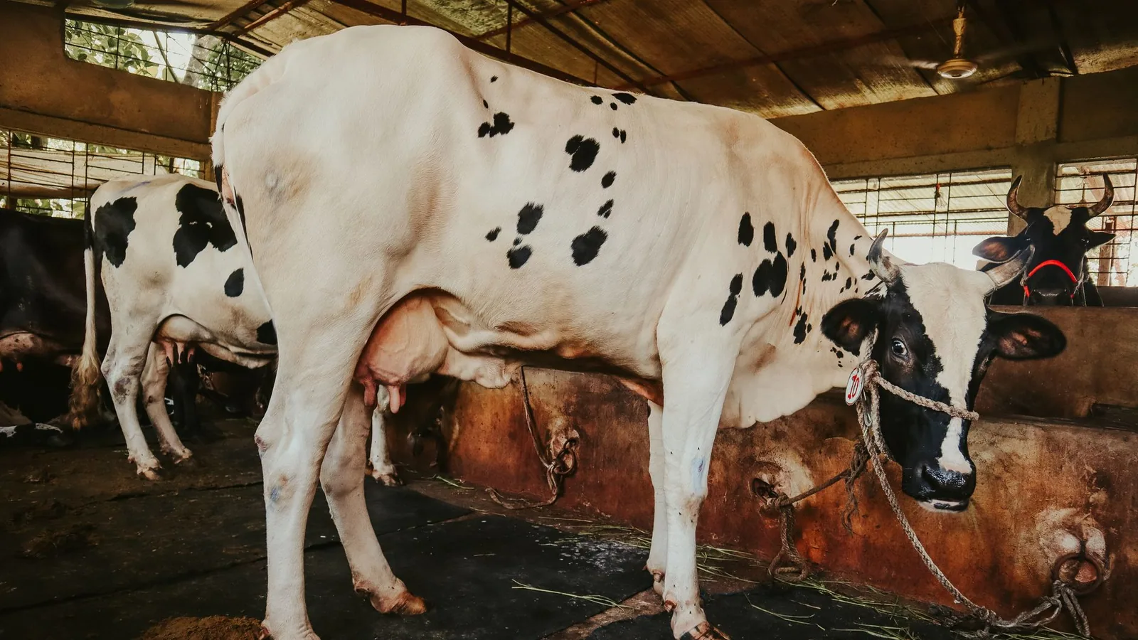
Bovine Respiratory Disease: Pathogenesis and Clinical Presentation
Bovine respiratory disease (BRD) is the most economically important disease of feedlot cattle, accounting for substantial morbidity, mortality, and treatment costs [4, 26]. Pasteurella multocida is one of the primary bacterial agents in the BRD complex, often acting as a secondary invader following viral infection or stress-induced immunosuppression.
Pathogenesis
The pathogenesis of BRD is multifactorial, involving a triad of host stress, viral infection, and bacterial colonization [27]. Stressors such as weaning, transport, commingling, and castration suppress innate immunity through glucocorticoid-mediated inhibition of neutrophil function and alveolar macrophage activity [28]. Viral pathogens including bovine respiratory syncytial virus (BRSV), bovine parainfluenza virus 3 (BPIV-3), bovine herpesvirus 1 (BHV-1), and bovine viral diarrhea virus (BVDV) damage the respiratory epithelium, impair mucociliary clearance, and upregulate adhesion receptors for bacterial binding [29, 30].
Pasteurella multocida colonizes the damaged tracheal and bronchial epithelium, adhering via fimbriae and OMPs [31]. The capsule and LPS resist phagocytosis and opsonization [17]. Bacterial proliferation in the lower respiratory tract triggers an intense neutrophilic inflammatory response, leading to fibrinosuppurative bronchopneumonia [32]. The release of LPS and bacterial DNA amplifies cytokine cascades, including tumor necrosis factor-alpha (TNF-alpha) and interleukin-8 (IL-8), resulting in tissue necrosis, fibrin deposition, and abscess formation [33].
Co-infection with Mannheimia haemolytica
Mannheimia haemolytica is the most frequently isolated bacterial pathogen from acute BRD cases, but P. multocida is often co-isolated, particularly in chronic or recurrent pneumonia [5, 34]. M. haemolytica produces a leukotoxin (LktA) that specifically kills bovine leukocytes and alveolar macrophages, releasing pro-inflammatory mediators that exacerbate tissue damage [35]. Co-infection with P. multocida and M. haemolytica is associated with more severe clinical disease and higher mortality compared to single-pathogen infections [36]. The synergistic mechanism involves M. haemolytica leukotoxin-mediated immune suppression, which facilitates P. multocida proliferation and dissemination [37].
Clinical Signs
Clinical signs of BRD include fever (rectal temperature exceeding 40.0 degrees Celsius), depression, anorexia, nasal discharge, ocular discharge, cough, tachypnea, and dyspnea [38]. Affected cattle often separate from the group and exhibit a drooped head and ears. Chronic cases may present with poor growth, intermittent cough, and exercise intolerance.
Pathology
Gross lesions in acute BRD consist of cranioventral consolidation of the lung lobes, with a firm, dark red to gray appearance. Fibrinous pleuritis and adhesions are common. Cut surface reveals purulent exudate in airways and necrotic foci [32]. Histologically, there is fibrinosuppurative bronchopneumonia with alveolar necrosis, neutrophil infiltration, and fibrin thrombi.
Antimicrobial Susceptibility and Resistance
Antimicrobial therapy is a cornerstone of BRD and fowl cholera management, but resistance is an increasing concern. Pasteurella multocida isolates from both avian and bovine sources have shown resistance to tetracyclines, sulfonamides, penicillins, and macrolides [39, 40].
Mechanisms of Resistance
Resistance mechanisms include enzymatic inactivation (beta-lactamases), target site modification (ribosomal protection proteins for tetracyclines), efflux pumps (resistance-nodulation-division family), and reduced outer membrane permeability [41]. Plasmid-mediated resistance genes such as tet(H), blaROB-1, sul2, and strA-strB are commonly identified in bovine and avian isolates [42].
Susceptibility Profiles
A summary of typical antimicrobial susceptibility patterns for P. multocida from livestock is presented in Table 1.
Table 1. Antimicrobial Susceptibility Profiles of Pasteurella multocida Isolates from Poultry and Cattle
| Antimicrobial Class | Agent | Poultry (Fowl Cholera) | Cattle (BRD) |
|---|---|---|---|
| Penicillins | Amoxicillin | Variable | Susceptible |
| Tetracyclines | Oxytetracycline | Resistant (frequent) | Variable |
| Macrolides | Tulathromycin | Not approved | Susceptible |
| Fluoroquinolones | Enrofloxacin | Susceptible | Susceptible |
| Phenicols | Florfenicol | Susceptible | Susceptible |
| Sulfonamides | Sulfadimethoxine | Resistant (frequent) | Variable |
Data compiled from references [39, 40, 43].
The emergence of multidrug-resistant (MDR) strains, particularly in poultry flocks with heavy antimicrobial use, complicates treatment protocols. Surveillance programs using broth microdilution and disk diffusion methods are essential for guiding empirical therapy [44].
Vaccination Strategies
Fowl Cholera Vaccines
Vaccination against fowl cholera has historically relied on inactivated bacterins and live attenuated vaccines. Bacterins, typically prepared from formalin-killed whole cells of serotype 1 or serotype 3,4, provide serotype-specific protection but limited cross-protection against heterologous strains [45]. Live attenuated vaccines, such as the CU strain (chicken origin) and the M-9 strain (turkey origin), induce stronger cell-mediated immunity and broader protection, but carry a risk of reversion to virulence in some hosts [46].
Recombinant subunit vaccines targeting OMPs, sialidases, and LPS core antigens are under development. These vaccines aim to provide cross-serotype protection and reduce reliance on live organisms [47].
Bovine Respiratory Disease Vaccines
Vaccination against BRD typically involves multivalent products containing inactivated P. multocida, M. haemolytica, and H. somni combined with viral antigens (BHV-1, BRSV, BPIV-3, BVDV) [48]. Modified-live viral vaccines are administered intranasally or parenterally to prime the mucosal immune system. Bacterial components are often formulated as bacterins or leukotoxin-enriched extracts [49].
Efficacy of BRD vaccines is variable, influenced by maternal antibody interference, stress at the time of vaccination, and antigenic diversity of circulating P. multocida strains [50]. Autogenous vaccines prepared from farm-specific isolates are sometimes used in herds with persistent BRD problems.
Diagnostic Approaches
Diagnosis of P. multocida infection relies on bacterial culture, molecular detection, and serological typing. A diagnostic workflow is presented in Figure 1.
flowchart TD
 A["Clinical Sample: Swab, Tissue, Blood"] --> B["Gram Stain: Gram-negative coccobacilli"]
 B --> C[Culture on Blood Agar or MacConkey Agar]
 C --> D["Colony Morphology: Gray, mucoid, non-hemolytic"]
 D --> E["Biochemical Confirmation: Oxidase +, Catalase +, Indole +"]
 E --> F[Capsular Typing Multiplex PCR]
 F --> G{Serogroup Identification}
 G --> H["Type A: Fowl cholera, BRD"]
 G --> I["Type B/E: Hemorrhagic septicemia"]
 G --> J["Type D: Atrophic rhinitis"]
 G --> K["Type F: Avian cholera"]
 E --> L[Antimicrobial Susceptibility Testing]
 L --> M[Broth microdilution or disk diffusion]
 E --> N[Virulence Gene Profiling by PCR]
 N --> O[Detection of toxA, ompH, nanB, etc.]
Figure 1. Diagnostic workflow for Pasteurella multocida isolation, capsular typing, and virulence profiling.
Molecular diagnostics, including multiplex PCR for capsular typing and real-time PCR for quantification of bacterial load in respiratory samples, offer rapid and specific identification [8, 51]. Whole-genome sequencing (WGS) is increasingly used for epidemiological surveillance, antimicrobial resistance gene detection, and phylogenetic analysis.
Conclusion
Pasteurella multocida remains a major bacterial pathogen in livestock, causing fowl cholera in poultry and contributing to the BRD complex in cattle. The bacterium's ability to switch from commensal to pathogen is driven by host stress, viral co-infection, and expression of a suite of virulence factors including capsule, LPS, OMPs, and sialidases. Capsular typing distinguishes strains associated with different disease presentations and host species. Antimicrobial resistance, particularly to tetracyclines and sulfonamides, is a growing concern that necessitates ongoing surveillance and judicious antimicrobial use. Vaccination strategies, while available for both fowl cholera and BRD, require refinement to achieve broader cross-protection and improved efficacy under field conditions. Continued research into the molecular mechanisms of pathogenesis and host-pathogen interactions will inform the development of next-generation diagnostics and control measures.
References
[1] Harper M, Boyce JD, Adler B. Pasteurella multocida pathogenesis: 125 years after Pasteur. FEMS Microbiol Lett. 2006;265(1):1-10.
[2] Wilkie IW, Harper M, Boyce JD, Adler B. Pasteurella multocida: diseases and pathogenesis. Curr Top Microbiol Immunol. 2012;361:1-22.
[3] Glisson JR, Hofacre CL, Christensen JP. Fowl cholera. In: Swayne DE, editor. Diseases of Poultry. 14th ed. Wiley-Blackwell; 2020. p. 807-823.
[4] Griffin D. Economic impact associated with respiratory disease in beef cattle. Vet Clin North Am Food Anim Pract. 1997;13(3):367-377.
[5] Rice JA, Carrasco-Medina L, Hodgins DC, Shewen PE. Mannheimia haemolytica and bovine respiratory disease. Anim Health Res Rev. 2007;8(2):117-128.
[6] Carter GR. Studies on Pasteurella multocida. I. A hemagglutination test for the identification of serological types. Am J Vet Res. 1955;16(60):481-484.
[7] Heddleston KL, Gallagher JE, Rebers PA. Fowl cholera: gel diffusion precipitin test for serotyping Pasteurella multocida from avian species. Avian Dis. 1972;16(4):925-936.
[8] Townsend KM, Boyce JD, Chung JY, Frost AJ, Adler B. Genetic organization of Pasteurella multocida cap loci and development of a multiplex capsular PCR typing system. J Clin Microbiol. 2001;39(3):924-929.
[9] Rhoades KR, Rimler RB. Capsular groups of Pasteurella multocida isolated from avian hosts. Avian Dis. 1987;31(4):895-898.
[10] Dabo SM, Taylor JD, Confer AW. Pasteurella multocida and bovine respiratory disease. Anim Health Res Rev. 2007;8(2):129-150.
[11] De Alwis MCL. Haemorrhagic septicaemia. Aust Vet J. 1992;69(10):245-248.
[12] Foged NT. Pasteurella multocida toxin. The characterisation of the toxin and its significance in the diagnosis and prevention of progressive atrophic rhinitis in pigs. APMIS Suppl. 1992;25:1-56.
[13] Wilson MA, Rimler RB, Hoffman LJ. Comparison of DNA fingerprints and somatic serotypes of Pasteurella multocida isolates from turkeys. Avian Dis. 1992;36(3):507-516.
[14] Carpenter TE, Snipes KP, Wallis D, Hird DW. Epidemiology of fowl cholera in California. Avian Dis. 1988;32(4):688-695.
[15] Samuel MD, Goldberg DR, Shadduck DJ, Price JI, Cooch EG. Pasteurella multocida serotype 1 isolated from a lesser snow goose. J Wildl Dis. 1997;33(1):155-157.
[16] Al-Haddawi MH, Jasni S, Zamri-Saad M, Mutalib AR, Sheikh-Omar AR. Ultrastructural pathology of the respiratory tract of chickens experimentally infected with Pasteurella multocida. J Comp Pathol. 2000;122(2-3):185-191.
[17] Boyce JD, Adler B. The capsule is a virulence determinant in the mouse model of Pasteurella multocida infection. Infect Immun. 2000;68(6):3463-3468.
[18] Harper M, Boyce JD, Adler B. The key surface components of Pasteurella multocida: capsule and lipopolysaccharide. Curr Top Microbiol Immunol. 2012;361:39-51.
[19] DeAngelis PL, Gunay NS, Toida T, Mao WJ, Linhardt RJ. Identification of the capsular polysaccharides of Type D and F Pasteurella multocida as unmodified heparin and chondroitin, respectively. Carbohydr Res. 2002;337(17):1547-1552.
[20] Harper M, Cox AD, St Michael F, Wilkie IW, Boyce JD, Adler B. A heptosyltransferase mutant of Pasteurella multocida produces a truncated lipopolysaccharide structure and is attenuated in virulence. Infect Immun. 2004;72(6):3436-3443.
[21] Luo Y, Glisson JR, Jackwood MW, Hancock RE, Bains M, Cheng IH, Wang C. Cloning and characterization of the major outer membrane protein gene (ompH) of Pasteurella multocida X-73. J Bacteriol. 1997;179(24):7856-7864.
[22] Steenbergen SM, Lichtensteiger CA, Caughlan R, Garfinkle J, Fuller TE, Vimr ER. Sialic acid metabolism and systemic pasteurellosis. Infect Immun. 2005;73(3):1284-1294.
[23] Lax AJ, Chanter N. Cloning of the toxin gene from Pasteurella multocida and its role in atrophic rhinitis. J Gen Microbiol. 1990;136(1):81-87.
[24] Olson LD, McCune EL, Bond RE. Comparison of commercial vaccines for fowl cholera. Avian Dis. 1969;13(2):268-276.
[25] Rhoades KR, Rimler RB. Fowl cholera. In: Calnek BW, editor. Diseases of Poultry. 10th ed. Iowa State University Press; 1997. p. 143-159.
[26] Smith RA. Impact of disease on feedlot performance: a review. J Anim Sci. 1998;76(1):272-274.
[27] Yates WD. A review of infectious bovine rhinotracheitis, shipping fever pneumonia and viral-bacterial synergism in respiratory disease of cattle. Can J Comp Med. 1982;46(3):225-263.
[28] Roth JA, Kaeberle ML. Effect of glucocorticoids on the bovine immune system. Am J Vet Res. 1982;43(2):239-244.
[29] Gershwin LJ, Gunther RA, Anderson ML, Woolums AR, McArthur-Vaughan K, Randel KE, Boyle GA, Friebertshauser KE, McInturff PS. Bovine respiratory syncytial virus-specific IgE is associated with interleukin-2 and -4 responses in cattle. Vet Immunol Immunopathol. 2000;75(1-2):1-15.
[30] Fulton RW, Confer AW, Burge LJ, Perino LJ, d'Offay JM, Payton ME, Ridpath JF. Antibody responses by cattle after vaccination with commercial viral vaccines containing bovine herpesvirus-1, bovine viral diarrhea virus, parainfluenza-3 virus, and bovine respiratory syncytial virus immunogens and subsequent revaccination at day 140. Vaccine. 1995;13(8):725-733.
[31] Confer AW, Panciera RJ, Mosier DA. Bovine pneumonic pasteurellosis: immunity to Pasteurella haemolytica. J Am Vet Med Assoc. 1988;193(10):1308-1316.
[32] Panciera RJ, Confer AW. Pathogenesis and pathology of bovine pneumonia. Vet Clin North Am Food Anim Pract. 2010;26(2):191-214.
[33] Yoo HS, Maheswaran SK, Lin G, Townsend EL, Ames TR. Induction of inflammatory cytokines in bovine alveolar macrophages following stimulation with Pasteurella haemolytica lipopolysaccharide. Infect Immun. 1995;63(2):381-388.
[34] Welsh RD, Dye LB, Payton ME, Confer AW. Isolation and antimicrobial susceptibilities of bacterial pathogens from bovine pneumonia: 1994-2002. J Vet Diagn Invest. 2004;16(5):426-431.
[35] Shewen PE, Wilkie BN. Cytotoxin of Pasteurella haemolytica acting on bovine leukocytes. Infect Immun. 1982;35(1):91-94.
[36] Mosier DA, Simons KR, Confer AW, Panciera RJ, Clinkenbeard KD. Pasteurella haemolytica and Pasteurella multocida in bovine respiratory disease. Vet Clin North Am Food Anim Pract. 1989;5(2):319-345.
[37] Clinkenbeard KD, Mosier DA, Confer AW. Effects of Pasteurella haemolytica leukotoxin on isolated bovine neutrophils. Am J Vet Res. 1989;50(6):982-988.
[38] Taylor JD, Fulton RW, Lehenbauer TW, Step DL, Confer AW. The epidemiology of bovine respiratory disease: what is the evidence for predisposing factors? Can Vet J. 2010;51(10):1095-1102.
[39] Watts JL, Yancey RJ Jr, Salmon SA, Case CA. A 4-year survey of antimicrobial susceptibility trends for isolates from cattle with bovine respiratory disease in North America. J Clin Microbiol. 1994;32(3):725-731.
[40] Singer RS, Hofacre CL. Potential impacts of antibiotic use in poultry production. Avian Dis. 2006;50(2):161-172.
[41] Kehrenberg C, Schwarz S. Occurrence and linkage of genes coding for resistance to sulfonamides, streptomycin, and chloramphenicol in bacteria of the genera Pasteurella and Mannheimia. FEMS Microbiol Lett. 2001;205(2):283-287.
[42] Schwarz S, Kehrenberg C, Salmon SA, Watts JL. Tetracycline resistance genes in Pasteurella multocida and Mannheimia haemolytica from cattle. Antimicrob Agents Chemother. 2004;48(2):636-638.
[43] Portis E, Lindeman C, Johansen L, Stoltman G. A ten-year (2000-2009) study of antimicrobial susceptibility of bacteria that cause bovine respiratory disease complex, Mannheimia haemolytica, Pasteurella multocida, and Histophilus somni, in the United States and Canada. J Vet Diagn Invest. 2012;24(5):932-944.
[44] Clinical and Laboratory Standards Institute (CLSI). Performance Standards for Antimicrobial Disk and Dilution Susceptibility Tests for Bacteria Isolated from Animals. 5th ed. CLSI supplement VET01S. Wayne, PA: CLSI; 2020.
[45] Bhasin JL, Biberstein EL. Fowl cholera in turkeys: the efficacy of adjuvant bacterins for the control of an experimental challenge. Avian Dis. 1968;12(1):159-168.
[46] Hofacre CL, Glisson JR, Kleven SH. Comparison of vaccination protocols of broiler breeder hens for Pasteurella multocida using enzyme-linked immunosorbent assay and virulent challenge. Avian Dis. 1987;31(2):260-263.
[47] Hatfaludi T, Al-Hasani K, Boyce JD, Adler B. Outer membrane proteins of Pasteurella multocida. Vet Microbiol. 2010;144(1-2):1-17.
[48] Fulton RW, Confer AW. Vaccination of cattle against bovine respiratory disease. Vet Clin North Am Food Anim Pract. 2012;28(1):1-17.
[49] Shewen PE, Carrasco-Medina L, McBey BA, Hodgins DC. Challenges in the development of effective vaccines for Mannheimia haemolytica and Pasteurella multocida infections in cattle. Anim Health Res Rev. 2007;8(2):131-140.
[50] Downey ED, Tait RG Jr, Mayes MS, Park CA, Ridpath JF, Garrick DJ, Reecy JM. An evaluation of circulating bovine viral diarrhea virus type 2 maternal antibody level and response to vaccination in calves. J Anim Sci. 2013;91(10):4585-4593.
Disclaimer: This article is for educational and informational purposes only. It is not intended to substitute for professional veterinary advice, diagnosis, treatment, or regulatory guidance. Always consult a licensed veterinarian or qualified specialist regarding animal health, disease diagnosis, and therapeutic decisions.