Mitosis Prophase: Stages and Key Events
By Dr. Zubair Khalid, DVM, MS, PhD ·
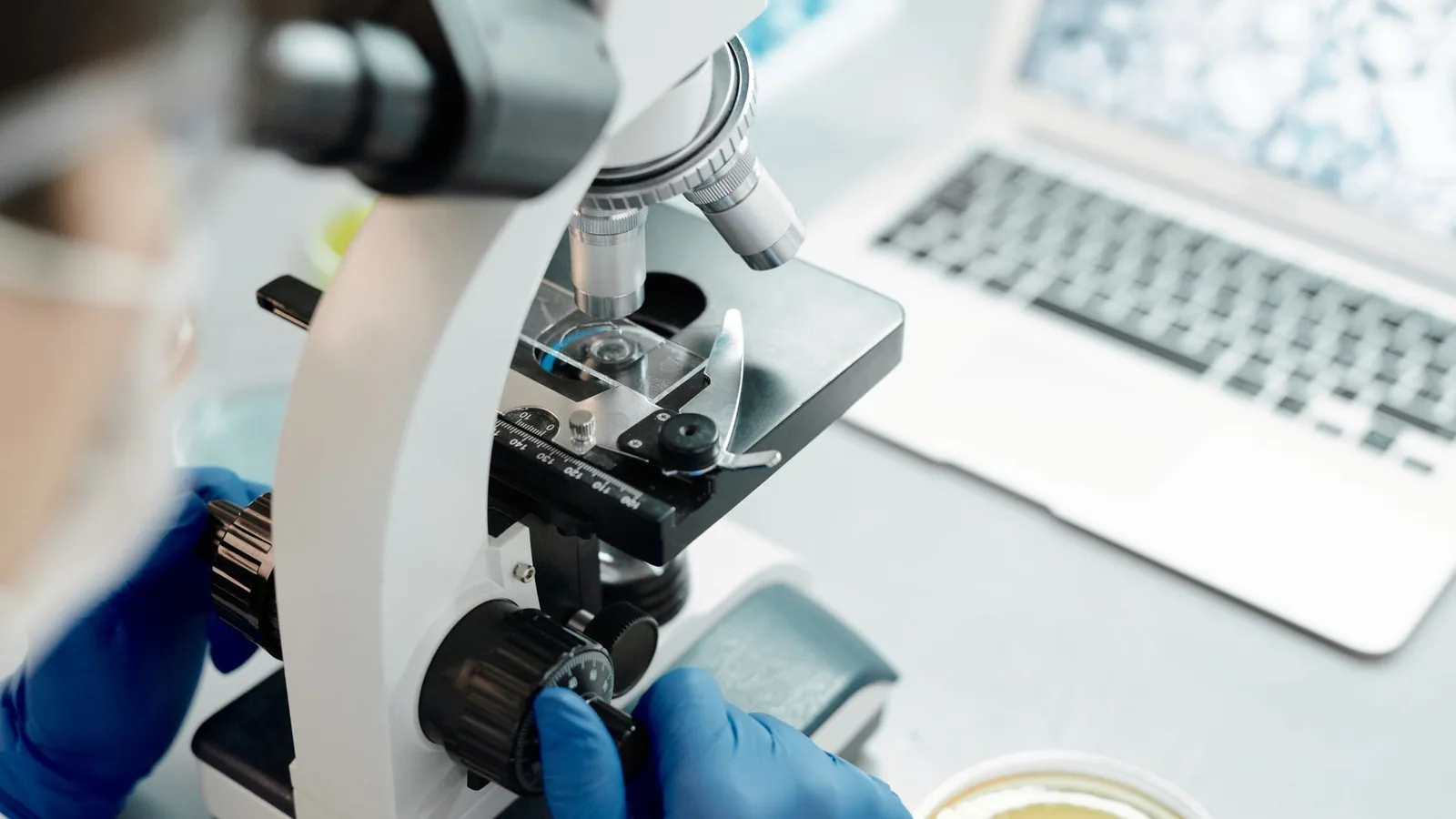
Mitosis prophase is the first phase of mitosis, the stage in which a cell converts its loose interphase chromatin into compact, individually resolvable chromosomes and begins building the machinery that will separate them. It follows interphase, ends when the nuclear envelope breaks down, and sets up every later step of chromosome segregation.
Prophase matters because it is the point of no return. Once a cell commits to prophase, transcription largely stops, the genome is physically reorganized, and the duplicated sister chromatids are prepared for a single, irreversible split. Errors that begin here, such as incomplete sister chromatid resolution or a poorly built spindle, propagate into anaphase and can produce aneuploid daughter cells. That is why prophase is studied so heavily in cancer biology, developmental biology, and any field that cares about genome stability.
What Prophase Is and Where It Sits in the Cell Cycle
The cell cycle has two broad halves. Interphase (G1, S, and G2) is when the cell grows and copies its DNA. M phase is when the copied genome is divided and the cell splits. M phase contains mitosis (nuclear division) and cytokinesis (cytoplasmic division). These are separate processes. A cell can complete mitosis and fail cytokinesis, producing a single cell with two nuclei, and that outcome is well documented in liver hepatocytes and in some cancer lines.
Mitosis itself runs in a fixed order: prophase, prometaphase, metaphase, anaphase, and telophase. Prophase is first. Prometaphase is second and is defined by nuclear envelope breakdown and the capture of chromosomes by spindle microtubules. The two are often merged in casual writing, but they are distinct. Prophase ends when the nuclear envelope disassembles. Prometaphase begins at that moment.
One clarification that prevents a lot of confusion: "prophase" in this article always means mitotic prophase. Meiosis has its own prophase I, which is far longer and includes homologous pairing, synapsis, and crossing over. Those events do not occur in mitosis, and this guide does not cover them.
The Five Key Events of Prophase, in Order
Prophase is not a single switch. It is a sequence of overlapping events that unfold over roughly 30 to 60 minutes in a typical mammalian tissue culture cell, though the exact window varies by species and cell type. The events below are listed in the order they typically begin, but they overlap substantially.
1. Chromatin Condensation
Interphase chromatin is a diffuse, largely decondensed polymer. During prophase it folds into the short, thick rods visible under a light microscope. Two protein complexes do the structural work. Condensin II acts first and is responsible for resolving sister chromatids from one another, meaning it separates the two replicated copies along their length. Condensin I acts later and drives the actual folding and compaction of each chromatid [1].
The timing of these two complexes is regulated by where they sit in the cell. Condensin II is nuclear during interphase and prophase. Condensin I is kept out of the nucleus until the nuclear envelope breaks down. When researchers forced condensin I into the nucleus early, chromosomes folded prematurely, at the same time as sister chromatid resolution, and resolution then failed in prometaphase and metaphase [1]. That experiment showed that the normal delay is not incidental. It is required for correct chromosome structure.
A second layer of control comes from the physical environment around the chromosomes. As cells progress from prophase to anaphase, the molecular density surrounding the chromosomes rises, and this crowding generates an attractive force called depletion attraction that helps pull chromatin into a compact state. When researchers made the surrounding medium hypotonic or hypertonic, chromosome condensation changed accordingly [2]. So condensation is not purely a protein-driven process. It also depends on the local physical chemistry of the nucleus.
Linker histones contribute as well. Phosphorylated histone H1 reaches its highest density during prophase and prometaphase, in parallel with a reduction in the heterochromatin mark H3K9me3 [3]. This histone modification pattern is part of how the cell remodels chromatin for division.
Condensation also deforms the nucleus itself. As chromatin pulls inward, the nuclear envelope develops centripetal shape fluctuations that begin in late G2 and early prophase, become unstable, and precede nuclear envelope breakdown [4]. The mechanical link between condensing chromatin and the envelope is one proposed trigger for the envelope's eventual disassembly.
2. Centrosome Separation
Animal cells have two centrosomes, each containing a pair of centrioles surrounded by pericentriolar material. These were duplicated during S phase. In prophase, the two centrosomes move apart along the nuclear envelope toward opposite sides of the nucleus.
Separation depends on motor proteins and on the growing plus-ends of microtubules pushing against each other. The result is two distinct microtubule-organizing centers positioned to build a bipolar spindle. Plant cells and many fungal cells lack centrosomes entirely and organize their spindles from diffuse microtubule-nucleating sites on the nuclear envelope, which is one of the clearest species differences in early mitosis.
3. Mitotic Spindle Assembly
Once the centrosomes are separated, they nucleate microtubules that grow and shrink rapidly. This dynamic instability lets the microtubules probe the space around them. Three classes of microtubules form.
- Astral microtubules point outward from each pole toward the cell cortex and help position the spindle.
- Kinetochore microtubules attach to chromosomes at their kinetochores.
- Interpolar microtubules from opposite poles overlap in the middle and push the poles apart.
The initial contact between a microtubule and a kinetochore is described by the search-and-capture model. A growing microtubule randomly encounters a kinetochore and is captured. This is where the Ndc80 complex becomes essential. Ndc80 is recruited to kinetochores during prophase and is required for forming stable kinetochore-microtubule attachments. Cells lacking Ndc80 show substantial defects in microtubule plus-end capture during prometaphase, and Ndc80 mutants that cannot bind microtubules fail to capture them properly [5]. The motor protein dynein works alongside Ndc80. Cells depleted of dynein alone can still make initial attachments, but cells depleted of both Ndc80 and dynein show severe capture defects, which indicates the two proteins cooperate [5].
4. Kinetochore Maturation
The kinetochore is the protein structure assembled on centromeric DNA that links a chromosome to spindle microtubules. It matures during prophase as more than 100 proteins assemble in layers. The inner layer contacts centromeric chromatin. The outer layer contains the Ndc80 complex and other microtubule-binding proteins.
Maturation is not just about adding parts. It is about timing. Ndc80 must be present and correctly phosphorylated for productive capture, and its phosphorylation status is critical for the attachment to hold [5]. A kinetochore that matures too early or too late will fail to capture microtubules efficiently, and the chromosome will lag behind during segregation.
A related prophase-specific safeguard involves the protein SRBD1. SRBD1 is a DNA-binding component of the mitotic chromatid axis, and its activity is most critical during prophase, when chromosome condensation is established. Loss of SRBD1 causes a severe sister chromatid segregation defect that resembles the phenotype seen when decatenation by topoisomerase II alpha is disrupted. The requirement for SRBD1 depends on condensin II but not condensin I [6]. In practical terms, SRBD1 helps ensure that the DNA entanglements between sister chromatids are resolved before the cell tries to pull them apart.
5. Nuclear Envelope Breakdown at Late Prophase
The nuclear envelope is a double membrane that separates the nucleus from the cytoplasm. During late prophase, it begins to disassemble. The nuclear lamina, a meshwork of lamin proteins just inside the inner membrane, is phosphorylated and depolymerizes. The membrane itself fragments and is absorbed into the endoplasmic reticulum.
The timing of this event is tied to chromatin condensation. The centripetal fluctuations of the nuclear envelope that begin in early prophase become unstable as the cell approaches breakdown [4]. Chromatin condensation and envelope breakdown are mechanically linked, not independent events.
Nuclear envelope breakdown is the boundary between prophase and prometaphase. Once the envelope is gone, spindle microtubules gain access to the chromosomes, and prometaphase begins. The protein RBR (retinoblastoma-related) illustrates how tightly this transition is controlled. In Arabidopsis thaliana root cells, RBR exits the nucleus before chromosomes are visibly in prophase, moving from dense nuclear aggregates to the cytoplasm in under two minutes, and it stays in the cytoplasm until telophase, when it returns to the reforming daughter nuclei [7].
Table: Mitosis Phase Order, Defining Event, and Duration
| Phase | Defining event | Typical duration (mammalian cultured cells) |
|---|---|---|
| Prophase | Chromatin condenses, centrosomes separate, spindle begins to form | 30 to 60 minutes |
| Prometaphase | Nuclear envelope breaks down, chromosomes attach to spindle | 10 to 20 minutes |
| Metaphase | Chromosomes align at the metaphase plate | 10 to 20 minutes |
| Anaphase | Sister chromatids separate and move to opposite poles | 5 to 10 minutes |
| Telophase | Chromosomes decondense, nuclear envelopes reform | 10 to 30 minutes |
| Cytokinesis | Cytoplasm divides (separate from mitosis) | Overlaps telophase |
Durations are approximate and vary widely. The table gives the order and the defining event for each phase, which is what exams and most practical work require.
How Prophase Is Observed and Measured
Prophase is studied with a small set of standard methods, and knowing them helps when reading primary literature.
Light microscopy with DNA stains. A DNA-binding dye such as DAPI or Hoechst reveals condensed chromosomes as distinct threads. Prophase cells show a nucleus full of visible chromosomes with an intact nuclear envelope. Prometaphase cells show chromosomes scattered without a clear envelope.
Live-cell imaging with fluorescently tagged proteins. Tagging a histone with GFP or mCherry lets researchers watch condensation in real time. Tagging tubulin shows spindle formation. Tagging a nuclear envelope protein shows exactly when the envelope breaks down. This approach was used to track RBR movement in living plant roots [7] and to monitor condensin localization and chromosome folding in human cells [1].
Orientation-independent differential interference contrast (OI-DIC). This technique maps optical path differences and estimates molecular density around chromosomes. It was used to show that the molecular density surrounding chromosomes rises from prophase to anaphase and falls again in telophase [2]. It measures the physical environment of the chromosome, not just its shape.
Immunostaining for specific marks. Antibodies against phosphorylated histone H1 reveal the prophase-specific phosphorylation pattern, with peak density in prophase and prometaphase and absence in anaphase and telophase [3].
Chromosome spread preparations. Cells are swollen in a hypotonic solution, fixed, and dropped onto a slide. This spreads the chromosomes so individual chromatids and sister chromatid resolution can be scored. It is the classic method for detecting condensation and resolution defects.
Comparative and Clinical Relevance
Prophase defects have direct consequences for human health. Aneuploidy, an abnormal number of chromosomes, is a hallmark of most solid tumors and is a leading cause of miscarriage and developmental disorders. Many aneuploidy-causing errors originate in prophase or the prophase-to-prometaphase transition.
Incomplete sister chromatid resolution is one such error. If condensin II fails to separate the two sister chromatids along their length, or if SRBD1 fails to safeguard decatenation, the entangled sisters can form ultrafine anaphase bridges, thin DNA threads that persist into anaphase. These bridges can break, causing chromosome missegregation [6][8]. The PICH protein, a DNA translocase, localizes to chromosome arms, centromeres, and ultrafine anaphase bridges, and helps resolve them [8]. When resolution fails, the risk of genome instability rises.
Kinetochore defects are another. Because Ndc80 is required for microtubule capture, mutations or misregulation that reduce Ndc80 function lead to chromosomes that cannot attach properly to the spindle [5]. The result is lagging chromosomes and nondisjunction.
Prophase is also relevant to cancer therapy. Many chemotherapy drugs target microtubules, and their effects depend on the spindle assembly that begins in prophase. Understanding the timing of prophase events helps explain why these drugs act when they do and why resistance can develop.
Species and cell-type variation matters here. Prophase duration in a rapidly dividing human cancer cell line is not the same as in a slow-dividing neuron or a plant root tip cell. Embryonic stem cells show distinct histone H1 phosphorylation sites compared with differentiated cells [3]. Plant cells organize spindles without centrosomes. Fungal cells, such as Sordaria macrospora, use meiosis-specific proteins like Hop1 in ways that differ from mitosis [9]. Any statement about "typical" prophase timing should be read as a range, not a fixed number.
Common Mistakes and Limitations
Conflating prophase with prometaphase. These are separate phases with a clear boundary: nuclear envelope breakdown. Prophase has an intact envelope. Prometaphase does not. Textbooks sometimes merge them, but for exams and for reading research papers, keep them distinct.
Thinking condensation is a single step. Condensin II resolves sister chromatids first. Condensin I folds them later. The order matters, and forcing condensin I to act early causes resolution failure [1].
Assuming prophase is purely protein-driven. Depletion attraction and molecular crowding contribute to condensation [2]. The physical environment of the chromosome is part of the mechanism.
Treating cytokinesis as part of mitosis. Cytokinesis is a separate process. Mitosis divides the nucleus. Cytokinesis divides the cytoplasm. They overlap in time but are genetically and mechanically distinct.
Assuming all cells look the same in prophase. Plant cells lack centrosomes. Embryonic stem cells have different histone modification patterns. Meiosis has an entirely different prophase I. Generalizations from one system do not always transfer.
Overlooking the nuclear envelope. The envelope is not a passive barrier. Its mechanical behavior is linked to chromatin condensation, and its breakdown is the event that defines the end of prophase [4].
Individual cases in a clinical or research setting require context-specific interpretation, and any diagnostic question belongs with a qualified professional.
Quick Review: 7 Points Worth Memorizing
- Prophase is the first phase of mitosis, after interphase and before prometaphase.
- The five key events are chromatin condensation, centrosome separation, spindle assembly, kinetochore maturation, and nuclear envelope breakdown.
- Condensin II resolves sister chromatids. Condensin I folds them. Condensin I is kept out of the nucleus until the envelope breaks down [1].
- Ndc80 is recruited to kinetochores in prophase and is required for microtubule capture, working with dynein [5].
- Nuclear envelope breakdown marks the end of prophase and the start of prometaphase.
- Mitosis phases in order are prophase, prometaphase, metaphase, anaphase, telophase. Cytokinesis is separate.
- Prophase duration varies by species and cell type, typically 30 to 60 minutes in mammalian cultured cells.
Frequently Asked Questions
What is mitosis prophase?
Mitosis prophase is the first phase of mitosis, in which chromatin condenses into visible chromosomes, centrosomes separate, the mitotic spindle begins to form, kinetochores mature, and the nuclear envelope prepares to break down. It ends when the envelope disassembles and prometaphase begins.
What are the phases of mitosis in order?
The phases of mitosis in order are prophase, prometaphase, metaphase, anaphase, and telophase. Cytokinesis, the division of the cytoplasm, is a separate process that overlaps with telophase but is not part of mitosis itself.
What is the main difference between prophase and prometaphase?
The main difference is the nuclear envelope. Prophase has an intact nuclear envelope. Prometaphase begins when the envelope breaks down, allowing spindle microtubules to reach the chromosomes. Prophase ends at that exact moment.
How long does prophase last?
In mammalian cultured cells, prophase typically lasts 30 to 60 minutes. Duration varies by species, cell type, and growth conditions. Embryonic cells tend to be faster, and some plant and fungal cells have different timing altogether.
What happens to chromatin during prophase?
Chromatin condenses from a diffuse interphase state into compact, rod-shaped chromosomes. Condensin II resolves sister chromatids first, then condensin I folds each chromatid. Molecular crowding around the chromosomes also contributes to compaction [2].
Is prophase the same in meiosis and mitosis?
No. Mitotic prophase is short and does not involve homologous pairing or crossing over. Meiotic prophase I is much longer and includes synapsis, recombination, and the formation of crossovers between homologous chromosomes. The two share a name but are different processes.
Related Articles
- Budding in Yeast: Mechanisms, Stages, and Key Experiments
- DNA Replication Before Mitosis: Why It Must Happen
- Biotech Networking Events vs. Online Communities
- Acute Inflammation: Vascular and Cellular Events
- Digital Pathology Conferences and Events: A Planning Guide
- Adverse Event Reporting in Clinical Trials
- Hog Feeding Guide: Rations, Stages, and Feed Conversion
Sources
- Nuclear exclusion of condensin I in prophase coordinates mitotic chromosome reorganization to ensure complete sister chromatid resolution.
- Orientation-independent-DIC imaging reveals that a transient rise in depletion attraction contributes to mitotic chromosome condensation.
- The Highest Density of Phosphorylated Histone H1 Appeared in Prophase and Prometaphase in Parallel with Reduced H3K9me3, and HDAC1 Depletion Increased H1.2/H1.3 and H1.4 Serine 38 Phosphorylation.
- Centripetal nuclear shape fluctuations associate with chromatin condensation in early prophase.
- Coordination between the Ndc80 complex and dynein is essential for microtubule plus-end capture by kinetochores during early mitosis.
- SRBD1, a highly conserved gene required for chromosome individualization.
- In vivo movement of retinoblastoma-related protein (RBR) towards cytoplasm during mitosis in Arabidopsisthaliana.
- Regulation of mitotic chromosome architecture and resolution of ultrafine anaphase bridges by PICH.
- Canonical and noncanonical roles of Hop1 are crucial for meiotic prophase in the fungus Sordaria macrospora.