
Menangle Virus
1. Discovery and Initial Characterization: The Emergence of a Novel Porcine Pathogen
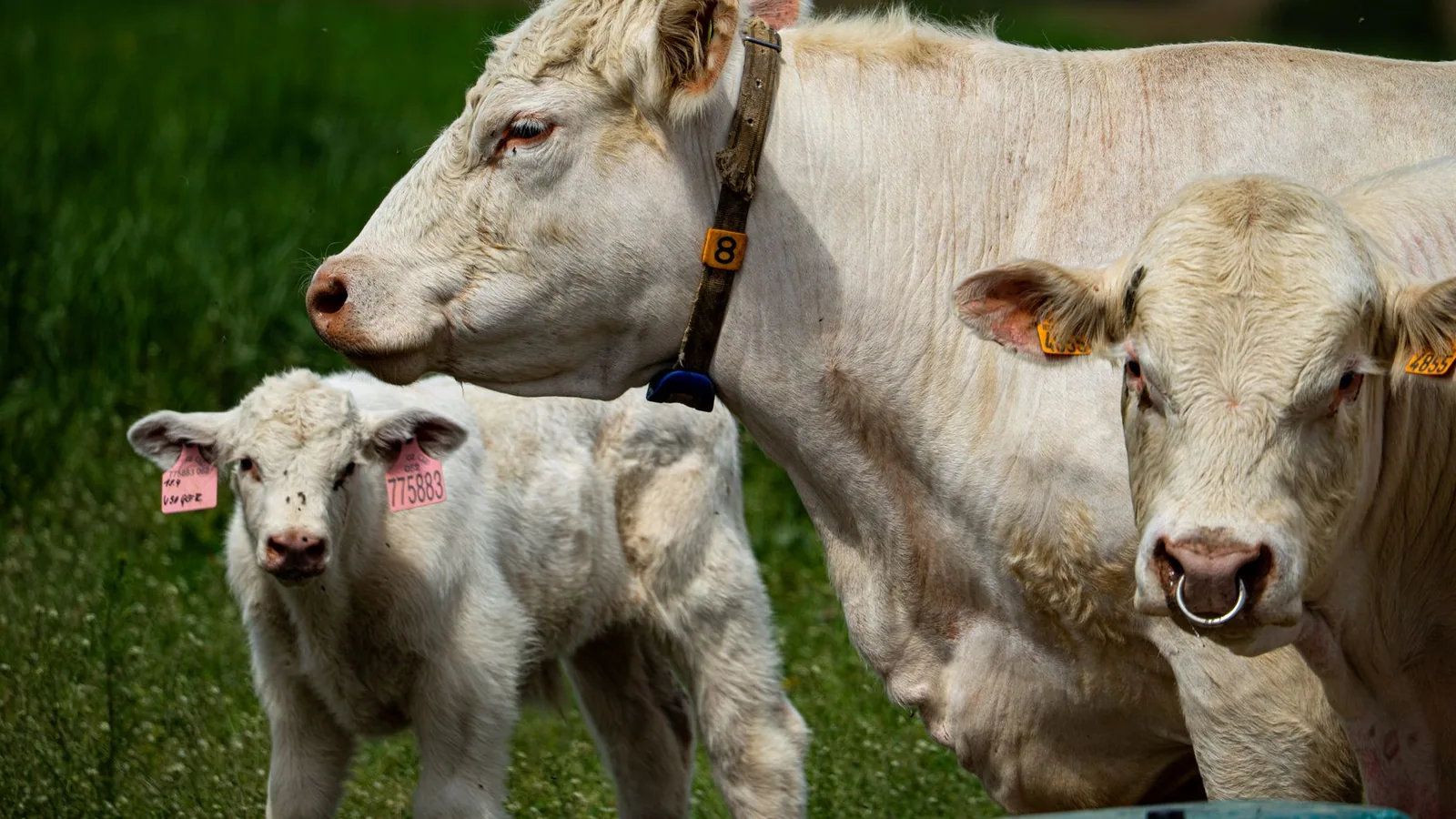
Menangle virus (MenV) first came to scientific attention in August 1997, when it was isolated from stillborn piglets exhibiting severe deformities during an explosive outbreak of reproductive disease at a large commercial piggery in New South Wales, Australia [1]. The index outbreak was clinically devastating: the pregnancy rate and litter size plummeted dramatically, while the proportion of mummified fetuses increased sharply [1]. This epizootic, occurring in a region already sensitized to emergent bat-borne viruses following the identification of Hendra virus in 1994, prompted immediate virological investigation. Initial characterization of the isolate by electron microscopy and biochemical assays revealed a virus with typical paramyxovirus morphology, yet serological testing demonstrated no cross-reactivity with any known paramyxovirus, including mumps virus, Newcastle disease virus, or the recently discovered Hendra virus [2, 1]. This lack of serological relatedness, combined with the unique clinical presentation in pigs and the serological evidence of infection in two of the piggery’s human staff who developed an influenza-like illness with rash, immediately classified MenV as a previously unrecognized agent within the Paramyxoviridae family [1]. The virus was provisionally named “Menangle virus” after the geographical location of the affected piggery, pending definitive molecular classification [1].
2. Taxonomic Classification Within the Family Paramyxoviridae
Definitive taxonomic classification of MenV was achieved through molecular characterization of its genome. Using a sophisticated cDNA subtraction method to isolate viral-specific cDNA from infected cells, Bowden et al. (2001) determined the complete sequences of the nucleocapsid (NP), phosphoprotein (P/V), matrix (M), fusion (F), and hemagglutinin-neuraminidase (HN) genes [2]. Comparative analysis of both nucleotide and deduced amino acid sequences, alongside determination of the P gene mRNA editing strategy, a hallmark feature of the subfamily Paramyxovirinae, unequivocally placed MenV within the genus Rubulavirus [2]. Phylogenetic analyses based on multiple gene sequences confirmed this assignment, showing MenV to be a distinct but clear member of this genus [2]. It is critical to note that the family Paramyxoviridae is a diverse group of enveloped, negative-sense, single-stranded RNA viruses that includes many significant human and animal pathogens. Within this family, the genus Rubulavirus (historically containing mumps virus, human parainfluenza virus types 2 and 4, and simian virus 5/parainfluenza virus 5) is characterized by a specific genome organization and a P gene that encodes multiple proteins via a co-transcriptional RNA editing mechanism. MenV shares these fundamental genetic and structural characteristics. More recent taxonomic refinements, reflecting the burgeoning diversity of bat-borne paramyxoviruses, have led to the establishment of the genus Pararubulavirus within the subfamily Rubulavirinae [3]. MenV, along with Tioman virus, Sosuga virus, and the Achimota viruses, is now classified within this genus, which encompasses many of the emerging bat-derived rubulaviruses with zoonotic potential [3, 4]. This reclassification underscores the distinct evolutionary trajectory and ecological niche of these viruses compared to classical rubulaviruses like mumps virus.
3. Genomic Architecture and Key Molecular Features
The genome of MenV follows the canonical paramyxovirus organization: 3′-N-P/V-M-F-HN-L-5′ [2]. However, several unique molecular details distinguish it from other rubulaviruses. The attachment protein, HN, is of particular evolutionary and functional interest. While the rubulavirus HN protein typically possesses both hemagglutinin (receptor-binding) and neuraminidase (receptor-cleaving) activities, the deduced amino acid sequence of MenV HN exhibits only limited homology to other paramyxovirus attachment proteins [2]. Critically, key amino acid residues considered essential for neuraminidase activity are divergent in MenV HN, strongly suggesting that this protein is unlikely to possess the same degree of neuraminidase function characteristic of other rubulaviruses and respiroviruses [2, 5]. This functional divergence has profound implications for viral entry, release, and host range, as the balance between receptor binding and cleavage is a major determinant of tissue tropism and pathogenicity.
The phosphoprotein (P protein), a non-catalytic subunit of the viral RNA-dependent RNA polymerase, has been extensively characterized at the structural and biophysical level for MenV. Webby et al. (2021) demonstrated that the MenV P protein forms a tetramer in solution but can dissociate into dimers at sub-micromolar concentrations [6]. The protein possesses a central coiled-coil oligomerization domain connected to the C-terminal binding domain by a highly dynamic, intrinsically disordered linker [6]. This linker, which can be modeled as a worm-like chain, is thought to be critical for polymerase translocation along the nucleocapsid template. Nuclear magnetic resonance (NMR) analysis revealed very weak local preferences for alpha-helical and extended beta conformation within this disordered linker, and X-ray crystallography showed a gradual disorder-to-order transition at the interface between the linker and the structured C-terminal binding domain, which wraps dynamically around the surface of the binding domain [6]. This structural plasticity is likely essential for coordinating the multiple molecular interactions required for viral RNA synthesis.
4. Phylogenetic Relationships and Serological Affinity with Other Bat-Borne Rubulaviruses
Phylogenetic analyses consistently demonstrate that MenV clusters robustly with other bat-derived rubulaviruses, forming a distinct clade within the genus Pararubulavirus [5, 7]. Its closest relatives include Tioman virus, isolated from Pteropus bats in Malaysia, and the Tuhoko viruses (ThkPV-1, -2, -3) discovered in Leschenault’s rousette bats (Rousettus leschenaultii) in China [5, 8]. Genome analysis of these Tuhoko viruses revealed a unique genomic feature, the presence of GA instead of AG at the 5th and 6th positions of the 3′-leader sequence, which sets them apart from MenV and Tioman virus [5]. The NEK (neuraminidase) active site residues implicated in neuraminidase activity, which are absent in MenV HN, are present in the Tuhoko virus attachment proteins, highlighting functional diversity even among closely related bat viruses [5]. Further afield, the Achimota viruses (AchPV1 and AchPV2), discovered in African straw-colored fruit bats (Eidolon helvum), also cluster with MenV and Tioman virus in phylogenetic analyses based on the polymerase (L) gene, underscoring the global distribution of these bat rubulaviruses and their common ancestry [4]. Molecular detection studies in Indonesia have identified additional novel paramyxovirus sequences in fruit bats that are phylogenetically related to rubulaviruses, including MenV, indicating that the diversity of this virus group is still far from fully catalogued [7].
Serological cross-reactivity has been observed among members of this group. Using a multiplex microsphere-based immunoassay targeting the MenV nucleocapsid protein and hemagglutinin-neuraminidase protein, Annand et al. (2020) demonstrated that sera from horses exposed to flying foxes in Australia contained antibodies that reacted not only against MenV but also against other related bat rubulaviruses, including Yeppoon virus and Grove virus [9]. This serological cross-reactivity complicates precise seroprevalence studies but also provides a powerful tool for broad surveillance of pararubulavirus spillover events.
5. Natural Reservoir Hosts and Geographic Distribution
Compelling evidence identifies fruit bats of the genus Pteropus (flying foxes) as the natural reservoir host for MenV. Neutralizing antibodies to MenV were detected in Pteropus poliocephalus, P. alecto, and P. scapulatus in Australia shortly after the initial outbreak [1, 8]. Subsequent serosurveys in Papua New Guinea confirmed the presence of rubulavirus antibodies (presumed to be against MenV, Tioman virus, or closely related viruses) in 56% of sampled Pteropus bats [10]. Critically, these seroprevalence rates can exhibit marked temporal and geographic variation; for instance, earlier studies in Papua New Guinea (1996-1999) found no antibodies against MenV in 103 bat samples, whereas later work (2009) found 56% seroprevalence, suggesting dynamic infection dynamics in bat populations over decadal timescales [10, 11]. The virus has been isolated from flying fox urine, confirming shedding and a mechanism for environmental contamination [9]. Beyond Australia and Papua New Guinea, the detection of related rubulaviruses in fruit bats across Southeast Asia (Malaysia, Indonesia, China) and Africa suggests that the geographic range of MenV-like viruses, or at least their close relatives, is far more extensive than initially appreciated, encompassing much of the global distribution of pteropodid and rousettine bats [5, 8, 4, 7]. The World Health Organization (WHO) and the World Organisation for Animal Health (WOAH) consider Menangle virus a significant, albeit not globally widespread, zoonotic pathogen, and its classification as a WOAH-listed pathogen for swine underscores its importance in international veterinary public health.
6. Inter-Species Transmission and Zoonotic Potential
MenV is distinguished by its demonstrated ability to cross species barriers, infecting not only its bat reservoir but also pigs and humans [1]. The 1997 outbreak in the New South Wales piggery provided the first documented evidence of porcine infection, characterized by severe reproductive failure (stillbirths, mummification, and congenital deformities) but without significant respiratory or neurological disease in adult pigs [1, 12]. Critically, two of the piggery workers seroconverted and developed an acute influenza-like illness accompanied by a rash, providing unequivocal evidence of zoonotic transmission [1]. The mechanism of spillover is presumed to involve direct or indirect contact with bat excreta (urine, feces, or saliva) contaminating pig feed or water, followed by amplification within the pig population and subsequent human exposure during farrowing or handling of infected animals [1, 12]. Serological evidence of rubulavirus exposure has also been documented in Australian horses, with some animals showing a greater-than-10-fold increase in antibody titers to MenV N protein in convalescent sera following severe acute respiratory illness, raising the possibility that horses may serve as sentinel species for bat rubulavirus activity [9]. While human infections appear to be mild and self-limiting, contrasting sharply with the high mortality seen with henipavirus infections, the potential for MenV to cause more severe disease in immunocompromised individuals or upon adaptation to new hosts cannot be discounted [3, 1, 13]. The Centers for Disease Control and Prevention (CDC) classifies Menangle virus as a biosafety level 2 (BSL-2) pathogen, reflecting its moderate risk to laboratory workers and the public, yet the virus remains a high-priority target for vaccine development and surveillance due to its zoonotic nature and capacity for future emergence [14, 3].
Molecular Virology and Genomic Organization
Menangle virus (MenV) is a non-segmented, negative-sense, single-stranded RNA virus classified within the genus Rubulavirus of the family Paramyxoviridae [2, 1]. This taxonomic assignment, confirmed shortly after its isolation during the 1997 outbreak of porcine reproductive disease in New South Wales, Australia, placed MenV alongside other zoonotic bat-borne paramyxoviruses, including Tioman virus (TioPV), Tuhoko viruses 1-3 (ThkPV-1-3), Achimota viruses 1 and 2 (AchPV1/2), and Sosuga virus [5, 4, 7]. Initial serological surveys found no cross-reactivity with any known paramyxoviruses, necessitating molecular characterization for definitive classification [2]. The genome of MenV, approximately 15.1-15.2 kilobases in length, adheres to the canonical rubulavirus gene order: 3′-leader, nucleocapsid (N), phosphoprotein (P/V), matrix (M), fusion (F), hemagglutinin-neuraminidase (HN), large polymerase (L), trailer-5′ [2, 5]. A distinctive feature of the MenV 3′-leader sequence, shared with ThkPV-1, -2, and -3 but absent from the archetypal rubulavirus mumps virus, is the presence of guanine-adenine (GA) dinucleotides at the fifth and sixth positions instead of the more common adenine-guanine (AG) motif, a variation that may influence viral polymerase recognition and replication efficiency [5].
Genomic Organization and Encoded Proteins
The N gene encodes the nucleocapsid protein (NP), which encapsidates the viral genomic RNA to form a nuclease-resistant helical ribonucleoprotein complex (RNP) that serves as the template for transcription and replication [15]. Biochemical characterization of bacterially expressed MenV NP has demonstrated its ability to self-assemble into nucleocapsid-like particles devoid of viral RNA, packaging bacterial nucleic acids instead, a property exploited to quantify RNA-to-protein stoichiometry via phosphorus content analysis using inductively coupled plasma mass spectrometry [15]. The NP protein contains a conserved N-terminal domain responsible for RNA binding and oligomerization, while its C-terminal tail interacts with the phosphoprotein (P protein) to recruit the viral polymerase to the RNP template [2].
P Gene RNA Editing and Phosphoprotein Architecture
The P gene of MenV undergoes co-transcriptional RNA editing via a pseudoknot structure within the editing site, a hallmark of rubulaviruses, resulting in the production of two distinct gene products: the phosphoprotein (P) and the V protein [2]. In MenV, the insertion of a single non-templated guanine (G) residue at the editing site shifts the reading frame downstream of a conserved sequence motif, generating the V protein, which shares an N-terminal region with P but possesses a unique, cysteine-rich C-terminal domain that functions as a zinc-binding motif and is critical for interferon antagonism [2, 5]. The P protein is the non-catalytic subunit of the viral RNA-dependent RNA polymerase complex (RdRp, composed of the P and L proteins), and it orchestrates multiple molecular interactions essential for viral RNA synthesis [6].
Recent structural and biophysical investigations have revealed that the MenV P protein exists as a homotetramer in solution, with a propensity to dissociate into dimers at sub-micromolar concentrations [6]. The central coiled-coil domain mediates oligomerization, while the C-terminal region of MenV P exhibits a remarkable degree of intrinsic disorder, a feature increasingly recognized as critical for paramyxovirus polymerase function. Using small-angle X-ray scattering (SAXS) and nuclear magnetic resonance (NMR) spectroscopy, researchers have demonstrated that the linker connecting the coiled-coil to the C-terminal X domain (XD) is globally disordered and can be accurately modeled as a worm-like chain [6]. Nevertheless, NMR analysis indicates very weak, transient local preferences for α-helical and extended β-strand conformations within this linker. Intriguingly, a gradual, "soft" disorder-to-order transition occurs at the interface between the disordered linker and the structured XD, with X-ray crystallography revealing a dynamic interfacial region that wraps asymmetrically across the surface of the X domain [6]. This built-in conformational flexibility is hypothesized to facilitate the translocation of the polymerase along the nucleocapsid template during RNA synthesis, allowing the catalytic L protein to traverse the encapsidated genome without steric impediment.
The Attachment Glycoprotein: A Unique HN
The hemagglutinin-neuraminidase (HN) glycoprotein of MenV is the least conserved structural protein relative to other paramyxoviruses, exhibiting only limited sequence homology to the HN proteins of rubulaviruses such as mumps virus and human parainfluenza virus 2 [2]. Phylogenetic analyses consistently place MenV HN in a distinct branch, corroborating the absence of serological cross-reactivity observed in early studies [2, 5]. Critically, key amino acid residues that are absolutely conserved in the neuraminidase (NA) active site of other rubulavirus and respirovirus HN proteins are absent in MenV HN [2]. In rubulaviruses, the canonical neuraminidase active site comprises a constellation of residues, including arginine, aspartic acid, and glutamic acid at specific positions; MenV HN exhibits substitutions at these positions that are predicted to severely compromise or abolish its sialidase activity [2, 5]. This suggests that MenV HN may function primarily as a receptor-binding protein with minimal, if any, receptor-destroying capability, a feature that fundamentally distinguishes MenV from most other rubulaviruses. This deficient neuraminidase activity may influence the virus's receptor preferences, potentially enabling it to exploit sialic acid linkages or alternative entry receptors distinct from those used by classical rubulaviruses, thereby influencing its tissue tropism and zoonotic potential. Indeed, comparative analysis with ThkPV-1, -2, and -3 demonstrated that these related viruses retain the key amino acids required for neuraminidase activity, underscoring the unusual nature of the MenV HN [5].
Fusion Protein and Membrane Fusion Machinery
The fusion (F) glycoprotein of MenV is a type I transmembrane protein responsible for mediating the merger of the viral envelope with the host cell membrane following receptor binding by HN [14, 2]. The F protein is synthesized as an inactive precursor, F0, which must be cleaved by host cell proteases into the disulfide-linked F1 (C-terminal, membrane-anchored) and F2 (N-terminal, external) subunits to render it fusion-competent [14]. The cleavage site sequence within F0 is a critical determinant of tissue tropism and pathogenicity, as it dictates which host proteases can activate the protein. For pararubulaviruses such as MenV, the F protein cleavage site typically contains a monobasic motif (a single arginine or lysine residue), suggesting activation by furin-like proprotein convertases in the trans-Golgi network or by extracellular proteases, which may restrict the range of susceptible cell types [14]. The F protein contains two heptad repeat (HR1 and HR2) domains that, upon activation, refold to form a six-helix bundle structure that brings the viral and cellular membranes into close apposition, driving membrane fusion [14].
Matrix Protein and the L Polymerase
The matrix (M) protein, encoded by the M gene, is the most abundant virion protein and is a structural bridge, lining the inner leaflet of the viral envelope and orchestrating the assembly and budding of progeny virions by coordinating interactions between the RNP, the envelope glycoproteins, and the host cell membrane [2]. The L gene encodes the large (L) protein, the catalytic subunit of the RNA-dependent RNA polymerase complex. The L protein harbors all enzymatic activities required for viral RNA synthesis, including the RNA-dependent RNA polymerase and the mRNA capping and polyadenylation activities [2, 13]. A broad-spectrum RT-PCR approach targeting the highly conserved motifs within the L protein, particularly the “GDNQ” polymerase motif within catalytic domain III, has been instrumental in the discovery of novel paramyxoviruses, including the Tuhoko viruses from Chinese bats that cluster phylogenetically with MenV [5, 7]. The L protein of MenV shares critical structural features with other rubulavirus polymerases, including conserved regions responsible for template binding, nucleotide addition, and processivity, though detailed structural data for the MenV L protein remain limited [2].
Molecular Pathogenesis and Host-Virus Interactions
Attachment and Entry: The Paramyxoviral Fusion Machinery
The initial stages of Menangle virus (MenV) infection are governed by the coordinated action of the viral attachment protein, hemagglutinin-neuraminidase (HN), and the fusion (F) glycoprotein. As a member of the genus Rubulavirus within the family Paramyxoviridae, MenV employs a canonical entry strategy that has been refined through molecular evolution. However, the MenV HN protein possesses a singular distinction among rubulaviruses: a significantly reduced neuraminidase activity. Classical rubulavirus and respirovirus HN proteins are bifunctional, mediating both receptor binding (hemagglutinin activity) and receptor cleavage (neuraminidase activity), which allows the virus to bud from the cell without being trapped by the sialic acid moieties on the host cell surface. Sequence analysis of the MenV HN gene, as elucidated by Bowden et al. [2], reveals key amino acid substitutions within the conserved active site residues that are critical for neuraminidase function. Specifically, residues considered essential for sialic acid hydrolysis are either absent or replaced in MenV, suggesting that the attachment protein has evolved toward a purely hemagglutinating function, relying instead on the host cellular machinery or other viral mechanisms for efficient egress. This functional divergence has profound implications for viral pathogenesis, as it may alter the kinetics of viral spread, the tropism for specific sialic acid linkages, and the nature of the host immune response directed against the viral surface.
The F protein, responsible for membrane fusion following receptor binding, is synthesized as an inactive precursor (F0) that must be cleaved by host cell proteases to become fusogenic. The precise cleavage site of MenV F protein, while not fully characterized in the available literature, is predicted to contain a basic amino acid motif typical of rubulaviruses, potentially rendering it susceptible to ubiquitous intracellular furin-like proteases. This would allow for systemic spread of the virus without restriction to tissues expressing trypsin-like enzymes. Following HN binding to an, as yet, unidentified sialic acid-containing receptor, a conformational cascade is triggered in F, culminating in the insertion of the fusion peptide into the target cell membrane and the formation of a six-helix bundle that drives membrane merger. The structural dynamics of this process are exquisitely regulated and represent a critical host-virus interface. The relatively mild clinical presentation in humans, typically an influenza-like illness with rash [1], compared to the devastating mortality of henipaviruses, may correlate with differences in the efficiency of F-mediated fusion or the distribution of functional receptors across human tissues. Computational vaccine design efforts now actively target these transmembrane proteins (F and HN) to elicit neutralizing antibodies, underscoring their primacy in the host-virus interaction [14].
Replication Complex and the Phosphoprotein (P) Hub
Once inside the host cell, the negative-sense single-stranded RNA genome of MenV must be transcribed and replicated by the viral RNA-dependent [RNA polymerase](/knowledge/bioinformatics/rna-polymerase-structure-transcription-mechanisms 2) (RdRp) complex. This complex is composed of the large (L) catalytic subunit and the phosphoprotein (P), which acts as a non-catalytic cofactor and a central organizing hub. The structural biology of the MenV P protein, investigated in meticulous detail by Webby et al. [6], reveals a sophisticated architecture essential for polymerase processivity. MenV P is a tetrameric protein in solution, organized around a central coiled-coil domain. This oligomeric state is critical for function, as it positions the downstream C-terminal domains (XD) to engage with the viral nucleocapsid (N protein) that encapsulates the genomic RNA. However, a striking feature of the MenV P protein is its ability to dissociate into dimers at sub-micromolar concentrations, suggesting a dynamic equilibrium that may be regulated during the viral life cycle to modulate polymerase assembly [6].
A defining aspect of MenV P is the presence of a long, intrinsically disordered linker region connecting the central oligomerization domain to the C-terminal XD. This linker is globally disordered and can be modeled as a worm-like chain, but nuclear magnetic resonance (NMR) analysis reveals subtle local preferences for alpha-helical and extended beta conformations [6]. This inherent flexibility is not merely a structural curiosity; it is a functional necessity for paramyxovirus transcription and replication. The linker is believed to act as a molecular tether, allowing the polymerase complex to translocate along the helical nucleocapsid template without steric hindrance. The "soft boundary" between disorder and order at the interface of the linker and the structured XD, visualized by X-ray crystallography as a dynamic interfacial structure that wraps the surface of the binding domain, suggests a controlled mechanism for regulating access to the nucleocapsid. This graded structural transition likely facilitates the precise stepwise movements required for RNA synthesis.
The P gene of rubulaviruses, including MenV, also encodes additional proteins through a co-transcriptional mRNA editing mechanism. During transcription, the viral polymerase stutters at a specific editing site within the P gene, inserting non-templated guanosine residues. This process yields a frameshifted mRNA that encodes the V protein, a conserved paramyxoviral accessory protein. The P gene mRNA editing strategy was confirmed for MenV by Bowden et al. [2]. The V protein is a major virulence factor that targets the host innate immune system, particularly the interferon (IFN) signaling cascade, through its C-terminal cysteine-rich domain. This domain coordinates zinc and possesses ubiquitin ligase activity, targeting STAT proteins (Signal Transducers and Activators of Transcription) for proteasomal degradation, thereby crippling the host's antiviral state. The balance between P and V expression, dictated by the editing frequency, is a critical determinant of viral pathogenesis.
Cytopathology and Cellular Effects
Infection of permissive cell lines, such as Vero cells (African green monkey kidney epithelial cells), with MenV results in a pronounced cytopathic effect (CPE) characterized by syncytium formation. This is a direct consequence of F protein-mediated cell-cell fusion, which is driven by the surface expression of viral glycoproteins on infected cells interacting with receptors on adjacent uninfected cells. This phenomenon allows the virus to spread directly from cell to cell, evading antibody neutralization. In the initial outbreak investigation, Philbey et al. [1] noted that the virus was isolated from stillborn piglets with severe deformities, indicating that in the porcine host, MenV has a strong tropism for rapidly dividing fetal tissues, particularly the brain and spinal cord. The resulting lesions, arthrogryposis (joint contractures), brachygnathia (shortened lower jaw), and hydranencephaly, are hallmarks of in utero viral infection that disrupts neuronal development and ossification.
The molecular basis for this profound teratogenicity likely involves the induction of apoptosis or necrosis in neural progenitor cells, compounded by a direct viral blockade of the host innate immune response. The V protein, by antagonizing STAT signaling, renders infected cells and potentially uninfected bystander cells highly susceptible to subsequent infection and less capable of initiating a type I IFN response. This immunosuppression at the maternal-fetal interface in pigs would allow unchecked viral replication, leading to the severe fetal pathologies observed. In humans, the clinical picture is significantly milder, with only two piggery workers developing an acute, self-limiting febrile illness with rash and antibodies detected post-recovery [1]. This stark difference in pathogenesis between the natural porcine host and the spillover human host suggests that species-specific differences in receptor distribution, innate immune signalling efficiency, or intracellular restriction factors play a decisive role in determining disease outcome.
Immune Evasion and Host Range Determinants
The ability of MenV to maintain a stable reservoir in pteropid bats (fruit bats of the genus Pteropus) without causing overt disease is a critical aspect of its ecology. Bats have evolved a unique relationship with viruses, exhibiting a dampened inflammatory response that allows high-level viral replication without immunopathology [16]. MenV appears to be exquisitely adapted to this niche, as serological surveys consistently demonstrate high prevalence of antibodies in flying fox populations across Australia, Papua New Guinea, and potentially Indonesia [2, 1, 8, 10, 7]. The lack of detectable disease in bats is likely a consequence of co-evolutionary adaptations that include a constitutively active IFN system that is rapidly mobilized but tightly regulated, preventing the deleterious effects of chronic inflammation.
Spillover from bats to pigs and humans represents a substantial host-jump event. The serological evidence of MenV or closely related rubulavirus infection in Australian horses, associated with severe respiratory illness and a greater-than ten-fold increase in antibody titers to MenV N protein [9], further expands the known host range and positions horses as potential sentinels for emerging rubulavirus activity. The molecular determinants that facilitate these cross-species transmissions are poorly understood but likely involve the HN attachment protein's specificity for sialic acid linkages. While the HN protein lacks robust neuraminidase activity, it must still bind with sufficient affinity to the sialic acid receptors present on swine and human respiratory epithelia. Minor changes in the receptor-binding pocket could dramatically alter host tropism, analogous to the well-documented switches in influenza virus host range. The presence of neutralizing antibodies to multiple rubulaviruses in bat sera [10] indicates that co-infection or sequential infection is common, which could drive antigenic drift or recombination, as can be detected with tools like RDP4 [17], though no evidence of MenV recombination is currently available.
The V protein's role in species-specific immune evasion is paramount. For MenV to replicate in a new host, the V protein must effectively antagonize the host's STAT proteins. The efficiency of this antagonism can vary significantly even between closely related species. A V protein optimized for bat STAT proteins may be less effective against porcine or human STATs, placing a bottleneck on replication. The fact that MenV causes only mild, self-limiting disease in humans suggests that the virus is under strong restriction by the human innate immune system, whereas in pigs, the blockade is more effective, leading to severe fetal pathology. This differential susceptibility highlights the fragile nature of the cross-species barrier and the potential for viral adaptation upon introduction to a new host population.
Pathogenesis in Pigs and Experimental Animal Models
The natural pathogenesis of MenV in pigs is characterized by a reproductive tragedy with subclinical infection in adult animals. The index outbreak in New South Wales, Australia, in 1997 saw a sharp drop in farrowing rates and a surge in mummified, stillborn, and deformed piglets [1]. Viremia in pregnant sows allows the virus to cross the placental barrier and infect the developing fetus. The fetal tissues, lacking a fully competent adaptive immune system and with high rates of cell division, provide an ideal environment for rapid viral replication. The resulting neural damage (hydranencephaly) and musculoskeletal deformities are consistent with infection targeting the germinal neuroepithelium.
Studies of related bat rubulaviruses, such as the Achimota viruses (AchPV1 and AchPV2), have provided a comparative model for understanding MenV pathogenesis. In experimental animal models, Achimota viruses were shown to infect ferrets and guinea pigs, causing seroconversion, viral shedding, and respiratory disease in ferrets [4]. Viral genome was detected across many tissues, but infectious virus could only be isolated from ferret tissues [4], indicating that tissue-specific restriction mechanisms exist even in susceptible species. These findings are directly translatable to MenV, suggesting that while the virus can disseminate widely, productive replication may be confined to specific cell types, such as respiratory epithelium and fetal neural tissue. The failure to infect mice in the Achimota studies [4] underscores the narrow host range and the importance of using appropriate animal models for pathogenesis and vaccine testing. For MenV, experimental infection studies in guinea pigs or ferrets would be invaluable for elucidating the precise sequence of events from respiratory entry to placental transfer, and for evaluating the efficacy of the multiepitope vaccines currently being designed in silico [14]. The World Organisation for Animal Health (WOAH) and the Food and Agriculture Organization of the United Nations (FAO) recognize the potential for rubulaviruses to impact swine production, underscoring the need for continued surveillance and research into their pathogenesis.
Epidemiology and Geographic Distribution
The epidemiological profile of Menangle virus (MenV) presents a compelling case study in the emergence of bat-borne zoonotic paramyxoviruses, characterized by a circumscribed geographic range, a discrete wildlife reservoir, and sporadic spillover events into domestic animal and human populations. Unlike the widespread endemicity observed for many paramyxoviruses, MenV’s documented distribution remains primarily confined to the Australasian region, yet serological and molecular evidence suggests a potentially broader, albeit cryptic, circulation among pteropid bat populations across the Indo-Pacific. Understanding this distribution requires a meticulous dissection of index outbreak data, reservoir host ecology, serosurveillance efforts, and the abiotic and biotic factors that govern spillover dynamics.
Discovery and Index Outbreak: New South Wales, Australia (1997)
The initial recognition of MenV as a distinct pathogenic entity occurred in August 1997, following an explosive outbreak of severe reproductive disease in a commercial piggery located in New South Wales, Australia [2, 1]. This index outbreak serves as the cornerstone of our epidemiological understanding. The affected facility experienced a dramatic and rapid decline in reproductive performance, characterized by a marked decrease in pregnancy rates and litter sizes, concomitant with a significant increase in the proportion of mummified fetuses and stillborn piglets exhibiting congenital deformities, most notably arthrogryposis and craniofacial malformations [1]. The temporal clustering and clinical presentation strongly indicated an acute, highly transmissible infectious agent.
Epidemiological investigation traced the likely source of the viral incursion to the presence of large numbers of fruit bats (flying foxes, genus Pteropus) roosting in trees immediately adjacent to the piggery [1]. This spatial association provided the initial, critical link to a wildlife reservoir. Concurrent with the porcine epizootic, two of the piggery’s human staff members developed an acute, self-limiting febrile illness accompanied by a maculopapular rash, and serological testing confirmed infection with MenV [1]. This contemporaneous occurrence of human disease alongside the porcine outbreak firmly established MenV as a zoonotic pathogen with the capacity for direct or indirect transmission from pigs to humans. The outbreak was effectively contained through the implementation of strict quarantine and biosecurity measures, but it illuminated the vulnerability of intensive livestock operations to virus spillover from nearby wildlife populations.
Reservoir Host Ecology and Geographic Range of Bat Seroprevalence
The identification of pteropid bats as the natural reservoir for MenV [2] has profound implications for its geographic distribution. The genus Pteropus is distributed across a vast swath of the Indo-Pacific, encompassing Australia, Papua New Guinea, Indonesia, and numerous island nations of the South Pacific. The geographic range of MenV is therefore theoretically limited only by the distribution of its primary reservoir hosts and the ecological conditions that facilitate viral maintenance within these populations.
Serosurveillance studies have provided critical data on the extent of MenV circulation. In the immediate aftermath of the Australian outbreak, serological evidence of MenV or a closely related rubulavirus was detected in fruit bats across the affected region. Subsequent, more extensive surveys have broadened this geographic picture. A pivotal study in Papua New Guinea (PNG) found a remarkably high seroprevalence of rubulavirus antibodies (potentially cross-reactive with MenV and Tioman virus) in pteropid bats. Breed et al. (2010) reported that 56% of 66 bats sampled at three locations in PNG harbored antibodies against rubulaviruses, with 36% of these bats showing concurrent seropositivity for both henipaviruses and rubulaviruses, suggesting active co-circulation [10]. However, intriguingly, earlier surveys from 1996-1999 in PNG failed to detect any neutralizing antibodies against MenV in 103 fruit bat samples, while later surveys (2009) showed high prevalence [11]. This temporal disparity is profound; Field et al. (2013) argued this likely reflects a major shift in infection dynamics within bat populations over a decade, possibly driven by ecological perturbations such as habitat loss, altered roosting behavior, or changes in bat population density and immunity [11]. This dynamic nature of viral prevalence in reservoir hosts is a critical epidemiological feature.
Beyond Australia and PNG, the search for MenV and related pararubulaviruses has extended northward. Molecular surveillance in Indonesia detected novel paramyxovirus RNA sequences in fruit bats that clustered phylogenetically within the rubulavirus clade, closely related to MenV and Tioman virus, from four locations across the archipelago [7]. Similarly, in China, the discovery of Tuhoko virus 1, 2, and 3 (ThkPVs) in Leschenault’s rousette bats (Rousettus leschenaultii) revealed viruses that are phylogenetically most closely related to MenV and Tioman virus [5]. Antibodies to these novel ThkPVs were detected in 48-60% of sampled rousette bats, indicating sustained enzootic transmission within these Chinese fruit bat populations [5]. This finding extends the potential geographic range of MenV-like rubulaviruses deep into Southeast and East Asia. The presence of these closely related viruses with distinct serological profiles confounds definitive geographic mapping of MenV sensu stricto, but strongly suggests that a diverse complex of MenV-related rubulaviruses circulates widely across the range of pteropid and rousette bats.
Spillover Events and Secondary Host Epidemiology
The epidemiology of MenV is defined by rare, sporadic spillover events into secondary hosts, primarily pigs and, less frequently, horses and humans. To date, no evidence suggests sustained transmission of MenV in non-reservoir hosts.
Porcine Epizootiology: The index 1997 outbreak remains the only documented epizootic of MenV in pigs. While the virus caused devastating reproductive loss, it did not establish endemicity in the swine population. The biological basis for this is unclear but may relate to the poor transmission efficiency of MenV in pigs compared to other swine viruses, or the rapid implementation of stamping-out policies. No subsequent outbreaks of MenV-associated reproductive disease in pigs have been reported in Australia or elsewhere, suggesting that spillover from bats into pig populations is a low-probability event dependent on specific ecological conditions (e.g., high-density bat roosts near piggeries, seasonal food stress driving bats into agricultural areas). The virus has been classified among the emerging and re-emerging swine viruses with zoonotic potential, but its impact on global swine health has been limited to this single, well-characterized event [12].
Equine Spillover: Perhaps the most significant recent epidemiological development is the demonstration of rubulavirus spillover into horses in Australia. Annand et al. (2020) conducted a serosurvey of Australian horses and found evidence of IgG antibodies to MenV nucleocapsid and hemagglutinin-neuraminidase proteins in 34% of horses with high perceived exposure to flying foxes, compared to negligible responses in horses without such exposure [9]. Critically, they documented two clinical cases of severe acute respiratory illness in young geldings in 2016. Convalescent sera from these horses showed a greater-than-10-fold increase in antibody titers against MenV N protein, and immunofluorescence assays confirmed specific antibodies to MenV, Yeppoon virus, and Grove virus [9]. This finding is of immense epidemiological and public health importance. It identifies horses as a sentinel species for rubulavirus activity and suggests that spillover from flying foxes into horses may be more frequent than previously recognized, potentially occurring on a regular but subclinical basis. The clinical significance of these infections in horses, and the risk of onward transmission to humans, remains to be fully elucidated.
Human Epidemiology: Human infection with MenV has been documented only in the two individuals directly exposed to infected pigs during the 1997 outbreak, and in no subsequent cases [1]. Both patients developed an influenza-like illness with skin rash and seroconverted. MenV is classified among zoonotic pararubulaviruses that typically induce mild symptoms or remain asymptomatic in humans, in stark contrast to the high mortality associated with henipaviruses like Hendra and Nipah [3]. There is no evidence of human-to-human transmission. The true prevalence of human infection, particularly among individuals with high occupational exposure to bats or bat habitats (e.g., ecotourists, wildlife researchers, fruit orchard workers), is unknown. A 2011 review by Wang highlighted that despite the surge in zoonotic virus discovery, the drivers for spillover remain poorly understood, and MenV is a prime example of a virus with demonstrated zoonotic potential that remains cryptic in its human epidemiology [18].
Geographic Distribution Synthesis and Future Considerations
Synthesizing the available data, the confirmed geographic distribution of MenV infection centers on eastern Australia (New South Wales and Queensland), with serological evidence of closely related or cross-reactive rubulaviruses extending into Papua New Guinea, Indonesia, and mainland China [5, 10, 7, 11]. The virus is enzootic in pteropid bat populations across this region, but its prevalence is dynamic and likely influenced by ecological factors such as bat population density, habitat fragmentation, and food availability [11].
The primary geographic risk for MenV spillover is therefore areas of high human-livestock-wildlife interface, particularly where intensive pig or horse production occurs in proximity to large Pteropus roosts. The World Organisation for Animal Health (WOAH) is aware of the potential risk posed by emerging paramyxoviruses from bats, and the Centers for Disease Control and Prevention (CDC) includes MenV in its list of potential zoonotic threats, though no travel advisories or surveillance programs specifically target this virus. The virus remains largely inconspicuous, but the detection of antibodies in horses [9] and the discovery of novel, related viruses across Asia [5, 7] suggest that the geographic and host range of MenV-like rubulaviruses is far from fully mapped. The epidemiology and distribution of MenV are thus characterized by a vast, enzootic reservoir in bat populations across the Indo-Pacific, punctuated by rare but significant spillover events that can cause disease in domestic animals and humans, underscoring the need for continued sentinel surveillance and enhanced cross-sectoral collaboration under a One Health framework.
Diagnostic Methods and Clinical Detection
The detection of Menangle virus (MenV) infection necessitates a multi-pronged diagnostic approach that integrates classical virology, molecular biology, and advanced serological platforms. The clinical presentation, ranging from severe reproductive failure in swine to mild influenza-like illness in humans, dictates that diagnostic algorithms must be both highly sensitive for acute infection and robust for retrospective surveillance [1, 19]. The zoonotic potential of MenV, coupled with its circulation in pteropid bat reservoirs, places a premium on diagnostic systems capable of differentiating this rubulavirus from other emerging paramyxoviruses such as Tioman virus, Hendra virus, and Nipah virus, which may co-circulate in overlapping geographic and host ranges [3, 10]. The World Organisation for Animal Health (WOAH) and the World Health Organization (WHO) have underscored the need for validated diagnostic capacity for emerging zoonotic paramyxoviruses, given the unpredictable nature of spillover events from bat reservoirs [3, 18].
Initial Detection and Clinical Context
The index identification of MenV in 1997 was a sentinel event in diagnostic virology, driven by an abrupt epizootic of porcine reproductive disease in New South Wales, Australia. Clinically, affected piggeries experienced a dramatic decrease in pregnancy rates, reduced litter sizes, and a marked increase in the proportion of mummified fetuses and stillborn piglets with congenital deformities, particularly arthrogryposis and craniofacial malformations [1]. These clinical signs are pathognomonic for a rubulavirus-induced reproductive syndrome in swine, but definitive diagnosis requires laboratory confirmation. In human cases, the clinical detection is more challenging, as the influenza-like illness with rash and malaise observed in two piggery workers is non-specific and could be attributed to numerous other viral etiologies [1]. Therefore, the diagnostic workup must be triggered by epidemiological linkage to affected swine herds or, in the case of bat reservoir surveillance, by targeted screening of high-risk populations [9, 19].
Virus Isolation and Classical Virology
Virus isolation remains a cornerstone for definitive diagnosis and for generating high-titer stocks for downstream characterization. MenV was originally isolated from the brain and lung tissues of stillborn piglets using cell culture systems. The virus replicates readily in Vero cells (African green monkey kidney epithelial cells), producing a characteristic cytopathic effect (CPE) within 3-5 days post-inoculation. The CPE is typified by syncytium formation, the fusion of adjacent cells into multinucleated giant cells, followed by progressive cell rounding, detachment, and lysis [1]. This fusogenic activity is mediated by the viral fusion (F) protein and is a hallmark of paramyxovirus infection in permissive cell lines. While Vero cells are the standard, primary porcine kidney cells or fetal porcine lung cells have also proven susceptible for primary isolation from field samples. For specimens derived from bat reservoirs or human cases, the use of a susceptible cell line is critical to avoid false negatives. However, virus isolation is time-consuming (often requiring 7-14 days), requires Biosafety Level 2 (BSL-2) or BSL-3 containment depending on the sample source and regional risk assessment, and is not amenable to high-throughput screening [1, 19]. Following isolation, the virus must be identified definitively, as CPE alone is insufficient given that other paramyxoviruses (e.g., Tioman virus, Nipah virus) can produce similar morphological changes. Confirmatory identification is achieved through electron microscopy, which reveals the characteristic “herringbone” nucleocapsid structure of rubulaviruses, or through specific immune-labeling [1]. Nonetheless, the labor-intensive nature of culture limits its utility for outbreak response, where rapid molecular detection is preferred.
Serological Diagnostics: From Classical to High-Throughput Platforms
Serology provides essential evidence of past or current infection, particularly for surveillance in reservoir hosts and for confirming zoonotic transmission. The original outbreak was investigated using a serum neutralization test (SNT), which remains a gold standard for specificity. The SNT measures the ability of patient or animal sera to neutralize the infectivity of a known MenV stock in cell culture, with a reciprocal titer of ≥5 considered indicative of prior exposure [1, 11]. This assay is highly specific, even among closely related rubulaviruses, as demonstrated by the absence of cross-neutralization with other paramyxoviruses in early studies [2, 1]. However, SNT is laborious, requires live virus, and is poorly suited for large-scale surveillance.
The development of recombinant protein-based assays has revolutionized serosurveillance. A multiplex microsphere-based immunoassay (MIA) has been developed using the MenV nucleocapsid (N) protein and the hemagglutinin-neuraminidase (HN) protein as antigens [9]. The N protein is highly immunogenic and conserved among rubulaviruses, making it an ideal target for screening, while the HN protein provides additional serotype-specific discrimination. In this MIA format, antigen-coupled microspheres are incubated with test sera, and bound IgG is detected using a fluorescent reporter system on a flow-cytometric platform (e.g., Luminex). This method allows for high-throughput processing of hundreds of samples per day and permits simultaneous detection of antibodies against multiple pathogens in a single well, a critical advantage for syndrome-based surveillance in horses and bats [9]. For example, in a study of Australian horses, a Bayesian latent class model estimated a prior prevalence of ~20% for MenV antibodies, with 34% of high-exposure horses showing significant IgG reactivity to MenV N protein [9].
Immunofluorescence assays (IFA) serve as a confirmatory tool, particularly when MIA results are equivocal. IFA uses Vero cells infected with MenV (or related rubulaviruses such as Yeppoon virus or Grove virus) as fixed antigen substrates. Positive sera produce a characteristic cytoplasmic and membrane-associated fluorescence pattern, confirming the presence of antibodies that recognize native, conformationally intact viral proteins [9]. IFA demonstrated that convalescent sera from two horses with severe acute respiratory illness exhibited a greater-than-10-fold increase in median fluorescence intensity (MFI) against MenV N protein, rising from 965 in acute sera to 12,886 in convalescent sera [9]. This seroconversion provides strong evidence of recent infection and highlights the utility of paired acute and convalescent sera for clinical diagnosis. The Food and Agriculture Organization of the United Nations (FAO) has recognized the importance of such sentinel surveillance in horses for early warning of bat-borne virus spillover [9].
Enzyme-linked immunosorbent assays (ELISA) using recombinant N protein have been employed extensively for bat serosurveillance. Studies in Papua New Guinea and Indonesia detected rubulavirus (Tioman/Menangle) antibodies in 56% of pteropid bats using recombinant N-protein-based ELISA [10, 11]. However, a critical caveat is that the high degree of N protein conservation among rubulaviruses can lead to serological cross-reactivity, meaning that a positive ELISA signal may indicate exposure to any related rubulavirus rather than MenV specifically. This was evident in studies where neutralizing antibody titers to Hendra virus exceeded those to Nipah virus, suggesting cross-neutralization by an unknown henipavirus [11]. For definitive species-level serodiagnosis, the virus neutralization test (VNT) remains the most specific method, though its limited throughput and requirement for infectious virus constrain its use.
Molecular Diagnostics: RT-PCR and Genomic Detection
Reverse transcription polymerase chain reaction (RT-PCR) is the primary modality for detecting acute MenV infection, particularly in clinical samples from stillborn piglets, nasopharyngeal swabs from humans, or urine/fecal samples from bats. The high RNA yield from rubulaviruses makes RT-PCR a sensitive method. The original molecular characterization of MenV utilized a cDNA subtraction method to obtain viral-specific cDNA from infected cells [2]. Today, real-time RT-PCR assays targeting conserved regions of the polymerase (L) gene or the nucleocapsid (N) gene are the standard. The L gene is preferred because it is the most conserved paramyxoviral gene, allowing for broad-spectrum detection of known and novel rubulaviruses in addition to specific MenV detection [7]. A semi-nested broad-spectrum RT-PCR targeting the paramyxovirus L gene was used to discover novel paramyxoviruses in fruit bats from Indonesia, detecting MenV-related sequences that were subsequently confirmed by phylogenetic analysis [7]. This approach is invaluable for pathogen discovery and for identifying spillover events from bats to livestock.
For specific MenV detection, single-plex or duplex real-time RT-PCR assays can be designed based on unique sequences within the HN or F genes. The HN gene of MenV exhibits limited sequence homology with other paramyxoviruses, including differences in key amino acid residues responsible for neuraminidase activity, which enables the design of highly specific primers that do not cross-react with Tioman virus or other rubulaviruses [2]. The cycling conditions typically involve an initial reverse transcription step at 50°C for 30 minutes, followed by 40 cycles of denaturation (95°C for 15 seconds) and annealing/extension (60°C for 60 seconds). A positive result is indicated by a cycle threshold (Ct) value < 38. Melting curve analysis using SYBR Green chemistry can provide additional confirmation by differentiating MenV-specific amplicons based on dissociation temperature. For quantitative applications, probe-based assays (e.g., TaqMan) offer greater sensitivity and specificity, with a limit of detection often below 10 RNA copies per reaction.
Genome sequencing and phylogenetic analysis provide the highest resolution for MenV diagnosis and epidemiological tracking. Complete genome sequencing (15,246 nucleotides) has been achieved for MenV, revealing its classification as a rubulavirus with the gene order 3′-N-P/V-M-F-HN-L-5′ [2, 5]. The discovery of mRNA editing in the P gene (adding two G residues to produce the V protein) is a molecular hallmark of rubulaviruses and can be used as a confirmatory diagnostic marker [2]. Sequencing of the L gene or complete genome allows for robust phylogenetic analysis to distinguish MenV from closely related rubulaviruses such as Tioman virus, Tuhoko virus 1, 2, and 3, and Achimota virus 1 and 2 [5, 4]. The high genetic diversity of these viruses, particularly in the hypervariable regions of the F and HN genes, means that a single negative RT-PCR result does not rule out infection, especially in novel geographic regions.
Differential Diagnosis and Interpretive Challenges
Diagnostic interpretation must account for the clinical and serological overlap with other swine and zoonotic pathogens. In pigs, MenV must be differentiated from other causes of reproductive failure such as porcine reproductive and respiratory syndrome virus (PRRSV), porcine circovirus type 2 (PCV2), classical swine fever virus (CSFV), and porcine parvovirus [12, 20]. In humans, the influenza-like illness and rash must be distinguished from infections with other zoonotic paramyxoviruses (Nipah, Hendra, Tioman) as well as from arboviruses (e.g., West Nile virus, Ross River virus) and enteroviruses [8, 9]. The lack of neuraminidase activity in MenV HN is a biochemical feature that can be exploited for differentiation from other rubulaviruses that possess this activity, but this is not typically used in routine diagnostic labs [2].
A significant interpretive challenge arises from serological cross-reactivity. The MIA and ELISA assays using N protein cannot always distinguish between MenV and other rubulavirus infections (e.g., Tioman virus, Yeppoon virus, Grove virus), as these proteins share conserved epitopes [9]. Consequently, serosurveillance studies often report antibodies to “rubulavirus” or “Tioman/Menangle virus” collectively, rather than to MenV alone [10, 11]. To resolve this, confirmatory testing with a VNT using MenV-specific virus particles is essential. The finding of antibodies in 36% of bats to both henipaviruses and rubulaviruses indicates concurrent or sequential infections, complicating the attribution of clinical signs to a single pathogen [10].
Another diagnostic pitfall is the detection of viral RNA in bat urine or feces without evidence of active infection or disease. Many paramyxoviruses, including MenV, are shed intermittently and at low levels in bat excreta, making RT-PCR detection sporadic even in seropositive animals [7]. The biological significance of such detection must be interpreted with caution, as it may represent transient shedding from a persistently infected host rather than an active outbreak. This is particularly relevant for surveillance programs aimed at predicting spillover risk, where the presence of viral RNA in environmental samples (e.g., urine collected under bat roosts) is a strong indicator of viral circulation but does not confirm a threat to human or livestock populations [9, 18]. The use of quantitative RT-PCR can help by providing a Ct value; lower Ct values (higher RNA copy numbers) are more suggestive of active replication and higher shedding, whereas high Ct values may indicate low-level or residual RNA.
Immunological Responses and Vaccine Development
The development of effective prophylactic and therapeutic interventions against Menangle virus (MenV) necessitates a comprehensive understanding of the host immunological responses elicited during infection and the subsequent identification of immunogenic targets for vaccine design. As a zoonotic paramyxovirus of the genus Rubulavirus, MenV presents a unique set of challenges and opportunities for vaccinology, particularly given its bat reservoir, its capacity for spillover into swine and humans, and the limited cross-reactivity observed with other paramyxoviruses [2, 1]. The immunological landscape of MenV infection is defined by the interplay between viral structural proteins and the host innate and adaptive immune systems, a relationship that is now being exploited through advanced computational and structural biology approaches to accelerate vaccine development.
Innate Immune Recognition and Evasion Mechanisms
The initial host defense against MenV infection is mediated by the innate immune system, which relies on pattern recognition receptors (PRRs) to detect viral pathogen-associated molecular patterns (PAMPs). While specific studies on MenV innate immune interactions are limited, insights can be drawn from related rubulaviruses and paramyxoviruses. The viral genome, a single-stranded negative-sense RNA, is recognized primarily by endosomal Toll-like receptors (TLR3, TLR7/8) and cytosolic RIG-I-like receptors (RLRs), such as RIG-I and MDA5. Upon infection, the viral nucleocapsid (N) protein, which encapsidates the genomic RNA, is a critical PAMP, as the RNA within the nucleocapsid is protected yet still accessible to innate sensors [15]. The phosphoprotein (P) protein, a non-catalytic subunit of the viral [RNA polymerase](/knowledge/bioinformatics/rna-polymerase-structure-transcription-mechanisms 2), plays a central role in coordinating RNA synthesis and has been structurally characterized for MenV, revealing a highly flexible, disordered linker region connecting an oligomerization domain to a C-terminal binding domain [6]. This intrinsic disorder is a common feature among paramyxoviral P proteins and is hypothesized to facilitate polymerase processivity and potentially contribute to immune evasion by sterically hindering access of host restriction factors to the replication complex [6].
A hallmark of paramyxovirus immune evasion is the inhibition of interferon (IFN) signaling. Many rubulaviruses, including MenV, encode the V protein, a product of P gene mRNA editing [2]. The V protein is a well-characterized antagonist of the host interferon response, targeting STAT proteins (Signal Transducers and Activators of Transcription) for proteasomal degradation, thereby blocking the JAK-STAT signaling pathway and preventing the establishment of an antiviral state. Although the specific mechanism for MenV V protein has not been fully elucidated, its homology to other rubulavirus V proteins strongly suggests a similar function. This ability to subvert the type I and type II interferon responses is a critical determinant of viral pathogenesis and host range, allowing MenV to replicate efficiently in bat reservoirs and spill over into immunologically naïve swine and human hosts [3, 13]. The bat immune system, which exhibits a unique "tolerance" to viral infections, often characterized by a dampened inflammatory response and constitutive expression of interferon-stimulated genes, is believed to be a key factor enabling persistent, asymptomatic infection in these reservoir hosts [16]. Understanding these bat-specific immunological adaptations is crucial for predicting spillover risk and designing vaccines that can overcome the virus's innate immune evasion strategies.
Humoral and Cellular Adaptive Immune Responses
The adaptive immune response to MenV infection is directed primarily against the surface glycoproteins, the hemagglutinin-neuraminidase (HN) protein and the fusion (F) protein, as well as the internal nucleocapsid (N) protein. The HN protein is the major target for neutralizing antibodies, as it mediates viral attachment to sialic acid receptors on host cells. However, MenV HN exhibits a unique characteristic: it possesses limited neuraminidase activity compared to other rubulaviruses, due to key amino acid substitutions in the active site [2]. This reduced enzymatic activity may influence viral entry, release, and the antigenic profile of the HN protein, potentially affecting the breadth of the neutralizing antibody response. The F protein, responsible for membrane fusion, is also a critical target for neutralizing antibodies, which typically block the conformational changes required for fusion. The internal N protein, while not a target for neutralizing antibodies, is highly immunogenic and elicits a strong T-cell response, making it a valuable antigen for diagnostic serology and for inclusion in T-cell-inducing vaccines [9].
Serological surveys have provided critical insights into the prevalence and dynamics of MenV infection in both reservoir and spillover hosts. In pteropid bats (Pteropus spp.), seroprevalence rates for rubulaviruses, including MenV and the closely related Tioman virus, can be remarkably high, ranging from 48% to 60% in some populations [5, 10]. In Papua New Guinea, a study found a 56% seroprevalence for rubulavirus antibodies in fruit bats, with 36% of bats showing concurrent antibodies to both henipaviruses and rubulaviruses, indicating frequent co-circulation and potential for co-infection [10, 11]. Early studies in Papua New Guinea (1996-1999) found no antibodies against MenV in bat samples, while later studies (2009) found a 56% prevalence, suggesting a dynamic and potentially recent emergence or expansion of MenV in these populations [11]. This temporal shift underscores the need for continuous surveillance.
In swine, the primary spillover host, infection is associated with reproductive disease, including stillbirths and mummified fetuses, but adult pigs typically seroconvert and clear the infection [1, 12]. The humoral response in pigs is robust, with neutralizing antibodies detectable post-infection. In humans, MenV infection has been documented in piggery workers, presenting as an influenza-like illness with rash and seroconversion [1]. A study of Australian horses, acting as sentinels for emerging bat-borne viruses, found that 34% of horses with high flying fox exposure had significant IgG antibodies against the MenV N protein, and convalescent sera from two horses with severe respiratory illness showed a greater than 10-fold increase in anti-MenV N antibody levels [9]. This finding demonstrates that MenV or closely related rubulaviruses can cause significant disease in horses and that serological monitoring of sentinel species is a powerful tool for public health surveillance, aligning with the One Health approach advocated by the World Health Organization (WHO) and the World Organisation for Animal Health (WOAH) [9, 18].
Vaccine Development: From In Silico Design to Multiepitope Constructs
Traditional vaccine development for emerging viruses like MenV is hampered by the time, cost, and biosafety constraints associated with live virus work. To overcome these barriers, a paradigm shift towards computational vaccinology has been pioneered. A landmark study by Naveed et al. (2025) employed a comprehensive in silico pipeline to design a novel multiepitope vaccine against MenV, targeting its transmembrane proteins [14]. This approach represents a rapid, safe, and cost-effective strategy for pandemic preparedness.
The methodology involved a multi-step process: (1) Proteome Mining and Epitope Prediction: The entire MenV proteome was screened using immunoinformatics tools to identify potential B-cell, T-cell (both CD4+ and CD8+), and MHC-binding epitopes. (2) Antigenicity, Allergenicity, and Toxicity Filtering: Predicted epitopes were rigorously evaluated to ensure they were antigenic (capable of eliciting an immune response), non-allergenic (to avoid hypersensitivity reactions), and non-toxic. (3) Construct Assembly: The selected epitopes were linked together using appropriate spacers (e.g., GPGPG, AAY, EAAAK) to form a single, chimeric polypeptide. This multiepitope construct was designed to maximize immunogenicity by incorporating multiple epitopes from different viral proteins (likely HN, F, and N), thereby targeting both humoral and cellular arms of the immune system. (4) Molecular Modeling and Docking: The 3D structure of the vaccine construct was modeled and validated. Molecular docking studies were performed to assess the binding affinity of the vaccine candidate with human Toll-like receptors (TLRs), particularly TLR3 and TLR4, which are critical for initiating innate immune signaling. (5) Molecular Dynamics (MD) Simulations: To evaluate the stability of the vaccine-TLR complexes, MD simulations were conducted, confirming stable interactions and suggesting a strong potential for robust immune activation [14].
The results of this in silico study are highly promising. The designed multiepitope vaccine demonstrated favorable physicochemical properties, high antigenicity, and strong, stable binding to human TLRs. This computational evidence provides a strong rationale for proceeding to preclinical validation. The vaccine construct is predicted to induce a comprehensive immune response, including the production of neutralizing antibodies against the HN and F proteins (blocking viral entry) and the activation of cytotoxic T lymphocytes (CTLs) against conserved internal proteins (eliminating infected cells). This dual mechanism is crucial for achieving both sterilizing immunity and long-term protection.
Future Directions and Challenges
The path from an in silico vaccine design to a licensed product is fraught with challenges. The next critical steps for the MenV multiepitope vaccine involve: (1) In Vitro Expression and Characterization: The synthetic gene encoding the vaccine construct must be cloned into an expression vector (e.g., bacterial, yeast, or mammalian system) and the recombinant protein produced and purified. (2) Immunogenicity Studies in Animal Models: The vaccine must be tested in appropriate animal models, such as mice, guinea pigs, or ferrets, which have been used successfully for related rubulaviruses like Achimota virus [4]. These studies will assess the magnitude and durability of the antibody and T-cell responses, determine the optimal adjuvant (e.g., alum, MF59, or novel TLR agonists), and evaluate the route of administration (e.g., intramuscular, subcutaneous). (3) Efficacy and Challenge Studies: The ultimate test is a viral challenge study in a susceptible animal model (e.g., pigs or ferrets) to determine if the vaccine protects against infection, disease, and viral shedding. (4) Safety and Toxicology: Extensive safety studies must be conducted to rule out any adverse effects, including autoimmunity or enhanced disease upon infection.
The development of other vaccine platforms, such as recombinant viral vectors (e.g., Modified Vaccinia Virus Ankara, MVA) or virus-like particles (VLPs), should be explored for MenV [21]. MVA-based vaccines have proven safe and immunogenic for a range of emerging viruses and could be rapidly adapted to express MenV glycoproteins. The establishment of a robust serological surveillance system, as demonstrated in horses [9], is also paramount. The Centers for Disease Control and Prevention (CDC) and the Food and Agriculture Organization (FAO) recognize the importance of such sentinel surveillance for early detection of zoonotic spillover events. While no MenV vaccine is currently available, the convergence of structural biology, immunoinformatics, and a One Health surveillance framework has laid a solid foundation for the rapid development of effective countermeasures against this emerging zoonotic threat.
Prevention, Control, and Public Health Impact
Public Health Impact of Menangle Virus
The public health significance of Menangle virus (MenV), while currently characterized by a low incidence of recognized human disease, is disproportionately high when considered within the broader context of emerging zoonotic paramyxoviruses [3, 13]. The initial and only documented human infections occurred concurrently with the 1997 epizootic in a New South Wales piggery, where two humans with intensive occupational exposure to infected swine developed an influenza-like illness characterized by fever, myalgia, and a distinctive maculopapular rash [1]. These individuals also demonstrated seroconversion to the virus, providing definitive evidence of cross-species transmission [2, 1]. Serological surveys conducted at the time further identified antibodies in two additional piggery workers who reported no clinical illness, suggesting that subclinical or mildly symptomatic infections may be more common than recognized [1]. Critically, no evidence of human-to-human transmission was documented during this outbreak, indicating that direct, high-dose exposure to infected porcine tissues or excretions is likely the primary route of zoonotic acquisition [1, 8].
The potential for MenV to cause more severe or widespread human disease, however, should not be dismissed. The virus belongs to the genus Rubulavirus within the Paramyxoviridae family, a taxonomic group that includes other zoonotic agents such as Sosuga virus, which has caused severe febrile illness in humans [4]. The identification of related rubulaviruses (e.g., Achimota viruses 1 and 2) in African bat populations, which have demonstrated the capacity to infect ferrets and guinea pigs with associated respiratory pathology, underscores the inherent spillover potential of this viral clade [4]. The absence of documented MenV human cases since 1997 may reflect a combination of factors: low-level circulation, misdiagnosis due to non-specific symptoms, and a genuine lack of further spillover events [3]. However, given the virus's presence in Pteropus bats across a wide geographic range spanning Australia, Papua New Guinea, and potentially Indonesia, the human populations living in proximity to bat roosts remain at risk [14, 5, 10, 7, 11]. The incubation period, infectious dose, and full clinical spectrum of MenV disease in humans remain incompletely defined, representing a critical gap in our understanding of the zoonotic threat.
From a global health security perspective, Menangle virus exemplifies the "One Health" challenge posed by bat-borne paramyxoviruses. The World Health Organization (WHO) and the World Organisation for Animal Health (WOAH) have increasingly emphasized surveillance for such pathogens, recognizing that their ability to cross species barriers is influenced by ecological disruption, habitat loss, and climate change [3, 16, 22]. Unlike henipaviruses (Hendra and Nipah), which carry high case-fatality rates, MenV is currently classified as a lower-risk zoonotic agent. However, its ability to cause reproductive catastrophe in swine, an economically critical livestock species, elevates its priority for national veterinary and public health agencies [12, 23].
Prevention Strategies: Surveillance, Biosecurity, and Vaccine Development
Surveillance in Reservoir and Spillover Hosts
The foundational pillar of MenV prevention is robust, integrated surveillance targeting its natural reservoir, fruit bats of the genus Pteropus (flying foxes) [14, 2, 1]. Seroprevalence studies have demonstrated that rubulavirus antibodies, including those reactive against MenV and the closely related Tioman virus, are widespread in bat populations. In Papua New Guinea, a 2010 study found a 56% seroprevalence for rubulaviruses among 66 bats sampled, with 36% of animals showing concurrent henipavirus and rubulavirus antibodies [10]. Earlier surveys in the same region (1996-1999) detected no MenV antibodies in 103 bats from Lae, suggesting temporal and spatial fluctuations in virus circulation that are poorly understood [11]. These dynamics may be driven by bat population density, migratory patterns, and stress from habitat fragmentation [16, 22]. Consequently, the Food and Agriculture Organization of the United Nations (FAO) and WOAH advocate for longitudinal, multi-site surveillance programs that combine serological assays with molecular detection (e.g., RT-PCR targeting the L gene) to map viral prevalence and identify emerging viral variants in bat populations [7].
Sentinels, such as horses and pigs, provide an additional layer of early warning. A landmark study of Australian horses demonstrated that 34% of animals with high perceived flying fox exposure had detectable IgG antibodies against MenV nucleocapsid protein, and in two cases, convalescent sera from horses with severe acute respiratory illness showed greater than 10-fold increases in antibody levels, confirming acute rubulavirus infection [9]. This finding positions horses as sentinel species for spillover events, enabling targeted surveillance in high-risk areas. In pig populations, clinical monitoring for reproductive failure, mummified fetuses, stillbirths, and reduced litter sizes, remains the primary indicator for suspicion of MenV introduction [1, 12].
Biosecurity and Risk Mitigation
Preventing the initial spillover from bats to pigs, and subsequently from pigs to humans, requires the implementation of strict biosecurity protocols. These include: (1) excluding bats from piggery premises through architectural modifications (e.g., netting, sealed eaves, and mesh over ventilation openings) to prevent direct contact with urine, feces, and birthing fluids; (2) ensuring feed and water sources are not accessible to bats; (3) employing personal protective equipment (PPE), including gloves, masks, and goggles, for workers handling potentially infected pigs or fetal tissues; and (4) implementing quarantine and testing protocols for newly introduced animals [1, 19]. The 1997 outbreak demonstrated that the greatest risk of human infection occurs during farrowing and necropsy of affected piglets, where the viral load in fetal tissues is likely extremely high [1].
In Silico Vaccine Design and Prophylactic Vaccination
Traditional vaccine development is both slow and resource-intensive, necessitating innovative approaches for a pathogen like MenV that may cause sporadic outbreaks. Recent breakthroughs in computational immunoinformatics have provided a powerful alternative. Naveed et al. (2025) employed a comprehensive in silico pipeline to design a multiepitope vaccine targeting the transmembrane proteins of MenV [14]. The process involved: (1) screening the entire viral proteome for potential B-cell, T-cell, and major histocompatibility complex (MHC)-binding epitopes; (2) rigorous filtering of these epitopes based on antigenicity, allergenicity, and toxicity profiles to ensure safety; (3) assembly of the selected epitopes into a single, optimized construct linked by appropriate spacers and adjuvants; and (4) in silico cloning and expression analysis to predict yield and stability [14].
Molecular docking and molecular dynamics (MD) simulations of the final vaccine construct with human Toll-like receptors (TLRs) demonstrated strong, stable binding interactions, indicative of a robust predicted immune response [14]. This computational approach is particularly advantageous for MenV because it bypasses the need to culture live virus, reduces development time from years to months, and allows for rapid modification of the vaccine if new viral variants emerge. While such an in silico vaccine remains in the computational design phase, it provides a compelling proof-of-concept for pandemic preparedness. Should MenV ever cause a larger human outbreak, this platform could be rapidly advanced to preclinical and clinical testing [14]. International bodies, including the WHO and the Coalition for Epidemic Preparedness Innovations (CEPI), have endorsed such platform-based vaccine strategies for emerging pathogens with pandemic potential. In parallel, future efforts must also explore the development of veterinary vaccines for pigs, which could prevent both economic losses and human exposure at the agricultural interface.
Control Measures: Outbreak Containment and One Health Integration
Veterinary Quarantine and Stamping Out
In the event of a MenV outbreak in swine, immediate and stringent control measures are essential to prevent viral dissemination. The current WOAH-recommended strategy for a highly pathogenic, emerging swine virus is "stamping out", the quarantine and humane destruction of all infected and potentially exposed animals, followed by thorough cleaning and disinfection of the premises [1, 19]. During the 1997 Australian outbreak, movement restrictions on pigs and pig products from the affected and associated farms were implemented, and the index piggery was depopulated to eliminate the source of infection for both other pigs and humans [1]. These measures proved effective, as no further cases were reported after the initial cluster. The absence of a licensed vaccine for swine currently makes culling the only reliable control option, underscoring the need for rapid diagnostic assays that can confirm infection within hours, rather than days [23].
Human Case Management and Infection Control
For suspected human cases of MenV infection, the primary control objectives are: (1) isolation of the patient to prevent potential (though unproven) person-to-person transmission; (2) immediate notification of local and national public health authorities; and (3) diagnostic confirmation via serology (ELISA or virus neutralization test) or RT-PCR [2, 1]. Healthcare workers should apply standard, contact, and droplet precautions, including the use of N95 respirators, gowns, and eye protection, until the diagnosis is excluded [3]. Currently, there are no specific antiviral therapies approved for MenV infection; treatment is supportive and symptomatic [13]. However, the structural characterization of the MenV phosphoprotein (P protein) and its role in RNA polymerase function has identified potential targets for broad-spectrum paramyxovirus inhibitors that could be repurposed [6].
One Health Surveillance and International Collaboration
The most effective long-term control strategy for Menangle virus is the operationalization of a One Health framework that integrates human, animal, and environmental health sectors [18]. This requires:
- Cross-Sectoral Data Sharing: Establishing real-time communication networks between veterinary diagnostic laboratories, wildlife biologists, and public health agencies to ensure that a new epizootic in pigs triggers immediate human health surveillance, and vice versa.
- Harmonized Diagnostic Protocols: Developing standardized serological and molecular assays that can be deployed across regions, enabling direct comparison of data from Australia, Papua New Guinea, Indonesia, and other at-risk countries [10, 7, 11].
- Ecological Monitoring: Tracking changes in bat habitat, roosting behavior, and migration patterns, which are increasingly influenced by deforestation and climate change, to predict high-risk periods for spillover [3, 16, 22].
- Strategic Vaccination Priorities: Prioritizing the development and eventual deployment of the multi-epitope vaccine [14] for at-risk human populations, such as piggery workers and veterinarians in endemic regions.
Given that MenV is one of a growing number of bat-borne rubulaviruses with zoonotic potential, the infrastructure and protocols developed for its prevention and control will directly benefit efforts against related threats, including Sosuga, Tioman, and Achimota viruses [3, 4]. The public health impact of MenV may be currently limited, but its capacity for future emergence, exacerbated by anthropogenic environmental change, mandates that it remain a high-priority target for proactive, science-based intervention. The integration of in silico vaccine design [14] with robust field surveillance [9] represents a paradigm shift in our ability to stay ahead of the next viral spillover.
References
[1] Philbey A, Kirkland P, Ross A, Davis RJ, Gleeson AB, Love R, et al.. An apparently new virus (family Paramyxoviridae) infectious for pigs, humans, and fruit bats.. Emerging Infectious Diseases. 1998. DOI: https://doi.org/10.3201/eid0402.980214
[2] Bowden T, Westenberg M, Wang L, Eaton B, Boyle D. Molecular characterization of Menangle virus, a novel paramyxovirus which infects pigs, fruit bats, and humans.. Virology. 2001. DOI: https://doi.org/10.1006/VIRO.2001.0893
[3] Branda F, Pavia G, Ciccozzi A, Quirino A, Marascio N, Matera G, et al.. Zoonotic Paramyxoviruses: Evolution, Ecology, and Public Health Strategies in a Changing World. Viruses. 2024. DOI: https://doi.org/10.3390/v16111688
[4] Barr J, Todd S, Crameri G, Foord A, Marsh G, Frazer L, et al.. Animal infection studies of two recently discovered African bat paramyxoviruses, Achimota 1 and Achimota 2. Scientific Reports. 2018. DOI: https://doi.org/10.1038/s41598-018-31193-z
[5] Lau S, Woo P, Wong BHL, Wong AYP, Tsoi H, Wang M, et al.. Identification and complete genome analysis of three novel paramyxoviruses, Tuhoko virus 1, 2 and 3, in fruit bats from China. Virology. 2010. DOI: https://doi.org/10.1016/j.virol.2010.03.049
[6] Webby MN, Herr N, Bulloch EM, Schmitz M, Keown J, Goldstone D, et al.. Structural Analysis of the Menangle Virus P Protein Reveals a Soft Boundary between Ordered and Disordered Regions. Viruses. 2021. DOI: https://doi.org/10.3390/v13091737
[7] Sasaki M, Setiyono A, Handharyani E, Rahmadani I, Taha S, Adiani S, et al.. Molecular detection of a novel paramyxovirus in fruit bats from Indonesia. Virology Journal. 2012. DOI: https://doi.org/10.1186/1743-422X-9-240
[8] Mackenzie J, Chua KB, Daniels P, Eaton BT, Field H, Hall RA, et al.. Emerging viral diseases of Southeast Asia and the Western Pacific.. Emerging Infectious Diseases. 2001. DOI: https://doi.org/10.3201/eid0707.017703
[9] Annand E, Barr J, Balasubramanian N, Reid PA, Boyd V, Burneikienė-Petraitytė R, et al.. Spillover of bat borne Rubulavirus in Australian horses, Horses as sentinels for emerging infectious diseases. International Journal of Infectious Diseases. 2020. DOI: https://doi.org/10.1016/J.IJID.2020.09.1051
[10] Breed AC, Yu M, Barr J, Crameri G, Thalmann C, Wang L. Prevalence of Henipavirus and Rubulavirus Antibodies in Pteropid Bats, Papua New Guinea. Emerging Infectious Diseases. 2010. DOI: https://doi.org/10.3201/eid1612.100879
[11] Field H, Jong CDd, Halpin K, Smith CS. Henipaviruses and Fruit Bats, Papua New Guinea. Emerging Infectious Diseases. 2013. DOI: https://doi.org/10.3201/eid1904.111912
[12] Meng X. Emerging and re-emerging swine viruses.. Transboundary and Emerging Diseases. 2012. DOI: https://doi.org/10.1111/j.1865-1682.2011.01291.x
[13] Aguilar HC, Lee B. Emerging paramyxoviruses: molecular mechanisms and antiviral strategies. Expert Reviews in Molecular Medicine. 2011. DOI: https://doi.org/10.1017/S1462399410001754
[14] Naveed M, Ali A, Aziz T, Fatima N, Amjad S, Fatima F, et al.. Development of a novel multiepitope vaccine against Menangle virus (MenV) using in-silico approaches by targeting its transmembrane proteins. Scientific Reports. 2025. DOI: https://doi.org/10.1038/s41598-025-98151-4
[15] Webby MN, Sullivan M, Yegambaram K, Radjainia M, Keown J, Kingston R. A method for analyzing the composition of viral nucleoprotein complexes, produced by heterologous expression in bacteria.. Virology. 2019. DOI: https://doi.org/10.1016/j.virol.2018.11.013
[16] Gupta P, Singh MP, Goyal K, Tripti P, Ansari MI, Rajendran VO, et al.. Bats and viruses: a death-defying friendship. VirusDisease. 2021. DOI: https://doi.org/10.1007/s13337-021-00716-0
[17] Martin D, Murrell B, Golden M, Khoosal A, Muhire B. RDP4: Detection and analysis of recombination patterns in virus genomes. Virus Evolution. 2015. DOI: https://doi.org/10.1093/ve/vev003
[18] Wang L. Discovering novel zoonotic viruses.. New South Wales Public Health Bulletin. 2011. DOI: https://doi.org/10.1071/NB10078
[19]. Menangle virus infection. CABI Compendium. 2022. DOI: https://doi.org/10.1079/cabicompendium.75651
[20] Ganges L, Crooke H, Bohórquez JA, Postel A, Sakoda Y, Becher P, et al.. Classical swine fever virus: the past, present and future.. Virus Research. 2020. DOI: https://doi.org/10.1016/j.virusres.2020.198151
[21] Volz A, Sutter G. Modified Vaccinia Virus Ankara. Advances in Virus Research. 2016. DOI: https://doi.org/10.1016/bs.aivir.2016.07.001
[22] L DMC. Modelos epidemiológicos y la pandemia COVID-19 en Chile.. 2020. DOI: https://doi.org/10.11565/ARSMED.V45I2.1712
[23] Killoran K, Larson K. Menangle virus. CABI Compendium. 2022. DOI: https://doi.org/10.1201/b16752-17
Disclaimer: This article is for educational and informational purposes only. It is not intended to substitute for professional veterinary advice, diagnosis, treatment, or regulatory guidance. Always consult a licensed veterinarian or qualified specialist regarding animal health, disease diagnosis, and therapeutic decisions.