Cyprinid Herpesvirus 1 (Carp Pox)
By Dr. Zubair Khalid, DVM, MS, PhD ·
Key Takeaways
- Cyprinid herpesvirus 1 (CyHV-1) is an enveloped DNA virus belonging to the Alloherpesviridae family, causing carp pox characterized by benign epidermal papillomas in common and koi carp.
- Diagnosis relies on molecular detection via conventional PCR targeting conserved genes like DNA polymerase or thymidine kinase, confirmed by Sanger sequencing, complemented by histopathological findings of epidermal hyperplasia and intranuclear inclusion bodies.
- The virus is transmitted horizontally through waterborne routes, with skin abrasions facilitating entry, and outbreaks are often linked to environmental stressors such as temperature fluctuations and poor water quality.
- While typically causing benign lesions, CyHV-1 has been associated with potential oncogenic roles, with DNA detected in cutaneous squamous cell carcinoma, and can lead to mortality events, particularly in juvenile fish.
- Control strategies focus on biosecurity measures, including quarantine and disinfection, as no specific antiviral treatment or commercial vaccine is currently available, though in silico vaccine development shows promise.
- CyHV-1 can infect cyprinid species beyond Cyprinus, with molecular evidence confirming its presence in barbel (Barbus barbus), expanding the known host range.
Etiology and Taxonomic Classification
Cyprinid herpesvirus 1 (CyHV-1) is the etiological agent of carp pox, a disease characterized by benign epidermal papillomas in cyprinid fish [<a href="#ref-1">1</a>, <a href="#ref-2">2</a>]. The virus is a member of the family Alloherpesviridae within the order Herpesvirales, a taxonomic grouping that encompasses herpesviruses infecting fish and amphibians [<a href="#ref-3">3</a>]. CyHV-1 is distinct from the other alloherpesviruses that infect cyprinids, namely Cyprinid Herpesvirus 2 (CyHV-2, the agent of goldfish hematopoietic necrosis) and Koi Herpesvirus (CyHV-3, the agent of koi herpesvirus disease) [<a href="#ref-3">3</a>]. Comparative genomic analyses have demonstrated that these three viruses share a conserved genomic core but possess distinct terminal repeat structures and species-specific open reading frames [<a href="#ref-3">3</a>].
Virion Morphology and Genomic Organization
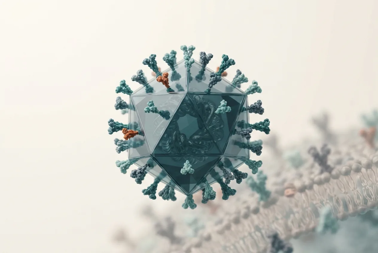
The CyHV-1 virion exhibits the characteristic herpesvirus architecture. The enveloped particle has a mean diameter of approximately 190 nm, with a capsid diameter of approximately 113 nm and surface projections measuring approximately 20 nm in length [<a href="#ref-4">4</a>]. The icosahedral nucleocapsid encloses a linear double-stranded DNA genome. The genome of CyHV-1 is approximately 290 kbp in size, making it one of the larger genomes among the alloherpesviruses [<a href="#ref-3">3</a>]. Comparative genomics with CyHV-2 and CyHV-3 has revealed a conserved set of core genes involved in DNA replication, capsid assembly, and envelope formation, alongside unique genes that may contribute to host specificity and pathogenesis [<a href="#ref-3">3</a>].
Host Range and Susceptibility
The primary host for CyHV-1 is the common carp (Cyprinus carpio) and its ornamental variety, the koi carp [<a href="#ref-1">1</a>, <a href="#ref-2">2</a>]. The virus has been historically considered to be restricted to carp species. However, molecular evidence has confirmed the presence of CyHV-1 DNA in barbel (Barbus barbus), a non-carp cyprinid, expanding the known host range [<a href="#ref-5">5</a>]. In barbel, CyHV-1 was detected in kidney tissue alongside a concurrent infection with barbel circovirus 1 (BaCV1), and the sequenced viral genes shared 98.7% nucleotide identity with carp isolates [<a href="#ref-5">5</a>]. This finding suggests that CyHV-1 can infect other cyprinid species beyond the genus Cyprinus [<a href="#ref-5">5</a>]. The virus has been reported in common carp and koi across Europe, Asia, and the Middle East, with molecular confirmations from Serbia, Iran, and Hungary [<a href="#ref-5">5</a>, <a href="#ref-6">6</a>, <a href="#ref-7">7</a>].
Clinical Signs and Pathology
The hallmark clinical sign of CyHV-1 infection is the development of carp pox, which manifests as raised, waxy, papillomatous growths on the skin, fins, and oral mucosa [<a href="#ref-2">2</a>, <a href="#ref-8">8</a>]. These lesions are typically benign and may slough off spontaneously [<a href="#ref-4">4</a>]. Histopathological examination of affected skin reveals severe epidermal hyperplasia with a marked reduction or absence of club cells and goblet cells [<a href="#ref-7">7</a>]. Degenerative changes such as spongiosis and single-cell necrosis are frequently observed, along with keratinocyte dysplasia and a moderate lymphocytic infiltration within the epidermis [<a href="#ref-7">7</a>]. In some cases, CyHV-1 DNA has been detected in cutaneous squamous cell carcinoma of koi carp, suggesting a potential oncogenic role for the virus [<a href="#ref-9">9</a>]. Experimental inoculation studies have demonstrated that tumor formation can occur more than five months post-infection, with virus particles observed in both the karyoplasm and cytoplasm of tumor cells [<a href="#ref-4">4</a>]. Mortality events associated with CyHV-1 have been reported, particularly in juvenile fish, where high mortality can accompany the characteristic papillomatous growths [<a href="#ref-6">6</a>, <a href="#ref-10">10</a>].
Molecular Virology and Replication
CyHV-1 replicates in the nucleus of infected cells, where it induces the formation of Cowdry type A intranuclear inclusion bodies [<a href="#ref-4">4</a>]. The virus can be propagated in cell lines such as FHM (fathead minnow) and MCT (carp tongue) cells, with optimal growth observed at 20 degrees Celsius [<a href="#ref-4">4</a>]. Viral titers can exceed 10^5.5 TCID50/mL after 14 days of incubation [<a href="#ref-4">4</a>]. The virus is sensitive to ether, acidic pH (pH 3), heating at 50 degrees Celsius, and treatment with 5-iodo-2'-deoxyuridine (IUdR), consistent with the properties of an enveloped DNA virus [<a href="#ref-4">4</a>]. Syncytium formation is not observed in infected cell cultures [<a href="#ref-4">4</a>]. The genome encodes a DNA polymerase that is a conserved target for molecular detection assays [<a href="#ref-6">6</a>, <a href="#ref-11">11</a>].
Diagnostic Approaches
Molecular Detection
Conventional polymerase chain reaction (PCR) targeting the DNA polymerase gene of cyprinid herpesviruses is a standard method for detecting CyHV-1 [<a href="#ref-6">6</a>, <a href="#ref-11">11</a>]. PCR products are typically visualized by agarose gel electrophoresis, and definitive confirmation is achieved through Sanger sequencing of the amplicon [<a href="#ref-6">6</a>]. A specific PCR assay for CyHV-1 has been developed for use in koi and common carp, providing a sensitive and specific diagnostic tool [<a href="#ref-11">11</a>]. The thymidine kinase (TK) gene has also been used as a PCR target, with amplicons showing high sequence identity (above 99%) among isolates within Cluster I and approximately 97% similarity to Cluster II isolates [<a href="#ref-7">7</a>].
Histopathology
Histological examination of skin lesions is a complementary diagnostic method. Key findings include severe epidermal hyperplasia, loss of club and goblet cells, spongiosis, single-cell necrosis, and lymphocytic infiltration [<a href="#ref-7">7</a>]. The presence of Cowdry type A intranuclear inclusion bodies in infected cells is a characteristic histopathological feature [<a href="#ref-4">4</a>].
Differential Diagnosis
The clinical presentation of carp pox must be differentiated from other proliferative skin conditions in cyprinids. Carp Edema Virus (CEV), the agent of koi sleepy disease, can cause skin lesions but is more commonly associated with lethargy and gill pathology [<a href="#ref-6">6</a>]. CyHV-3 (koi herpesvirus) causes acute gill necrosis and high mortality without the chronic papillomatous growths typical of CyHV-1. Spring Viremia of Carp Virus (SVCV) presents with hemorrhagic signs rather than proliferative skin lesions. Co-infections with other pathogens, such as barbel circovirus 1, have been documented and may complicate the clinical picture [<a href="#ref-5">5</a>].
Epidemiology and Transmission
CyHV-1 is distributed across Europe, Asia, and the Middle East [<a href="#ref-1">1</a>, <a href="#ref-6">6</a>, <a href="#ref-7">7</a>]. The virus is believed to be transmitted horizontally through waterborne routes, with skin abrasions likely facilitating entry [<a href="#ref-2">2</a>]. The long-term persistence of the viral genome in infected fish has been demonstrated, with detection of CyHV-1 DNA in tumorous tissues and internal organs for extended periods [<a href="#ref-12">12</a>]. Latency, a hallmark of herpesvirus infections, is presumed to occur, though the specific sites of latency in cyprinids have not been fully characterized. Outbreaks are often associated with environmental stressors such as temperature fluctuations, poor water quality, and handling [<a href="#ref-8">8</a>]. The disease is most commonly observed in juvenile and young adult fish, though all age classes are susceptible [<a href="#ref-6">6</a>, <a href="#ref-10">10</a>].
Immune Response and Vaccine Development
The host immune response to CyHV-1 involves both innate and adaptive components. Histopathological evidence of lymphocytic infiltration in affected epidermis indicates a cell-mediated immune response [<a href="#ref-7">7</a>]. To date, no commercial vaccine is available for CyHV-1 [<a href="#ref-13">13</a>]. However, immunoinformatics approaches have been employed to design multi-epitope subunit vaccines targeting both CyHV-1 and CyHV-3 [<a href="#ref-13">13</a>]. These in silico studies identified highly antigenic epitopes from critical viral proteins, assessed for antigenicity, allergenicity, and toxicity, and constructed vaccine proteins incorporating suitable adjuvants and linkers [<a href="#ref-13">13</a>]. Molecular docking studies demonstrated binding affinity between the vaccine constructs and Toll-like receptors (TLR3 and TLR5), with molecular dynamics simulations suggesting that one vaccine construct (V1) exhibited greater stability with both TLR3 and TLR5 based on RMSD analysis [<a href="#ref-13">13</a>]. MM-PBSA analysis indicated that both vaccine constructs had a better affinity for TLR5 compared to TLR3 [<a href="#ref-13">13</a>]. These computational findings suggest that multi-epitope vaccines hold promise for disease management, though in vivo trials are required to validate efficacy [<a href="#ref-13">13</a>].
Control and Prevention
Control of CyHV-1 relies on biosecurity measures, including quarantine of new fish, disinfection of equipment, and maintenance of optimal water quality [<a href="#ref-2">2</a>]. There is no specific antiviral treatment approved for use in fish. Affected fish may recover spontaneously, particularly if environmental conditions are optimized and secondary infections are prevented [<a href="#ref-2">2</a>]. The benign papillomas often regress when water temperatures are elevated, though the virus may persist in a latent state [<a href="#ref-12">12</a>]. Regulatory oversight varies by jurisdiction, and CyHV-1 is not currently listed by the World Organisation for Animal Health (WOAH), though it is considered a pathogen of concern in aquaculture.
Diagnostic Workflow
The following Mermaid diagram illustrates a recommended diagnostic workflow for suspected CyHV-1 infection in cyprinid fish.
flowchart TD
 A["Clinical Signs: Papillomatous skin/fin lesions"] --> B{"Histopathology"}
 B --> C["Epidermal hyperplasia, loss of club/goblet cells, lymphocytic infiltration"]
 B --> D["Intranuclear inclusion bodies (Cowdry type A)"]
 A --> E{"Sample Collection"}
 E --> F["Skin lesion biopsy"]
 E --> G["Kidney/gill tissue (for co-infection screening)"]
 F --> H["DNA Extraction"]
 G --> H
 H --> I{"Conventional PCR: DNA pol or TK gene"}
 I --> J["Positive: Amplicon of expected size"]
 I --> K["Negative: No amplicon"]
 J --> L["Sanger Sequencing"]
 L --> M["Sequence identity >98% with CyHV-1"]
 K --> N["Consider alternative diagnoses: CEV, CyHV-3, SVCV"]
 M --> O["Confirmed CyHV-1 infection"]
 O --> P["Report and implement biosecurity measures"]
Frequently Asked Questions
What is the primary host of Cyprinid Herpesvirus 1?
The primary host is the common carp (Cyprinus carpio) and its ornamental variety, the koi carp [<a href="#ref-1">1</a>, <a href="#ref-2">2</a>].
Can CyHV-1 infect fish species other than carp?
Yes, CyHV-1 DNA has been molecularly confirmed in barbel (Barbus barbus), demonstrating that the virus can infect other cyprinid species [<a href="#ref-5">5</a>].
What are the characteristic clinical signs of carp pox?
The characteristic clinical signs are raised, waxy, papillomatous growths on the skin, fins, and oral mucosa [<a href="#ref-2">2</a>, <a href="#ref-8">8</a>].
How is CyHV-1 diagnosed in a laboratory setting?
Diagnosis is primarily achieved through conventional PCR targeting the DNA polymerase or thymidine kinase genes, followed by Sanger sequencing for confirmation [<a href="#ref-6">6</a>, <a href="#ref-7">7</a>, <a href="#ref-11">11</a>]. Histopathology of skin lesions provides supportive evidence [<a href="#ref-7">7</a>].
Is there a vaccine available for CyHV-1?
No commercial vaccine is currently available, though multi-epitope subunit vaccines have been designed using immunoinformatics approaches and are awaiting in vivo validation [<a href="#ref-13">13</a>].
What is the difference between CyHV-1 and CyHV-3?
CyHV-1 causes benign epidermal papillomas (carp pox) with relatively low mortality, while CyHV-3 (koi herpesvirus) causes acute gill necrosis and high mortality [<a href="#ref-3">3</a>]. They are genetically distinct but related alloherpesviruses.
Can CyHV-1 cause mortality in fish?
Yes, mass mortality events have been reported, particularly in juvenile common carp, where high mortality can accompany the characteristic papillomatous growths [<a href="#ref-6">6</a>, <a href="#ref-10">10</a>].
How is CyHV-1 transmitted?
Transmission is believed to occur horizontally through water, with skin abrasions facilitating viral entry [<a href="#ref-2">2</a>].
What histopathological findings are typical of CyHV-1 infection?
Typical findings include severe epidermal hyperplasia, loss of club and goblet cells, spongiosis, single-cell necrosis, keratinocyte dysplasia, and lymphocytic infiltration [<a href="#ref-7">7</a>]. Cowdry type A intranuclear inclusion bodies are also characteristic [<a href="#ref-4">4</a>].
Is CyHV-1 considered an oncogenic virus?
The virus has been associated with papilloma formation, and CyHV-1 DNA has been detected in cutaneous squamous cell carcinoma of koi carp, suggesting a potential oncogenic role [<a href="#ref-4">4</a>, <a href="#ref-9">9</a>].