Lymphosarcoma in Dogs: Types and Prognosis
By Dr. Zubair Khalid, DVM, MS, PhD ·
Lymphosarcoma, also called lymphoma, is the most common hematopoietic cancer in dogs. It is not one disease. It is a family of lymphoid cancers that differ by where they start, which lymphocyte they arise from, and how aggressively they behave. The anatomic form and the immunophenotype (whether the cancer is a B-cell or T-cell tumor) are the two facts that most change a prognosis.
The most common form, multicentric lymphoma, accounts for roughly 80 to 85 percent of canine cases. It usually appears as painless, enlarged peripheral lymph nodes in a dog that otherwise seems well. Dogs with multicentric B-cell lymphoma treated with a CHOP-based protocol (a rotating combination of chemotherapy drugs) commonly achieve a first remission measured in months, and median survival in the range of about a year is realistic for many patients. T-cell disease, alimentary lymphoma, and cutaneous lymphoma generally carry shorter expectations. Staging and immunophenotyping are what separate a dog likely to do well from one that needs a different plan.
Owner triage summary. Firm, enlarged lymph nodes under the jaw, in front of the shoulders, or behind the knees, especially if they are painless and the dog is still eating, are the classic first sign and warrant a veterinary visit within days, not months. Difficulty breathing, a swollen face or neck, collapse, or a distended abdomen are emergencies. Vomiting, diarrhea, weight loss, or chronic itching and skin plaques can also be lymphoma. None of these signs prove cancer on their own, and none can be diagnosed at home.
This article is educational and is not a substitute for veterinary diagnosis or treatment.
What Lymphosarcoma Actually Is
Lymphocytes are white blood cells that patrol the body for infection and abnormal cells. They mature in the bone marrow, get educated in the thymus (for T cells) or in lymphoid tissue (for B cells), and then circulate through blood, lymph nodes, spleen, gut, skin, and almost every other organ. Lymphosarcoma begins when one lymphocyte lineage acquires mutations that let it multiply without normal controls.
Because lymphocytes live everywhere, lymphoma can appear almost anywhere. That is why veterinarians classify it by anatomy first. The World Health Organization classification system, adapted from human medicine, sorts canine lymphoma by cell lineage, cell size, and growth pattern, but in everyday practice the anatomic form and the B versus T immunophenotype do most of the clinical work.
Two features matter for prognosis above all others:
- Immunophenotype. B-cell lymphomas generally respond better to chemotherapy and stay in remission longer than T-cell lymphomas. This is a consistent finding across the veterinary literature [1][2].
- Clinical stage and substage. Stage describes how widely the cancer has spread. Substage describes whether the dog is clinically sick. A dog with no clinical signs (substage a) does better than a dog with fever, vomiting, weight loss, or lethargy (substage b) [3].
The Five Main Anatomic Forms
Veterinarians divide canine lymphosarcoma into five anatomic forms. Each has a different typical patient, a different set of symptoms, and a different prognosis.
Multicentric Lymphoma
Multicentric lymphoma is the default form, representing about 80 to 85 percent of cases. Malignant lymphocytes populate the peripheral lymph nodes, and the disease often involves the spleen, liver, and bone marrow as it progresses.
Typical presentation. A middle-aged to older dog (often 6 to 9 years old) arrives with enlarged, rubbery, painless lymph nodes. The mandibular (under the jaw), prescapular (in front of the shoulder), and popliteal (behind the knee) nodes are easiest to feel. Many dogs feel completely normal at first. Others show vague signs: lethargy, reduced appetite, weight loss, or increased drinking and urination if calcium is elevated.
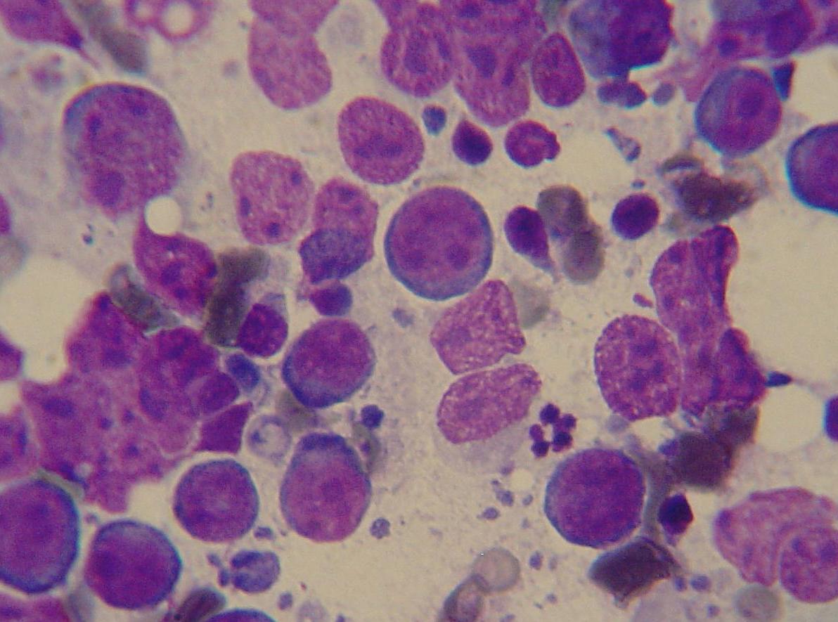
Diagnosis. Fine needle aspiration of an enlarged node with cytology is the first step. A lymph node aspirate showing a monotonous population of large lymphoid cells supports the diagnosis. Immunophenotyping by flow cytometry or immunocytochemistry separates B-cell from T-cell disease [4][3].
Treatment and prognosis. CHOP-based protocols are the standard of care. Median survival around 12 months is a reasonable expectation for multicentric B-cell lymphoma, with a subset of dogs living considerably longer. T-cell multicentric lymphoma responds less durably. A randomized trial of dogs with peripheral nodal T-cell lymphoma found median progression-free survival of only 103 days in the placebo-plus-L-CHOP arm [2]. A separate study of naive non-indolent T-cell lymphoma treated with a lomustine, vincristine, procarbazine, and prednisolone protocol reported a median overall survival of 202 days [5].
Alimentary (Gastrointestinal) Lymphoma
Alimentary lymphoma arises in the stomach, small intestine, or colon. It is the second most common anatomic form in dogs.
Typical presentation. Chronic vomiting, diarrhea, weight loss, and a poor appetite. Some dogs have a palpable abdominal mass or thickened bowel loops. Because the signs mimic inflammatory bowel disease, many dogs are treated for a gastrointestinal problem before cancer is suspected.
Diagnosis. Abdominal ultrasound shows thickened bowel wall or enlarged mesenteric lymph nodes. Definitive diagnosis usually requires full-thickness surgical biopsy or endoscopic biopsy with histopathology and immunohistochemistry. Cytology from ultrasound-guided aspirates of mesenteric nodes can help but may miss the diagnosis.
Treatment and prognosis. Alimentary lymphoma carries a poorer prognosis than multicentric B-cell disease. Many cases are T-cell in origin, which is part of why outcomes are shorter. Chemotherapy can produce remissions, but median survival is typically measured in months rather than a year.
Cutaneous Lymphoma
Cutaneous lymphoma starts in the skin. It is uncommon, but it is the form owners most often mistake for an allergy or skin infection.
Two subtypes matter:
- Epitheliotropic (also called mycosis fungoides). Malignant T cells infiltrate the epidermis and adnexal structures. Lesions often start as scaly, red, itchy patches that progress to plaques, nodules, and ulcers. It can mimic atopic dermatitis, flea allergy, or a bacterial skin infection for months.
- Non-epitheliotropic. The cancer sits in the dermis and subcutis without invading the epidermis. It usually appears as firm nodules or plaques without the intense surface scaling and itching of the epitheliotropic form.
Typical presentation. Cutaneous lymphoma in dogs often appears on the face, muzzle, and trunk, but it can occur anywhere. Pruritus (itching) can be severe in the epitheliotropic form. Lesions that fail to respond to antibiotics, antifungals, or steroids should prompt a biopsy.
Treatment and prognosis. Cutaneous lymphoma is difficult to cure. Treatment options include prednisolone, lomustine, retinoids, and other chemotherapy agents. A study of recombinant canine interferon-gamma in dogs with cutaneous epitheliotropic T-cell lymphoma found no significant survival difference compared with prednisolone alone, though the interferon group showed improvement in ulcers, bleeding, pruritus, sleep, appetite, and body weight [6]. A retrospective study of oclacitinib, a Janus kinase inhibitor, in eight dogs with cutaneous epitheliotropic lymphoma found only one dog showed symptomatic improvement, and median survival after diagnosis was 228.5 days [7]. Median survival for cutaneous lymphoma is generally measured in months.
Mediastinal Lymphoma
Mediastinal lymphoma forms a mass in the chest cavity, usually in the cranial mediastinum (the space in front of the heart).
Typical presentation. Labored breathing, exercise intolerance, a cough, or a swollen face and neck from compression of the cranial vena cava. Some dogs have no respiratory signs and the mass is found on routine chest radiographs. Hypercalcemia is common, causing increased drinking and urination.
Diagnosis. Thoracic radiographs or CT show the mass. Ultrasound-guided aspiration or biopsy provides the diagnosis. Immunophenotyping often reveals a T-cell lymphoma, frequently CD4-positive [8].
Treatment and prognosis. Mediastinal lymphoma is often T-cell and responds less well to chemotherapy than multicentric B-cell disease. A case report described a young dog with mediastinal T-cell lymphoma that achieved complete remission on an L-CHOP protocol but relapsed at week 8 and progressed to multicentric disease [8]. Prognosis is guarded.
Extranodal Lymphoma
Extranodal lymphoma arises in organs outside the lymph nodes. This category includes lymphoma of the central nervous system, eyes, kidneys, bone, muscle, and other sites. It is rare.
Typical presentation. Signs depend entirely on the organ involved. A dog with renal lymphoma may present with kidney failure. A dog with ocular lymphoma may have a red, cloudy, or painful eye. A dog with muscular lymphoma may have lameness and a soft tissue mass [9].
Treatment and prognosis. Extranodal lymphoma is generally treated with systemic chemotherapy, sometimes combined with local radiation. Prognosis varies widely by site. Primary muscular T-cell lymphoma with cutaneous involvement has been reported as a very rare presentation with a poor prognosis [9].
At a Glance: Form, Presentation, Treatment, and Survival
| Form | Typical presentation | First-line treatment | Median survival (approximate) |
|---|---|---|---|
| Multicentric B-cell | Enlarged painless lymph nodes, often well otherwise | CHOP-based chemotherapy | About 12 months |
| Multicentric T-cell | Enlarged nodes, often with clinical signs | CHOP or LOPP-based protocols | 3 to 7 months |
| Alimentary | Vomiting, diarrhea, weight loss, abdominal mass | Chemotherapy, sometimes surgery | Months |
| Cutaneous epitheliotropic | Scaly, itchy, red patches progressing to plaques and nodules | Prednisolone, lomustine, interferon-gamma, oclacitinib | About 7 to 8 months |
| Cutaneous non-epitheliotropic | Firm dermal nodules without surface scaling | Chemotherapy, sometimes radiation | Variable, often months |
| Mediastinal | Breathing difficulty, facial swelling, hypercalcemia | CHOP-based chemotherapy, radiation | Guarded, often months |
| Extranodal | Organ-specific signs | Systemic chemotherapy, sometimes radiation | Variable by site |
Survival figures are population medians. An individual dog can do better or worse, and the numbers should never be used to make a decision without a veterinarian.
How Staging and Diagnostics Work

Staging tells you how far the cancer has spread. It guides treatment and refines prognosis. The standard workup includes:
- Physical examination. The veterinarian palpates all peripheral lymph nodes, the abdomen, and the oral cavity, and listens to the chest. Lymph node size, symmetry, and consistency are recorded.
- Complete blood count (CBC). Anemia, low platelet count, or abnormal white cell counts can indicate bone marrow involvement or a leukemia phase.
- Serum chemistry panel. Elevated calcium is common in lymphoma and carries prognostic weight. Liver and kidney values guide drug selection.
- Lymph node cytology or biopsy. Fine needle aspiration is minimally invasive and often diagnostic. Excisional biopsy with histopathology and immunohistochemistry is the gold standard when cytology is equivocal.
- Imaging. Thoracic radiographs and abdominal ultrasound look for mediastinal masses, organ involvement, and lymph node enlargement inside the body. Advanced imaging such as CT is used when radiation therapy is planned.
- Flow cytometry or PARR. Flow cytometry identifies cell surface markers (such as CD3 for T cells and CD20 for B cells) and gives a rapid immunophenotype. PARR (PCR for antigen receptor rearrangements) detects clonal lymphocyte populations and confirms the diagnosis when cytology is ambiguous [4][3].
- Bone marrow evaluation. Indicated when the CBC is abnormal or when the dog is a candidate for certain protocols.
A study comparing minimally invasive techniques found that cell block immunocytochemistry confirmed lymphoma in 35 of 38 dogs, identifying 80 percent as B-cell and 20 percent as T-cell [4]. This shows that a well-performed minimally invasive workup can often replace more invasive procedures.
The following flowchart shows the main decision path from a suspected case to a treatment plan.
flowchart TD
    A[Enlarged lymph nodes or suspicious signs] --> B[Physical exam and blood work]
    B --> C[Lymph node cytology or biopsy]
    C --> D{Diagnosis confirmed}
    D -->|No| E[PARR or repeat biopsy]
    D -->|Yes| F[Immunophenotype by flow cytometry]
    F --> G[Staging with imaging and bone marrow]
    G --> H{Anatomic form and stage}
    H --> I[Multicentric B cell]
    H --> J[Multicentric T cell]
    H --> K[Alimentary or cutaneous or other]
    I --> L[CHOP based protocol]
    J --> L
    K --> M[Form specific plan]
Causes and Risk Factors
The cause of lymphoma in dogs is not known in most cases. Several factors are associated with increased risk:
- Age. Most forms occur in middle-aged to older dogs, though mediastinal lymphoma has been reported in young dogs [8].
- Breed. Certain breeds, including Boxers, Golden Retrievers, Bernese Mountain Dogs, and Bulldogs, are overrepresented in some studies. Breed alone does not predict whether a dog will get lymphoma.
- Immune status. Chronic immune stimulation and immunosuppression have been proposed as contributors, but the evidence is not definitive.
- Environmental exposures. Herbicides, pesticides, and certain household chemicals have been studied as possible risk factors. The evidence is mixed and no single exposure has been proven to cause lymphoma in dogs.
There is no vaccine or proven preventive measure for lymphoma in dogs.
Differential Diagnoses
Many conditions mimic lymphoma. A veterinarian will consider:
- Reactive lymphadenopathy. Lymph nodes enlarge in response to infection or inflammation. The nodes are usually painful and the underlying cause is identifiable.
- Inflammatory bowel disease. Can mimic alimentary lymphoma closely. Biopsy is often needed to distinguish them.
- Allergic or infectious skin disease. Can mimic cutaneous lymphoma. A biopsy is required when lesions do not respond to standard treatment.
- Other cancers. Mast cell tumors, histiocytic sarcomas, and other round cell tumors can look similar on cytology.
Evidence-Based Management
Treatment depends on the form, the immunophenotype, the stage, and the owner's goals.
Chemotherapy
CHOP-based protocols remain the standard first-line treatment for multicentric lymphoma. The acronym stands for cyclophosphamide, doxorubicin (hydroxydaunorubicin), vincristine (Oncovin), and prednisone, often with L-asparaginase added at the start (L-CHOP). Protocols typically run 19 to 25 weeks.
A retrospective study compared L-CHOP alone with L-CHOP plus sequential half-body radiotherapy in dogs with intermediate- to high-grade multicentric lymphoma. The combined group had median progression-free survival of 532 days and median overall survival of 752 days, significantly longer than the L-CHOP-alone group (296 days and 457 days, respectively) [10]. This is an investigational approach and not standard everywhere, but it shows that intensifying treatment can improve outcomes in selected dogs.
A response-based modification of CHOP has been studied in dogs with B-cell lymphoma. Dogs that responded well to the first cycle had median progression-free survival of 210 days and overall survival of 354 days. Dogs that failed to respond by week 3 and then to doxorubicin by week 5 had median progression-free survival of only 34 days and overall survival of 80.5 days [11]. Early response is a strong prognostic signal.
A novel afucosylated anti-canine CD20 antibody combined with CHOP was tested in 13 dogs with high-grade B-cell lymphoma. All 13 achieved complete response, with median progression-free survival of 340 days and median overall survival of 458 days. One- and two-year survival rates were 69.2 percent and 38.9 percent [12]. This is an investigational therapy and not yet widely available.
For T-cell lymphoma, outcomes are shorter. The randomized AT-005 trial in peripheral nodal T-cell lymphoma found median progression-free survival of 103 days in the placebo-plus-L-CHOP group and 64 days in the antibody group [2]. The LOPP protocol (lomustine, vincristine, procarbazine, prednisolone) is an alternative for naive non-indolent T-cell lymphoma, with median progression-free survival of 118 days and median overall survival of 202 days in one study [5].
An oral protocol of procarbazine, prednisolone, and cyclophosphamide (PPC) has been studied in dogs that failed or could not tolerate maximum-tolerated-dose chemotherapy. The overall response rate was 70 percent, with 24 percent partial and 46 percent complete responses. The protocol was well tolerated, with only one dog requiring discontinuation due to grade 4 thrombocytopenia [13]. This is a reasonable rescue option when injectable chemotherapy is not feasible.
Radiation Therapy
Radiation is used for localized disease, for mediastinal masses, and as part of combined protocols. The half-body radiotherapy study described above used low-dose-rate radiation at 6 Gy [10]. Radiation can also palliate painful bone lesions or obstructive masses.
Supportive Care
Supportive care matters as much as the chemotherapy itself. It includes anti-nausea drugs, appetite stimulants, fluids, pain control, and nutritional support. Weight loss during the first five weeks of chemotherapy is a negative prognostic sign. In one study, dogs that lost more than 5 percent of body weight had significantly shorter progression-free survival than dogs that maintained or gained weight [14].
Monitoring
Response is assessed by physical examination, lymph node measurement, and repeat imaging. The Veterinary Cooperative Oncology Group criteria are used to define complete response, partial response, stable disease, and progressive disease [12][2].
Prognostic Factors Beyond Form and Phenotype
Several additional factors refine prognosis:
- Clinical substage. Dogs with substage a (no clinical signs) have longer overall survival than dogs with substage b. In a study of 441 dogs with multicentric B-cell lymphoma, substage a dogs had median overall survival of 186 days versus 140 days for substage b dogs [3].
- Neutrophil-to-lymphocyte ratio (NLR). In small dogs (10 kg or less) with multicentric lymphoma, an NLR above 3.764 was associated with significantly increased risk of early progression and decreased survival [15].
- Monocytosis. High-grade multicentric lymphoma with monocytosis at diagnosis has been associated with shorter survival in dogs treated with COP or L-COP protocols [16].
- DNA methylation patterns. Genome-wide methylation analysis has identified CpG sites associated with longer survival in dogs with high-grade B-cell lymphoma treated with CHOP. A methylation level below 40 percent at one candidate site was associated with a favorable outcome [17].
- Serum microRNA profiles. A three-microRNA model differentiated B-cell, T-cell, and control samples with 83 percent accuracy, and another model separated B-cell cases into groups with hazard ratios ranging from 0.44 to 3.5 for overall survival [1].
- Metabolic profiling. NMR-based serum metabolomics has been shown to differentiate dogs with lymphoma from healthy dogs and to distinguish immunophenotypes [18].
- Lymphocyte-to-monocyte ratio at relapse. In dogs with relapsed diffuse large B-cell lymphoma, this ratio has prognostic value at the time of relapse [19].
These markers are not yet standard in every practice, but they point toward a future where treatment is tailored more precisely.
Unsafe Home Remedies and What Not to Do
No home remedy treats lymphoma. Several common practices are actively harmful:
- Do not give human chemotherapy drugs. Doses and formulations are not interchangeable, and accidental exposure is dangerous.
- Do not use high-dose steroids without a diagnosis. Steroids can shrink lymphoma temporarily and make a biopsy or cytology non-diagnostic, delaying accurate diagnosis.
- Do not rely on supplements, herbs, or dietary changes to treat cancer. Some supplements interact with chemotherapy drugs and reduce their effectiveness.
- Do not delay veterinary care because the dog seems comfortable. Lymphoma can progress quickly, and early treatment gives the best chance of a good response.
Prevention
There is no proven way to prevent lymphoma in dogs. Responsible breeding, routine veterinary care, and avoiding known carcinogens are reasonable general measures. No diet, supplement, or vaccine has been shown to prevent lymphoma.
Emergency Red Flags
Seek immediate veterinary care if your dog has:
- Difficulty breathing or open-mouth breathing
- A swollen face, neck, or front legs
- Collapse or extreme weakness
- A rapidly distending abdomen
- Severe vomiting or diarrhea with blood
- Inability to urinate
- A temperature above 103.5 F (39.7 C) or below 99 F (37.2 C)
Clinical Relevance, Limitations and Common Mistakes
Lymphosarcoma in dogs is a diagnosis that requires a veterinarian. The form, the immunophenotype, and the stage cannot be determined at home. The most common mistake owners make is waiting. A dog with enlarged lymph nodes that feels fine may still have aggressive disease, and early treatment produces better outcomes.
The second common mistake is assuming that a diagnosis of lymphoma is an immediate death sentence. Many dogs with multicentric B-cell lymphoma live a year or more with good quality of life on chemotherapy. The third mistake is treating a dog with steroids before a diagnosis is confirmed. This can mask the cancer and make accurate classification impossible.
A fourth mistake is comparing a dog's prognosis to a human's. Canine lymphoma protocols are designed for dogs, and human non-Hodgkin lymphoma regimens are not used in veterinary medicine.
Individual cases vary. A veterinarian who has examined your dog and reviewed the test results is the only person who can give you a meaningful prognosis.
Frequently Asked Questions
What are the first signs of lymphosarcoma in dogs?
The most common first sign is painless enlargement of the lymph nodes under the jaw, in front of the shoulders, or behind the knees. Many dogs feel normal at this stage. Other signs include lethargy, reduced appetite, weight loss, vomiting, diarrhea, and skin lesions.
Is lymphosarcoma in dogs curable?
Most forms are not curable, but many are treatable. Multicentric B-cell lymphoma often goes into remission with chemotherapy, and some dogs live a year or more. Cutaneous and alimentary forms are harder to control.
What is the difference between B-cell and T-cell lymphoma in dogs?
B-cell lymphoma arises from B lymphocytes and generally responds better to chemotherapy. T-cell lymphoma arises from T lymphocytes and tends to be more resistant, with shorter remission and survival times.
Can skin lymphoma in dogs be mistaken for allergies?
Yes. Cutaneous epitheliotropic lymphoma often looks like severe itching, redness, and scaling that does not respond to allergy treatment. A skin biopsy is needed to tell the difference.
How is lymphoma diagnosed in dogs?
Diagnosis usually starts with fine needle aspiration of an enlarged lymph node and cytology. Immunophenotyping by flow cytometry or immunocytochemistry identifies the cell type. PARR can confirm clonality when cytology is unclear.
What is the median survival for multicentric lymphoma in dogs?
For multicentric B-cell lymphoma treated with CHOP-based chemotherapy, median survival is approximately 12 months. T-cell disease has shorter median survival, often in the range of 3 to 7 months.
Can lymphoma in dogs be treated without chemotherapy?
Palliative treatment with prednisolone alone can improve quality of life for a short time, but it does not produce durable remissions. Oral chemotherapy protocols are an option for dogs that cannot tolerate injectable drugs.
When should I take my dog to the vet for possible lymphoma?
Take your dog to the vet within days if you notice enlarged lymph nodes, especially if they are painless and your dog seems otherwise well. Seek emergency care immediately for breathing difficulty, facial swelling, collapse, or a rapidly distending abdomen.
Related Articles
- Koi Fish Parasites: Common Types, Symptoms, and Treatment
- Feline Feline Lymphoma: Diagnosis and Management
- Flow Cytometry Immunophenotyping of Canine Lymphoma
- Osteosarcoma in Large Breeds: Bone Amputation, Chemotherapy and Prognosis
- Aortic Thromboembolism FATE in Cats: Saddle Thrombus Pain and Prognosis
- Calf Septicemia: Early Recognition, Blood Sampling, Treatment, and Prognosis
- Mange in Cats: Symptoms, Types, and Treatment
- Dog Digestive Issues: Causes, Symptoms, and When to Worry
- What Is Anthrax Disease? Causes and Symptoms
Sources
- Classification and Prognostication of B-Cell and T-Cell Multicentric Lymphoma in Dogs Using Serum MicroRNAs.
- Randomised trial evaluating chemotherapy alone or chemotherapy and a novel monoclonal antibody for canine T-cell lymphoma: A multicentre US study.
- Correlation between clinical variables and overall survival time in dogs with multicentric B-cell lymphoma: a retrospective study in Taiwan.
- Comparison of the accuracy of minimally invasive techniques (cytology, cell block, immunocytochemistry and clonality assay) in the diagnosis of canine multicentric lymphoma.
- Prognostic indicators for naïve canine non-indolent T-cell lymphoma treated with combination lomustine, vincristine, procarbazine and prednisolone chemotherapy.
- Clinical efficacy of recombinant canine interferon-gamma therapy in dogs with cutaneous epitheliotropic T-cell lymphoma.
- Short-term administration of oclacitinib with concomitant medications in canine epitheliotropic lymphoma: A retrospective study of eight dogs.
- Progression of primary mediastinal T-cell lymphoma to a multicentric form in a young dog.
- Primary multifocal muscular T-cell lymphoma with cutaneous involvement in a dog: A case report and review of the literature.
- Survival outcomes of dogs with multicentric lymphoma treated with sequential half-body radiotherapy and L-CHOP without maintenance chemotherapy.
- Response-based modification of CHOP chemotherapy for canine B-cell lymphoma.
- Afucosylated anti-canine CD20 antibody combined with cyclophosphamide, doxorubicin, vincristine, and prednisone chemotherapy in dogs with B-cell lymphoma.
- Procarbazine, prednisolone and cyclophosphamide oral combination chemotherapy protocol for canine lymphoma.
- Association between weight change during initial chemotherapy and clinical outcome in dogs with multicentric lymphoma.
- Blood Neutrophil-to-Lymphocyte Ratio as a Potential Prognostic Marker in Dogs ≤10 kg With Multicentric Lymphoma.
- Hematological and blood biochemistry parameters as prognostic indicators of survival in canine multicentric lymphoma treated with COP and L-COP protocols.
- Use of genome-wide DNA methylation analysis to identify prognostic CpG site markers associated with longer survival time in dogs with multicentric high-grade B-cell lymphoma.
- Application of (1)H NMR Metabolic Profiling of Serum in Canine Multicentric Lymphoma.
- Monocyte and Lymphocyte Count, and Lymphocyte/Monocyte Ratio as Prognostic Factors at the Time of First Relapse in Canine Diffuse Large B-Cell Lymphoma Patients Receiving Chemotherapy.