DIC Coagulopathy: Pathogenesis and Lab Findings
By Dr. Zubair Khalid, DVM, MS, PhD ·
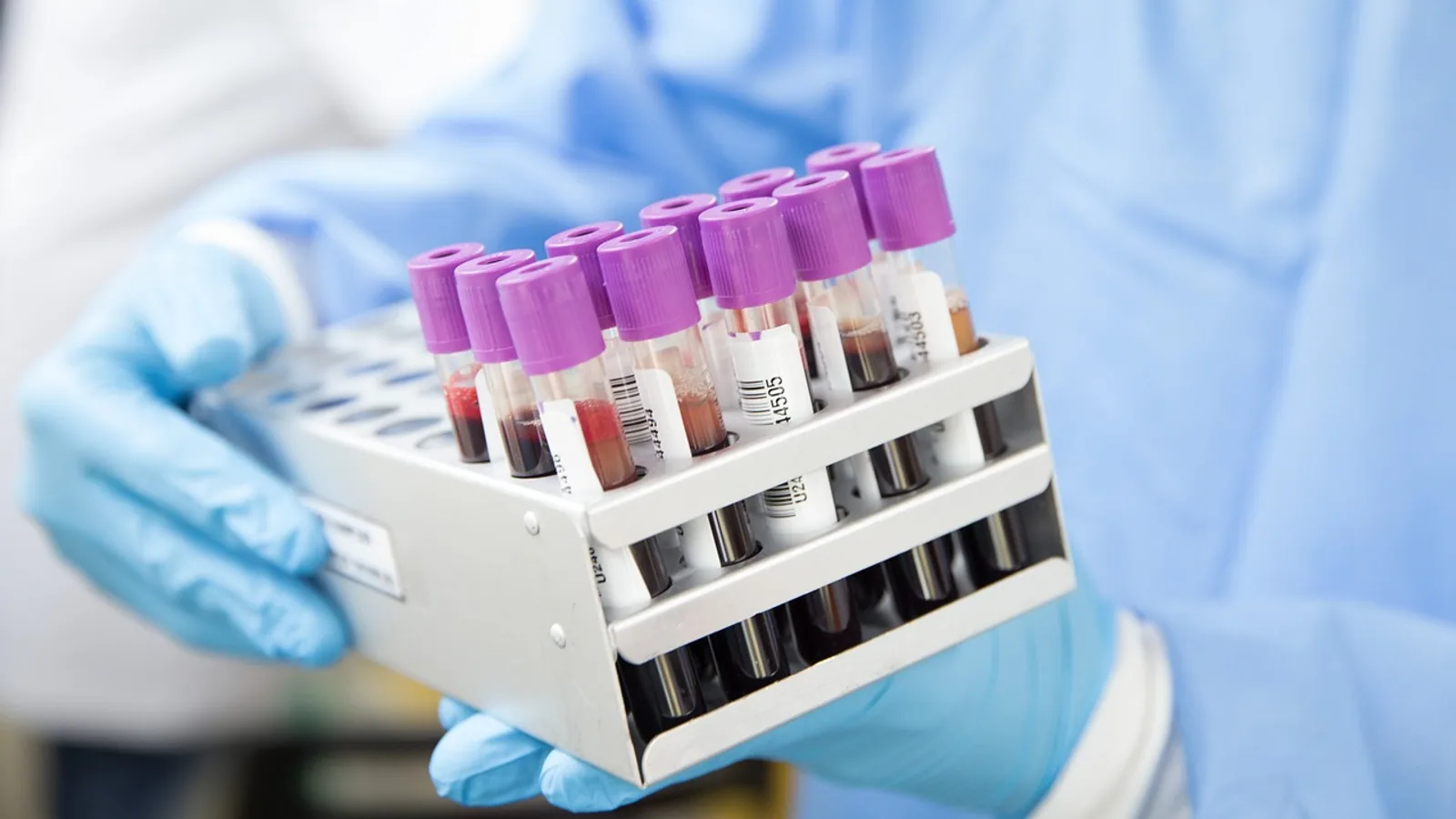
Disseminated intravascular coagulation (DIC) is a systemic thrombohemorrhagic syndrome in which widespread, uncontrolled activation of the coagulation cascade generates microthrombi throughout the microvasculature while simultaneously consuming platelets and clotting factors, so that bleeding and thrombosis occur together. It is not a single disease but a final common pathway that complicates sepsis, trauma, neoplasia, hemolysis, and obstetric catastrophes, and it is one of the few hematologic diagnoses in veterinary medicine that can kill a patient within hours if the underlying trigger is not controlled.
DIC matters because it sits at the intersection of inflammation, endothelial injury, and hemostasis. A dog with parvovirus, a cat with sepsis, or a horse with African horse sickness can all develop the same laboratory signature even though the initiating insult is completely different [1]. Recognizing that signature early changes how a clinician monitors the patient, what blood products are available, and whether the primary disease is being treated aggressively enough. DIC is a diagnosis of pattern, not of any single test, and that distinction is the single most important concept for students to internalize.
This article is educational and is not a substitute for veterinary diagnosis or treatment.
What DIC Actually Is
The classic teaching phrase is "consumption coagulopathy." That term captures the depletion of platelets and clotting factors, but it understates the thrombosis. In DIC, the coagulation cascade is activated systemically rather than locally. Thrombin is generated far beyond what a controlled hemostatic plug requires. Fibrin is deposited in small vessels of the lung, kidney, liver, and brain. At the same time, the fibrinolytic system is activated to break down that fibrin, and the breakdown products themselves interfere with normal clotting.
The result is a paradox: the patient is forming clots in organs while bleeding from venipuncture sites, mucous membranes, and surgical wounds. A dog with pancreatic adenocarcinoma and DIC presented with hematuria and progressive anemia alongside fibrinohaemorrhagic peritonitis, illustrating how bleeding and microthrombosis coexist in the same patient [2]. A horse experimentally infected with African horse sickness virus showed submucosal petechiae and prolonged bleeding after venipuncture, consistent with hypocoagulation from DIC [1].
Acute versus chronic DIC
DIC is often divided into acute and chronic forms. Acute DIC is decompensated: the coagulation system is overwhelmed, fibrinogen falls, platelets drop sharply, and bleeding dominates. Chronic DIC is compensated: the bone marrow and liver keep pace with consumption, so platelet count and fibrinogen may remain near normal or even elevated, and thrombosis or low-grade bleeding may be the only clue. A dog with metastatic nasal adenocarcinoma was diagnosed with chronic DIC and concurrent paraneoplastic secondary hyperfibrinolysis after a year of progressive epistaxis [3]. That case shows how chronic DIC can smolder for months before it becomes clinically obvious.
DIC versus other coagulopathies
Students frequently confuse DIC with rodenticide toxicity, immune-mediated thrombocytopenia, or inherited factor deficiencies. The table below separates these entities by their core mechanism and laboratory pattern.
| Condition | Core mechanism | Platelets | PT/aPTT | Fibrinogen | FDPs/D-dimer |
|---|---|---|---|---|---|
| Acute DIC | Systemic coagulation activation plus fibrinolysis | Low | Prolonged | Low | High |
| Chronic DIC | Compensated consumption | Low to normal | Normal to prolonged | Normal to high | High |
| Rodenticide toxicity | Vitamin K-dependent factor deficiency | Normal | Prolonged | Normal | Normal |
| Immune-mediated thrombocytopenia | Platelet destruction | Very low | Normal | Normal | Normal |
| Inherited factor deficiency (e.g., hemophilia A) | Single factor absent | Normal | aPTT prolonged, PT normal | Normal | Normal |
The key discriminator is that DIC is the only condition in this list that combines thrombocytopenia, prolonged clotting times, low fibrinogen, and elevated fibrin degradation products in the same patient.
Pathogenesis: How DIC Unfolds
DIC develops through a sequence of overlapping events. Understanding the sequence explains why the laboratory findings look the way they do.
Step 1: Tissue factor exposure and coagulation activation
Tissue factor (TF) is the physiologic initiator of the extrinsic coagulation pathway. It is normally sequestered from circulating blood. When endothelial cells are damaged, when monocytes and macrophages are activated by inflammatory mediators, or when tumor cells express TF, the cascade fires systemically. In a rat model of sepsis-induced DIC, knockdown of Wilms tumor 1-associated protein reduced TF activity and improved coagulation parameters, confirming that TF regulation is a central node in DIC pathogenesis [4]. A separate study showed that miR-19a-3p directly targets TF and that restoring this microRNA reduced TF procoagulant activity in a rat DIC model [5].
Bacterial endotoxin (lipopolysaccharide, or LPS) is a potent trigger of TF expression. LPS activates caspase-11 in monocytes, which forms gasdermin D pores and exposes phosphatidylserine on the cell surface. Phosphatidylserine exposure accelerates TF-dependent coagulation, and blocking any step in this pathway prevented LPS-induced DIC in experimental models [6]. This mechanism links innate immune recognition of Gram-negative bacteria directly to coagulation activation.
Step 2: Thrombin generation and microthrombus formation
Once TF is exposed, the common pathway generates thrombin, which converts fibrinogen to fibrin. In DIC, thrombin generation is diffuse and sustained. Fibrin strands deposit in the microvasculature of multiple organs. A dugong that died of systemic Achromobacter xylosoxidans infection had fibrin thrombi in multiple organ systems at necropsy, along with hemorrhage, hepatocellular degeneration, and neuronal damage [7]. Those findings are the histologic footprint of DIC: fibrin deposition plus ischemic and hemorrhagic tissue injury occurring simultaneously.
Step 3: Platelet consumption and activation
Platelets are consumed as they aggregate on fibrin and as they are activated by thrombin and inflammatory mediators. In sepsis, platelets undergo a form of cell death called PANoptosis, which combines pyroptosis, apoptosis, and necroptosis. Platelet PANoptosis is associated with the onset and progression of DIC, and inhibiting it with myricetin improved DIC in a sepsis model [8]. Complement activation also contributes to thrombocytopenia in sepsis-associated DIC. In a study of 49 adult sepsis patients, complement activation through the alternative pathway was associated with lower platelet counts and higher mortality when combined with DIC [9].
Step 4: Fibrinolysis and the bleeding phase
As fibrin is deposited, plasminogen activators convert plasminogen to plasmin, which degrades fibrin into fibrin degradation products (FDPs), including D-dimer. This is a protective response meant to clear microthrombi, but in DIC it becomes excessive. The FDPs themselves have anticoagulant properties: they interfere with fibrin polymerization and platelet function. The result is the bleeding tendency that characterizes decompensated DIC.
The balance between coagulation and fibrinolysis varies with the trigger. A study comparing LPS-induced and TF-induced DIC in rats found that both models had identical degrees of hemostatic activation, measured by thrombin-antithrombin complex levels, but the TF-induced model had markedly higher D-dimer concentrations and more bleeding symptoms, while the LPS-induced model had more severe inflammation and organ dysfunction [10]. This distinction matters clinically: the trigger determines whether a patient presents primarily with thrombosis or with hemorrhage.
Step 5: Endothelial injury and amplification
Endothelial injury is both a cause and a consequence of DIC. Damaged endothelium exposes subendothelial collagen, loses its anticoagulant surface (thrombomodulin, heparan sulfate), and expresses adhesion molecules that recruit leukocytes and platelets. Thrombomodulin is a marker of endothelial injury. In patients with sepsis-induced DIC, plasma thrombomodulin was significantly higher in non-survivors than in survivors and correlated with the Sequential Organ Failure Assessment score [11]. This finding supports the concept that DIC is not just a clotting problem but a vascular problem.
The complement system amplifies endothelial injury. In the sepsis study mentioned above, patients with both DIC and complement activation had a 60-day mortality of 66 percent, compared with 14 percent in the DIC-alone group and 0 percent in the complement-activation-alone group [9]. That is a striking demonstration of how inflammation and coagulation reinforce each other.
flowchart TD
    A[Trigger] --> B[Tissue factor exposure]
    B --> C[Thrombin generation]
    C --> D[Fibrin deposition]
    C --> E[Platelet consumption]
    D --> F[Microthrombi in organs]
    E --> G[Thrombocytopenia]
    D --> H[Fibrinolysis]
    H --> I[Elevated D dimer and FDPs]
    I --> J[Bleeding tendency]
    F --> K[Organ dysfunction]
    G --> J
    K --> L[Poor outcome]
    J --> L
Triggers of DIC in Veterinary Patients
DIC is always secondary to another condition. The trigger list is long, but the major categories are consistent across species.
Sepsis
Sepsis is the most common trigger in small animal practice. Gram-negative bacteria release LPS, which activates TF through the caspase-11 and gasdermin D pathway [6]. Gram-positive organisms and fungi can trigger DIC through different mechanisms, including capsular polysaccharides and exotoxins. A comprehensive review of Klebsiella pneumoniae and DIC described how capsular polysaccharides, lipopolysaccharides, siderophores, and other virulence factors interact with the host thrombo-inflammatory response to produce endothelial dysfunction and coagulation activation [12]. In the dugong case, A. xylosoxidans was isolated from multiple organs, and the authors concluded that septicemia-associated DIC was the cause of death [7].
Trauma and tissue injury
Major trauma releases TF from damaged tissue into the circulation. Large burns, crush injuries, and surgical complications can all trigger DIC. A human case report described recalcitrant postoperative bleeding after a minor eyelid procedure in a patient with undiagnosed chronic DIC attributed to a large burden of endovascular stents [13]. While that case is human, the principle applies: any condition that exposes blood to abnormal surfaces or damaged tissue can initiate DIC.
Neoplasia
Cancer cells frequently express TF and other procoagulant molecules. In dogs, pancreatic adenocarcinoma has been associated with DIC even when the tumor is not visible on imaging or gross examination [2]. Metastatic nasal adenocarcinoma in a dog produced chronic DIC with secondary hyperfibrinolysis and severe epistaxis [3]. Paraneoplastic hyperfibrinolysis is a recognized complication of certain cancers and should be considered in any dog with a known malignancy and unexplained bleeding.
Hemolysis and intravascular red cell destruction
Intravascular hemolysis releases red cell membrane fragments and ADP, which activate platelets and the coagulation cascade. Immune-mediated hemolytic anemia, babesiosis, and leptospirosis are all potential triggers in veterinary patients. The melarsomine case in a dog with Dirofilaria immitis illustrates that even treatment-related lysis of parasites or microfilariae can release procoagulant material and precipitate DIC [14].
Obstetric complications
In human obstetrics, DIC is a leading cause of maternal mortality. Risk factors include placental abruption, amniotic fluid embolism, preeclampsia, and retained dead fetus [15]. A retrospective study of 91 women with intrauterine fetal death found that the degree of fetal maceration did not correlate with DIC scores, suggesting that retention time alone is not the primary determinant of coagulopathy risk [16]. In veterinary medicine, pyometra, dystocia with fetal maceration, and placental abnormalities can trigger DIC through similar mechanisms.
Other triggers
Severe inflammatory conditions such as pancreatitis, immune-mediated disease, and heatstroke can all activate coagulation. Thyroid storm has been reported as a rare trigger of DIC in humans, mediated by uncontrolled release of pro-inflammatory cytokines [17]. The common thread is that any condition causing widespread endothelial injury, cytokine release, or TF exposure can initiate DIC.
Laboratory Findings in DIC
No single test confirms DIC. The diagnosis rests on a combination of history, clinical signs, and laboratory abnormalities that together form a recognizable pattern. Scoring systems such as the International Society on Thrombosis and Haemostasis (ISTH) DIC score and the veterinary-modified scoring systems use these parameters to assign a probability.
Thrombocytopenia
Platelet consumption is a hallmark of DIC. In acute DIC, platelet counts fall rapidly and may reach severely low levels. In chronic DIC, the bone marrow can compensate, and platelet counts may be low-normal or only mildly decreased. The melarsomine case showed persistent thrombocytopenia despite multiple transfusions [14]. In the sepsis and complement study, complement activation was independently associated with thrombocytopenia [9].
Prolonged PT and aPTT
Prothrombin time (PT) measures the extrinsic and common pathways. Activated partial thromboplastin time (aPTT) measures the intrinsic and common pathways. In DIC, both are prolonged because multiple clotting factors are consumed. The melarsomine case had a PT of 84.2 seconds (reference interval 7.0 to 9.3 seconds) and an aPTT greater than 140 seconds (reference interval 10.4 to 12.9 seconds), with fibrinogen below 50 mg/dL (reference interval 109 to 311 mg/dL) [14]. Those values are extreme, but they illustrate the direction and magnitude of change.
Low fibrinogen
Fibrinogen is an acute-phase protein, so it can be elevated in early or chronic DIC due to inflammation. As consumption outpaces production, fibrinogen falls. A low fibrinogen in a bleeding patient with prolonged clotting times is strong evidence of DIC. The horse with African horse sickness had fibrinogen measured as part of a comprehensive hemostatic panel [1].
Elevated D-dimer and FDPs
D-dimer is a specific fragment produced when plasmin degrades cross-linked fibrin. FDPs are a broader category that includes D-dimer and other fragments. Both are elevated in DIC because fibrinolysis is active. The TF-induced rat DIC model had markedly elevated D-dimer concentrations and prominent bleeding symptoms [10]. D-dimer is more specific than FDPs for DIC because it confirms that fibrin was formed and then degraded, rather than just that fibrinogen was broken down.
Schistocytes on blood smear
Schistocytes are fragmented red blood cells. They form when red cells are sliced by fibrin strands in the microvasculature. Finding schistocytes on a blood smear in a patient with thrombocytopenia and prolonged clotting times supports the diagnosis of DIC. Schistocytes are not specific to DIC (they also occur in hemangiosarcoma and other conditions), but in the right clinical context they are a useful clue.
Other laboratory findings
Antithrombin (AT) is consumed in DIC and often decreased. The horse with African horse sickness had AT activity measured as part of the hemostatic panel [1]. Thromboelastography (TEG) provides a global assessment of clot formation and lysis. In the same horse study, TEG showed increased clot initiation time (R), decreased alpha-angle, decreased maximum amplitude (MA), and decreased clot strength (G), consistent with hypocoagulability [1]. TEG is increasingly used in veterinary referral settings because it captures the balance between coagulation and fibrinolysis in a single assay.
| Lab parameter | Expected change in DIC | Clinical interpretation |
|---|---|---|
| Platelet count | Decreased | Consumption exceeds production. Severe thrombocytopenia increases bleeding risk. |
| PT | Prolonged | Extrinsic and common pathway factors consumed. |
| aPTT | Prolonged | Intrinsic and common pathway factors consumed. |
| Fibrinogen | Decreased in acute DIC, normal or increased in chronic DIC | Low fibrinogen indicates decompensated consumption. |
| D-dimer | Increased | Confirms fibrin formation and degradation. |
| FDPs | Increased | Broader marker of fibrinolysis. Less specific than D-dimer. |
| Schistocytes | Present | Red cell fragmentation by fibrin strands. Supports microangiopathic hemolysis. |
| Antithrombin | Decreased | Consumption of natural anticoagulant. |
| TEG | Prolonged R, decreased alpha-angle, decreased MA | Global hypocoagulability. Useful for monitoring. |
Species Differences
DIC manifests differently across species, and those differences affect clinical presentation and monitoring.
Dogs
Dogs with DIC often present with bleeding. Petechiae, ecchymoses, hematuria, melena, and prolonged bleeding from venipuncture sites are common. The pancreatic adenocarcinoma case presented with hematuria and progressive anemia [2]. The melarsomine case presented with limb swelling and coagulopathy that worsened despite plasma transfusion [14]. The nasal adenocarcinoma case presented with severe epistaxis [3]. In dogs, the combination of bleeding and thrombocytopenia should always prompt consideration of DIC.
Cats
Cats with DIC are more likely to present with thrombosis than with bleeding. Aortic thromboembolism, portal vein thrombosis, and disseminated microthrombosis are recognized complications of DIC in cats. The clinical picture may be dominated by organ dysfunction (azotemia, elevated liver enzymes, neurologic signs) rather than by overt hemorrhage. This difference is not absolute, but it is a useful rule of thumb for clinicians and students.
Horses
Horses with DIC often present with petechiae, prolonged bleeding after venipuncture, and signs of systemic illness. The African horse sickness study documented overt DIC with prolonged PT and aPTT, low fibrinogen, and elevated D-dimer in experimentally infected horses [1]. TEG was useful for characterizing the hypocoagulable state.
Other species
DIC has been reported in many species, including marine mammals. The dugong case is the first report of septicemia-associated DIC in that species [7]. The principles of pathogenesis and laboratory diagnosis are similar across species, but reference intervals for coagulation parameters vary, and species-specific normal values must be used.
Treatment Principles
Treatment of DIC follows three principles: treat the underlying cause, support the patient with blood products when bleeding is severe, and consider anticoagulant therapy when thrombosis dominates. Specific drug doses are beyond the scope of this article and should be determined by the attending veterinarian based on the individual patient.
Treat the trigger
The single most effective intervention in DIC is to control the underlying disease. Source control for sepsis, surgical removal of a necrotic intestine, or initiation of appropriate antimicrobial therapy can halt the coagulation activation. In the thyroid storm case, treatment of the underlying endocrine emergency led to recovery [17]. In the melarsomine case, the trigger was a drug reaction, and the patient required intensive support [14].
Blood product support
Fresh frozen plasma (FFP) replaces consumed clotting factors and antithrombin. Packed red blood cells correct anemia from hemorrhage or hemolysis. Platelet transfusions are rarely available in veterinary medicine but may be considered in severe thrombocytopenia with life-threatening bleeding. The melarsomine case received multiple units of FFP and packed red blood cells [14].
Anticoagulant therapy
Heparin is sometimes used in DIC to inhibit thrombin and prevent further microthrombus formation. The rationale is strongest in chronic DIC or when thrombosis is the dominant clinical problem. In the human case of chronic DIC with postoperative bleeding, anticoagulation with apixaban plus tranexamic acid achieved lasting hemostasis [13]. That case illustrates the counterintuitive principle that anticoagulation can stop bleeding in DIC by interrupting the cycle of coagulation activation and consumption.
Antifibrinolytic therapy
Antifibrinolytic drugs such as aminocaproic acid or tranexamic acid are used when hyperfibrinolysis is the dominant problem. The dog with nasal adenocarcinoma and secondary hyperfibrinolysis was treated with anti-fibrinolytic medications [3]. The melarsomine case also received aminocaproic acid [14]. Antifibrinolytics should be used with caution in DIC because they can worsen thrombosis if fibrinolysis is not the primary problem.
Monitoring
Patients with DIC require serial monitoring of platelet count, PT, aPTT, fibrinogen, and D-dimer. TEG can provide additional information about clot strength and fibrinolysis. The goal is to detect worsening coagulopathy before it becomes clinically catastrophic.
Clinical Relevance, Limitations and Common Mistakes
DIC is a medical emergency. It signals that a patient has a severe underlying disease, and it independently increases mortality. In the sepsis and complement study, patients with both DIC and complement activation had a 66 percent 60-day mortality [9]. In the horse with African horse sickness, DIC was part of a disease process that is often fatal [1]. Recognizing DIC early gives the clinician a chance to intervene before the patient decompensates.
The most common mistake students make is trying to diagnose DIC with a single test. A low platelet count alone is not DIC. A prolonged PT alone is not DIC. The diagnosis requires a pattern: thrombocytopenia, prolonged PT and aPTT, low fibrinogen, and elevated D-dimer or FDPs, ideally with schistocytes on the blood smear. Scoring systems exist precisely because no single parameter is sufficient.
Another common mistake is assuming that DIC always causes bleeding. In cats, thrombosis may be the dominant clinical sign. In chronic DIC, the patient may have normal platelet count and fibrinogen. The clinician must maintain a high index of suspicion and interpret laboratory results in the context of the whole patient.
A third mistake is failing to treat the underlying cause. Supportive care with blood products can temporize, but it cannot cure DIC. The trigger must be identified and controlled.
Individual cases require veterinary assessment. This article provides a framework for understanding DIC, but it cannot replace clinical judgment.
Quick Review
- DIC is simultaneous widespread coagulation activation and fibrinolysis.
- Triggers include sepsis, trauma, neoplasia, hemolysis, and obstetric complications.
- The core laboratory pattern is thrombocytopenia, prolonged PT and aPTT, low fibrinogen, and elevated D-dimer or FDPs.
- Schistocytes on the blood smear support the diagnosis.
- No single test confirms DIC. Use scoring systems and the full clinical picture.
- Dogs often present with bleeding. Cats often present with thrombosis.
- Treatment is directed at the underlying cause, with blood product support and sometimes anticoagulant or antifibrinolytic therapy.
Frequently Asked Questions
What is the most common trigger of DIC in dogs?
Sepsis is the most common trigger. Other important triggers include neoplasia, trauma, hemolysis, and obstetric complications.
Can a dog survive DIC?
Yes, some dogs survive if the underlying cause is controlled and supportive care is provided. Survival depends on the severity of the trigger and how quickly treatment is initiated.
Why do cats with DIC sometimes not bleed?
Cats with DIC often have a thrombotic phenotype, meaning microthrombi form and cause organ dysfunction rather than overt hemorrhage. The balance between coagulation and fibrinolysis determines the clinical presentation.
What is the difference between D-dimer and FDPs?
D-dimer is a specific fragment produced when plasmin degrades cross-linked fibrin. FDPs are a broader category that includes D-dimer and other fibrinogen breakdown products. D-dimer is more specific for DIC.
Can DIC be diagnosed with a single blood test?
No. DIC is diagnosed by recognizing a pattern of abnormalities across multiple tests. Scoring systems combine platelet count, PT, fibrinogen, and D-dimer or FDPs to assign a probability.
Is DIC always fatal?
No. DIC is a serious condition with high mortality, but some patients recover if the underlying cause is treated effectively. Early recognition and aggressive management improve the odds.
Related Articles
- Equine Hematologic Disorders: Anemia and Coagulopathies
- Anesthesia for Patients with Hematologic Disease: Coagulopathy and Anemia
- Understanding Research Implications: From Findings to Impact
- Finding a Reputable Hamster Breeder: Questions to Ask
- Adopt a Pet: Complete Guide to Pet Adoption and Finding Your Perfect Companion
- Cat Adoption: Complete Guide to Finding Your Perfect Feline Friend
- Lab Shepherd Mix: Traits, Health, and Care Guide
- Maltose Binding Protein: Structure and Lab Uses
Sources
- Experimental infection of horses with African horse sickness virus results in overt disseminated intravascular coagulation.
- Unveiling the Invisible: A Case of Canine Diffuse Microscopic Pancreatic Adenocarcinoma With Normal Imaging and Gross Appearance of the Pancreas Complicated by Sterile Peritonitis and Disseminated Intravascular Coagulation.
- Case report: Chronic disseminated intravascular coagulopathy with concurrent paraneoplastic secondary hyperfibrinolysis in a dog with metastatic nasal adenocarcinoma.
- Wilms Tumor 1-associated Protein Promotes Sepsis-induced Disseminated Intravascular Coagulation by Stabilizing Intercellular Adhesion Molecule 1 Via N6-methyladenosine Modification.
- miR-19a-3p downregulates tissue factor and functions as a potential therapeutic target for sepsis-induced disseminated intravascular coagulation.
- Bacterial Endotoxin Activates the Coagulation Cascade through Gasdermin D-Dependent Phosphatidylserine Exposure.
- Novel Insight into Dugong Mortality: First Report of Systemic Achromobacter xylosoxidans Infection, Disseminated Intravascular Coagulation, and Associated Pathogenesis.
- Myricetin reduces platelet PANoptosis in sepsis to delay disseminated intravascular coagulation.
- Complement system activation through the alternative pathway associates with disseminated intravascular coagulation to increase mortality in sepsis.
- Detailed exploration of pathophysiology involving inflammatory status and bleeding symptoms between lipopolysaccharide- and tissue factor-induced disseminated intravascular coagulation in rats.
- Increased Level of Thrombomodulin is Associated with Endothelial Injury in Patients with Sepsis-Induced Disseminated Intravascular Coagulation.
- Klebsiella pneumoniae and Disseminated Intravascular Coagulation: A Comprehensive Review of Pathogenesis, Clinical Manifestations, and Management Strategies.
- Recalcitrant postoperative bleeding after ectropion repair in the setting of undiagnosed chronic disseminated intravascular coagulation.
- Case report: Disseminated intravascular coagulation in a dog following treatment with melarsomine for Dirofilaria immitis.
- Risk Factors Associated with Development of Disseminated Intravascular Coagulation (DIC) in Obstetrical Cases.
- Impact of fetal maceration grade on risk of maternal disseminated intravascular coagulation after intrauterine fetal death - A retrospective cohort study.
- Thyroid Storm-Induced Acute Liver Dysfunction and Disseminated Intravascular Coagulation.