Vertebrae Lumbales: Lumbar Vertebrae Anatomy
By Dr. Zubair Khalid, DVM, MS, PhD ·
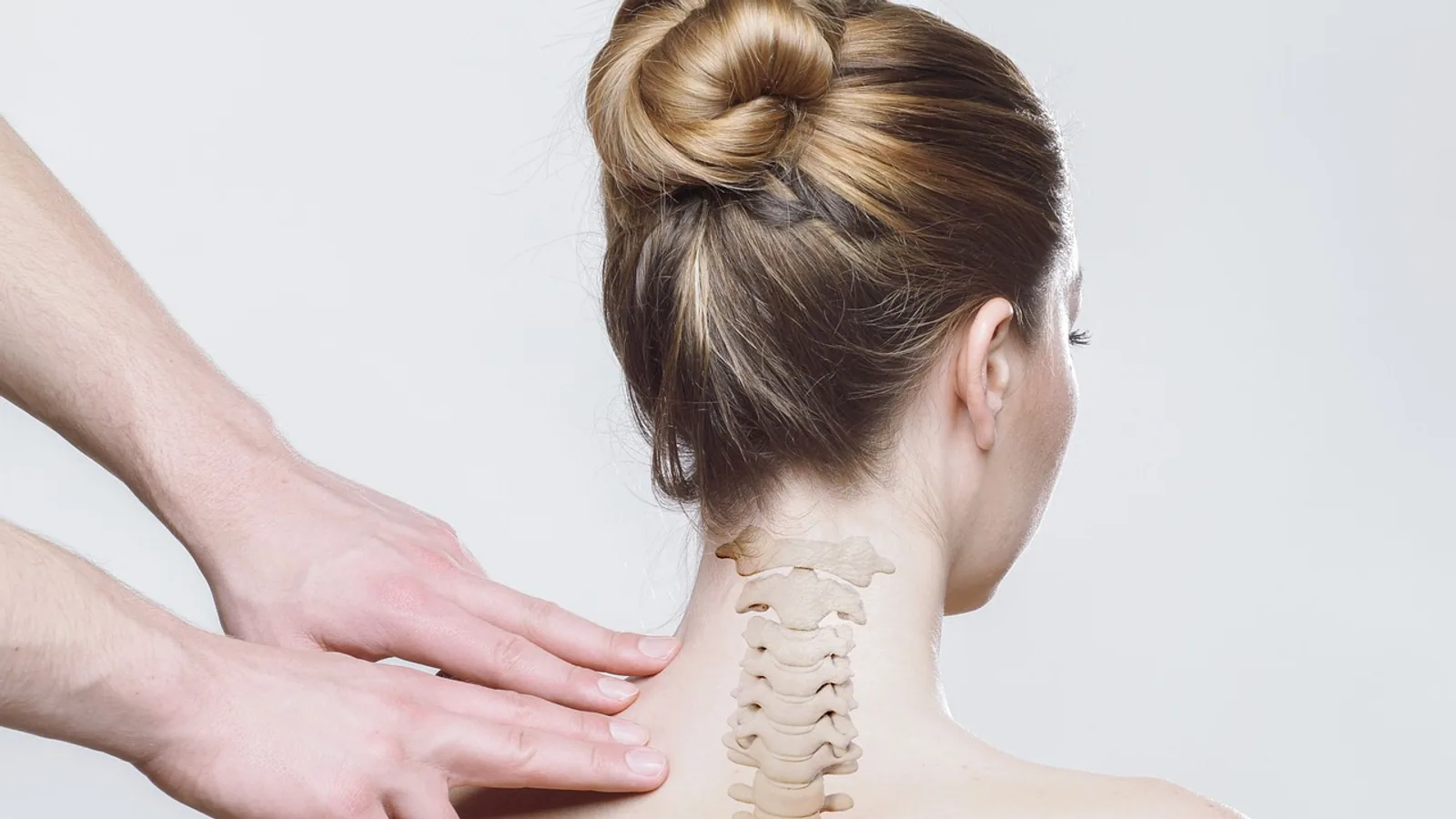
The vertebrae lumbales (lumbar vertebrae) are the series of presacral vertebrae that sit between the thoracic spine and the sacrum, and they are the most massive and most mobile of the presacral vertebrae in most domestic mammals. Each lumbar vertebra has a large cylindrical body, a short spinous process, broad transverse processes, and well-developed articular (zygapophyseal) processes that lock adjacent vertebrae together while still allowing sagittal bending.
Lumbar anatomy matters in practice because this is where the spine carries the greatest compressive load, where the largest epaxial and hypaxial muscle masses attach, and where a large share of clinically significant vertebral and spinal cord disease appears in dogs, cats, and horses. Getting the numbering and the shape of the transverse processes right is the difference between reading a radiograph correctly and mislabeling a lesion.
This article is educational and is not a substitute for veterinary diagnosis or treatment.
What "Vertebrae Lumbales" Means and Where the Region Sits
Vertebrae lumbales is the Latin anatomical term for the lumbar vertebrae. In the standard vertebral formula, the spine is written as cervical (C), thoracic (T), lumbar (L), sacral (S), and caudal or coccygeal (Cd or Cy) counts, in that order. The lumbar region is defined by position, not by a fixed number: it is the segment between the last rib-bearing vertebra and the first vertebra that contributes to the sacrum.
Lumbar vertebrae are identified by two negative features and two positive ones. They do not bear ribs, and they do not have transverse foramina (the holes in the cervical transverse processes that transmit the vertebral artery). They do have large, flat-ended bodies and long transverse processes. Those transverse processes are the single most useful landmark for numbering lumbar vertebrae on a radiograph, because their shape and angle change in a predictable craniocaudal sequence.
The lumbosacral junction, where the last lumbar vertebra meets the sacrum, is a focal point of pathology in large-breed dogs. The lumbosacral region is described as the most susceptible part of the canine spine to disease, with intervertebral disc disease, vertebral distortion, narrowing of the lumbosacral canal, and congenital spinal defects among the common problems [1].
The Standard Lumbar Vertebral Plan
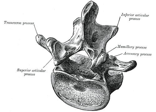

Every lumbar vertebra, regardless of species, is built on the same basic plan. Learn the plan once and you can identify a lumbar vertebra from any mammal.
The vertebral body (centrum)
The body is the ventral, cylindrical mass of bone. Lumbar bodies are wider and taller than thoracic bodies and roughly equal in craniocaudal length across the region in most species. The cranial and caudal faces are flat or slightly concave and are separated from the next vertebra by an intervertebral disc. In the dog, vertebral body height and length follow a consistent mathematical relationship with position along the lumbosacral spine, and the proportions hold across small, medium, and large breeds even though absolute size varies enormously [1]. That consistency is what makes ratio-based radiographic assessment possible.
The vertebral arch and vertebral canal
The arch rises from the dorsolateral edges of the body and encloses the vertebral canal. The canal carries the spinal cord and, more caudally, the cauda equina. In cats, the height, width, and cross-sectional area of the lumbar vertebral canal and spinal cord are significantly greater in males than in females [2]. The ratio of spinal cord area to vertebral canal area stays stable between sexes and does not correlate with age or body weight in that species [2].
The spinous process
Lumbar spinous processes are short, thick, and directed dorsally or slightly caudally. They are much shorter than the tall, overlapping thoracic spines. This shortening is functional: it removes the bony lever arms that would block extension and gives the epaxial muscles a broad surface for attachment. In horses, the spinous processes of the most caudal lumbar vertebrae can contact each other, and contact was detected in 21 of 40 post-mortem specimens (54%) with outright fusion in 6 of 40 (15%) [3].
The transverse processes
Lumbar transverse processes are broad, flat, and plate-like. They are homologous to ribs in the thoracic region, which is why they are sometimes called costal processes. They project laterally and slightly ventrally and provide leverage for the iliopsoas and quadratus lumborum muscles. Their shape changes along the series, and that change is the standard way to number lumbar vertebrae radiographically.
In the orange-rumped agouti, a fast-running rodent, the lumbar vertebrae are described as robust with well-developed transverse processes that extend cranioventrally, a configuration that supports powerful sagittal (dorsoventral) movement [4]. The same principle applies across cursorial mammals: big transverse processes equal big sagittal muscle leverage.
The articular (zygapophyseal) processes
Each vertebra carries a pair of cranial articular processes and a pair of caudal articular processes. Together these form the zygapophyseal joints. In the lumbar region the articular surfaces are oriented so that they resist rotation and shear but permit flexion and extension. This is a shift from the thoracic region, where the facets and ribs together limit motion, and from the cervical region, where the facets are oriented for rotation and lateral bending.
The agouti study puts this in comparative terms: articular facets are horizontal in the anterior vertebral series and oblique in the posterior series, which allows both lateral and sagittal movement during locomotion [4]. The lumbar facets are the posterior, oblique type.
The intervertebral foramina
Between each pair of adjacent vertebrae, a notch on the caudal edge of one and a matching notch on the cranial edge of the next form an intervertebral foramen. Each foramen transmits the spinal nerve, its accompanying vessels, and the recurrent meningeal nerve. The lumbar spinal nerves exit through these foramina, and any narrowing of the foramen (from disc extrusion, facet hypertrophy, or spondylosis) can compress the nerve root.
The anticlinal vertebra
The anticlinal vertebra is the vertebra at which the spinous process changes from a caudal to a cranial tilt, marking the transition between the thoracic and lumbar patterns of spinal muscle pull. In the greater cane rat, the anticlinal vertebra is mainly the 11th thoracic vertebra [5]. In the black-striped capuchin, it is always the penultimate thoracic vertebra [6]. In the agouti, it is T12 [4]. The anticlinal vertebra is a useful radiographic landmark because it is constant within a species and helps anchor a vertebral count.
Vertebral Formulas and Numbering Across Species
The lumbar count is not fixed across mammals. The table below summarizes the standard counts and the key comparative features.
| Species | Lumbar count | Typical formula (C-T-L-S-Cd) | Transverse process shape | Anticlinal vertebra |
|---|---|---|---|---|
| Human | 5 (L1 to L5) | 7-12-5-5-3 to 5 | Broad, flat, no true transverse foramina | Last thoracic (T12) |
| Dog | 7 (L1 to L7) | 7-13-7-3-20 to 23 | Long, flat, angled cranioventrally | T11 or T12 (breed variable) |
| Cat | 7 (L1 to L7), sometimes 6 | 7-13-7-3-20 to 23 | Long, slender, angled cranioventrally | T11 to T13 |
| Horse | 6 (L1 to L6) | 7-18-6-5-15 to 21 | Broad, thick, with intertransverse joints | T15 or T16 |
| Ruminants (cattle, sheep, goats) | 6 (L1 to L6) | 7-13-6-5-18 to 20 | Broad, flat, with prominent mammillary processes | T13 |
| Rabbit | 7 (L1 to L7) in most | 7-12-7-4-15 to 16 | Narrow, rod-like | T12 |
| Agouti | 7 (L1 to L7) | 7-12-7-5-5 to 6 | Well developed, cranioventral extension | T12 |
| Capuchin monkey | 5 or 6 | 7-13 or 14-5 or 6-2 or 3-23 or 24 | Bifurcated spinous process | Penultimate thoracic |
Humans: five lumbar vertebrae
Humans have five lumbar vertebrae, L1 through L5. Human lumbar vertebrae are adapted for bipedal locomotion, and their morphology changes measurably from late infancy to full adulthood. As modern humans grow, the lumbar vertebrae increase in size and change shape, and the vertebral canal becomes relatively shorter dorsoventrally but wider mediolaterally, so juveniles have relatively narrower canals than adults [7]. That ontogenetic detail is a reminder that "normal lumbar anatomy" is age-dependent, not just species-dependent.
Dogs and cats: seven lumbar vertebrae
Dogs have seven lumbar vertebrae. Cats usually have seven as well, but six is a recognized variant. In a review of abdominal radiographs from 674 cats, 102 cats (about 15%) had six lumbar vertebrae and 572 had seven [8]. Cats with six lumbar vertebrae had significantly longer vertebral bodies and lower height-to-length ratios at L1 through L5, while L6 was longer in the seven-vertebra group, and vertebral body height was consistently lower across lumbar levels in the six-vertebra group [8]. Vertebral length and height correlated positively with body weight in both groups, and males had larger vertebrae than females [8]. Transitional vertebra prevalence did not differ between the two groups, but hypoplasia of the 13th rib was more common in cats with six lumbar vertebrae [8].
This matters clinically because vertebral body length and height are standard radiographic references for assessing abdominal organ size. If a cat has six lumbar vertebrae, ratio-based assessments built on a seven-vertebra assumption can be off. The same caution applies to dogs, where lumbosacral vertebral height follows a predictable relationship to position but the absolute values scale with breed [1].
Horses and ruminants: six lumbar vertebrae
Horses have six lumbar vertebrae in the standard formula, and the standard equine formula of 18 thoracic, 6 lumbar, and 5 sacral vertebrae was found in 78% of Konik horses but only 53% of Warmblood horses and 38% of Shetland ponies [9]. Shetland ponies showed a higher tendency toward thoracoization (a thoracic vertebra behaving like a lumbar one) and lumbarization (a lumbar vertebra behaving like a sacral one), plus more variation in vertebral number and rib pairs [9]. Ankylosed intertransverse joints between lumbar transverse processes were most common between the second-to-last and last lumbar vertebra [9].
In horses, the four most caudal lumbar vertebrae are often described from a caudal reference point at the lumbosacral junction, numbered L(i) through L(iv), because the cranial end of the series is harder to define when counts vary [3]. Lumbar spondylosis, a bony proliferation of the vertebral margins, was seen in 17 of 40 horses (42.5%) and was more common in older horses (p < 0.001) [3]. The highest prevalence of bony change was at the intertransverse joints between L(ii) and L(i), found in 28 specimens (97%) on the left and 22 (96%) on the right [3].
Ruminants, including cattle, sheep, and goats, also have six lumbar vertebrae. In the Egyptian Baladi goat, the lumbar enlargement of the spinal cord spans L5 to S1, and the dura mater attaches caudally at the fourth and fifth lumbar vertebrae, with an epidural space height of 2 mm in all lumbar spaces and 3 mm at the lumbosacral space [10]. Those measurements are directly relevant to epidural anesthesia and to cerebrospinal fluid sampling in small ruminants.
The sacrum fuses caudal vertebrae
The sacrum is not a separate category of bone. It is a fusion of caudal vertebrae (specifically, vertebrae that were originally part of the caudal series in the evolutionary sense, now incorporated into the pelvis). In the greater cane rat, the sacrum consists of four fused sacral vertebrae [5]. In the agouti, the formula includes five sacral vertebrae [4]. In the capuchin monkey, two or three sacral vertebrae are reported, with three different sacral morphologies observed across specimens [6]. The practical point is that the boundary between "last lumbar" and "first sacral" is a fusion boundary, and fusion boundaries vary.
Species with unusual lumbar counts
The rabbit is a common laboratory model for lumbar fusion, and the majority of New Zealand White rabbits have seven lumbar vertebrae (620 of 868, or 71.4%) [11]. The greater cane rat has mainly six lumbar vertebrae, with 13 thoracic vertebrae and an anticlinal vertebra at T11 [5]. The agouti has seven lumbar vertebrae with 12 thoracic vertebrae [4]. The capuchin monkey has five or six lumbar vertebrae, and its lumbar spinous processes are bifurcated [6]. These variations are not trivia. They determine which vertebra you are operating on, which level you are radiographing, and which spinal cord segment sits at which bony level.
Function: Load Bearing, Flexion and Extension, and Muscle Attachment
Load bearing
The lumbar spine is the primary weight-bearing segment of the presacral vertebral column. In quadrupeds, the lumbar region sits between the relatively rigid thoracic cage and the rigid pelvis, so it must transmit the entire propulsive force from the hindlimbs to the trunk while remaining flexible. The large cylindrical bodies and the thick intervertebral discs are the structural answer to that demand. The lumbar bodies are the largest presacral bodies in most species for exactly this reason.
Flexion and extension
The lumbar spine's dominant motion is sagittal: flexion (ventral bending) and extension (dorsal bending). Rotation and lateral bending are limited by the orientation of the zygapophyseal facets and by the transverse processes. The agouti study describes the lumbar region as adapted for powerful sagittal or dorsoventral movement, with robust vertebrae and well-developed transverse processes extending cranioventrally [4]. The same description fits the dog, cat, horse, and ruminant, with species-specific tuning.
Attachment of epaxial and hypaxial muscles
The epaxial muscles (the longissimus, iliocostalis, and transversospinalis systems) lie dorsal to the transverse processes and fill the space between the spinous processes and the transverse processes. The hypaxial muscles (the psoas major and minor, quadratus lumborum, and iliacus) lie ventral to the transverse processes. The transverse processes are the bony shelves that these muscles pull against. In the horse, the intertransverse joints between adjacent lumbar transverse processes are true synovial joints, and they are the site of the most common bony change in the caudal lumbar region [3].
Protection of the spinal cord and cauda equina
The lumbar vertebral canal protects the spinal cord in its cranial portion and the cauda equina caudally. In the goat, the lumbar enlargement of the spinal cord (the swelling that supplies the pelvic limbs) spans L5 to S1 [10]. In the dog, the spinal cord typically ends around L6 or L7, and the cauda equina occupies the remaining lumbar canal. This is why lumbosacral disease in dogs often produces cauda equina signs rather than upper motor neuron signs.
How Lumbar Vertebrae Are Examined in Practice
Radiography
Survey radiographs remain the first-line imaging method. The standard approach is a lateral view plus a ventrodorsal or dorsoventral view. On the lateral view, count lumbar vertebrae from the last rib-bearing vertebra, then use the shape and angle of the transverse processes to confirm the numbering. On the ventrodorsal view, the transverse processes are seen end-on or in profile depending on the level.
Computed tomography
CT gives cross-sectional measurements of the vertebral canal and spinal cord. In healthy Korean Shorthair cats, CT measurements of canal and cord height, width, and area were significantly greater in males than females, while the cord-to-canal area ratio did not differ between sexes and did not correlate with age or body weight [2]. That means the ratio is a more stable reference than absolute dimensions when you are comparing across cats.
Magnetic resonance imaging
MRI is the standard for soft tissue evaluation of the lumbar spine. In a study of degenerative lumbar scoliosis in humans, MRI was used to measure corridor angles and distances, psoas muscle cross-sectional areas, and segmental artery positions at L2-3, L3-4, and L4-5 [12]. The left-sided corridor was consistently larger across all levels regardless of curve direction, and psoas muscle areas were larger on the concave side of the curve [12]. Those findings are human surgical anatomy, but they illustrate the principle that lumbar anatomy is asymmetric and that side matters.
Post-mortem and comparative methods
Post-mortem CT is used in horses to describe the caudal lumbar region, and it revealed spinous process contact in 54% of specimens and fusion in 15% [3]. In research settings, the rabbit lumbar spine is a standard fusion model, and the majority of New Zealand White rabbits have seven lumbar vertebrae [11]. Skeletal maturity in that model is confirmed by radiographic closure of the hindlimb physes before surgery [11].
Comparative Species Notes
Dog
The dog is the species most often presented for lumbar spine disease. The lumbosacral region is the most susceptible part of the canine spine to pathology, and the common problems include intervertebral disc disease, vertebral distortion, lumbosacral canal narrowing, and congenital spinal defects [1]. The morphology of the lumbosacral region shows similar proportions across small, medium, and large breeds, which is what allows a single mathematical model to describe vertebral height as a function of position for the whole species [1].
Cat
Cats usually have seven lumbar vertebrae, but about 15% have six, and that variant changes vertebral body length and height in a predictable way [8]. Cats with six lumbar vertebrae also have a higher rate of 13th rib hypoplasia [8]. If you are using vertebral body length as a reference for kidney size or another organ, check the lumbar count first.
Horse
Horses have six lumbar vertebrae in the standard formula, but breed variation is substantial. The standard formula was seen in 78% of Konik horses, 53% of Warmblood horses, and 38% of Shetland ponies [9]. Shetland ponies show more thoracoization, lumbarization, and variation in vertebral number and rib pairs [9]. Intertransverse joint ankylosis is most common between the second-to-last and last lumbar vertebrae [9], and lumbar spondylosis is more common in older horses [3].
Ruminants
Cattle, sheep, and goats have six lumbar vertebrae. In the goat, the lumbar spinal cord enlargement spans L5 to S1, and the epidural space is 2 mm at all lumbar levels and 3 mm at the lumbosacral space [10]. The dura mater attaches caudally at L4 and L5 [10]. These values guide epidural needle placement and cerebrospinal fluid collection.
Rabbit
Most New Zealand White rabbits have seven lumbar vertebrae, making the rabbit a useful model for single-level lumbar fusion at L4-L5 [11]. Skeletal maturity is confirmed radiographically before surgery [11].
Rodents and other species
The greater cane rat has mainly six lumbar vertebrae and an anticlinal vertebra at T11 [5]. The agouti has seven lumbar vertebrae, 12 thoracic vertebrae, and five sacral vertebrae, with lumbar vertebrae adapted for powerful sagittal movement [4]. The capuchin monkey has five or six lumbar vertebrae with bifurcated spinous processes [6]. In the Syrian hamster, the kidneys sit opposite the first to third lumbar vertebrae, and the second lumbar vertebral body is used as a radiographic length reference [13].
Clinical Relevance, Limitations and Common Mistakes
Lumbar vertebral anatomy is directly relevant to disc disease, lumbosacral stenosis, vertebral fracture, and surgical planning. In dogs, the lumbosacral region is the most common site of spinal pathology in large breeds [1]. In horses, intertransverse joint disease and spondylosis are common findings in the caudal lumbar region, especially in older animals [3]. In cats, a six-vertebra lumbar spine changes the radiographic reference values you would otherwise use [8].
Common mistakes students and clinicians make:
- Counting lumbar vertebrae from the sacrum cranially without confirming the count from the last rib. This is unreliable when vertebral number varies, as it does in horses and cats.
- Assuming seven lumbar vertebrae in every dog and cat. Six occurs in cats, and the count should be verified radiographically before any ratio-based measurement [8].
- Confusing the anticlinal vertebra with the first lumbar vertebra. The anticlinal vertebra is in the thoracic region in most species [5][6][4].
- Treating the transverse processes as uniform along the series. Their shape and angle change, and that change is the numbering tool.
- Ignoring breed variation in horses. The standard formula is not universal, and Shetland ponies in particular deviate frequently [9].
- Forgetting that the sacrum is fused caudal vertebrae, not a distinct bone category.
Individual animals vary, and any clinical decision about a specific patient requires a veterinarian who can examine that patient.
Quick Review
- Vertebrae lumbales are the presacral vertebrae between the last rib-bearing vertebra and the sacrum.
- The standard lumbar vertebra has a large cylindrical body, short spinous process, broad transverse process, well-developed zygapophyseal processes, and paired intervertebral foramina.
- Lumbar counts: human 5, dog 7, cat 7 (sometimes 6), horse 6, ruminants 6, rabbit 7, agouti 7, capuchin 5 or 6.
- The anticlinal vertebra is the point where spinous process tilt reverses, and it is thoracic in most species.
- The lumbar spine bears the greatest compressive load and permits mainly flexion and extension.
- Transverse processes are the attachment shelves for epaxial and hypaxial muscles.
- Vertebral count and shape vary within species, so confirm numbering radiographically before measuring.
Frequently Asked Questions
How many lumbar vertebrae does a dog have?
A dog has seven lumbar vertebrae, L1 through L7. The count is consistent across breeds, though the absolute size of each vertebra scales with body size [1].
How many lumbar vertebrae does a cat have?
A cat usually has seven, but about 15% of cats have six [8]. The six-vertebra variant is associated with longer vertebral bodies at L1 through L5 and a higher rate of 13th rib hypoplasia [8].
How many lumbar vertebrae does a horse have?
A horse has six lumbar vertebrae in the standard formula. Breed variation is common, and the standard count was seen in only 38% of Shetland ponies in one study [9].
What is the anticlinal vertebra?
The anticlinal vertebra is the vertebra where the spinous process changes from tilting caudally to tilting cranially. It is usually a thoracic vertebra, such as T11 in the greater cane rat [5] or T12 in the agouti [4].
Why are lumbar transverse processes so large?
They provide leverage for the epaxial and hypaxial muscles that flex and extend the spine. In cursorial species like the agouti, well-developed transverse processes with cranioventral extension support powerful sagittal movement [4].
Do all mammals have the same number of lumbar vertebrae?
No. Counts range from five in humans to seven in dogs and cats, with six in horses and ruminants. Even within a species, counts can vary, as in cats and horses [8][9].
Related Articles
- Comparative Anatomy of the Mammalian Kidney
- Comparative Anatomy of the Mammalian Heart
- Gastrointestinal Adaptations in Exotic Herbivores
- Canine Respiratory System: Anatomy and Physiology
- Feline Cardiovascular Anatomy and Physiology
- Bovine Female Reproductive Anatomy and Physiology
- Cervical Spine Anatomy: Vertebrae Explained for Students
- Spinal Column Vertebrae: Regions and Structure
- Vertebral Column: Vertebra Anatomy and Regions Explained
Sources
- An approximate mathematical model binding the height and position of lumbar vertebrae in the canine spine.
- Morphometric Evaluation of the Thoracic and Lumbar Vertebral Canal and Spinal Cord Using Computed Tomography in Healthy Korean Shorthair Cats.
- Post-Mortem Computed Tomographic Features of the Most Caudal Lumbar Vertebrae, Anatomical Variations and Acquired Osseous Pathological Changes, in a Mixed Population of Horses.
- Anatomy of the vertebral column, ribs and sternum in orange rumped agouti (Dasyprocta leporina Linnaeus, 1758): Structural and Functional perspectives.
- Thoracic and Abdominal Radiologic Anatomy of the Greater Cane Rat (Thryonomys swinderianus).
- Comparative morphofunctional analysis of axial skeleton excluding the skull of primates based on the anatomical, radiographic, and tomographic description of the black-striped capuchin (Sapajus libidinosus Spix, 1823).
- Three-dimensional geometric morphometric analysis of Homo erectus lumbar vertebrae, with a focus on the Dmanisi specimen D2672.
- Radiographic comparison of lumbar vertebral morphology between cats with six and seven lumbar vertebrae.
- A comparative study of breed differences in the anatomical configuration of the equine vertebral column.
- Morphology and morphometry of the spinal cord and meninges in Egyptian Baladi goat (Capra hircus): Stereomicroscopy of blue-stained gray matter.
- Single level posterolateral lumbar fusion in a New Zealand White rabbit (Oryctolagus cuniculus) model: Surgical anatomy, operative technique, autograft fusion rates, and perioperative care.
- Morphometric Variations in Oblique Lumbar Interbody Fusion Corridors in Degenerative Lumbar Scoliosis: A Comparative Study of the Apex Direction.
- Assessment of Renal Measurements and Position in the Syrian Hamster (Mesocricetus auratus) Using Survey Radiography and In Situ Macroscopic Anatomy.