Spinal Column Vertebrae: Regions and Structure
By Dr. Zubair Khalid, DVM, MS, PhD ·
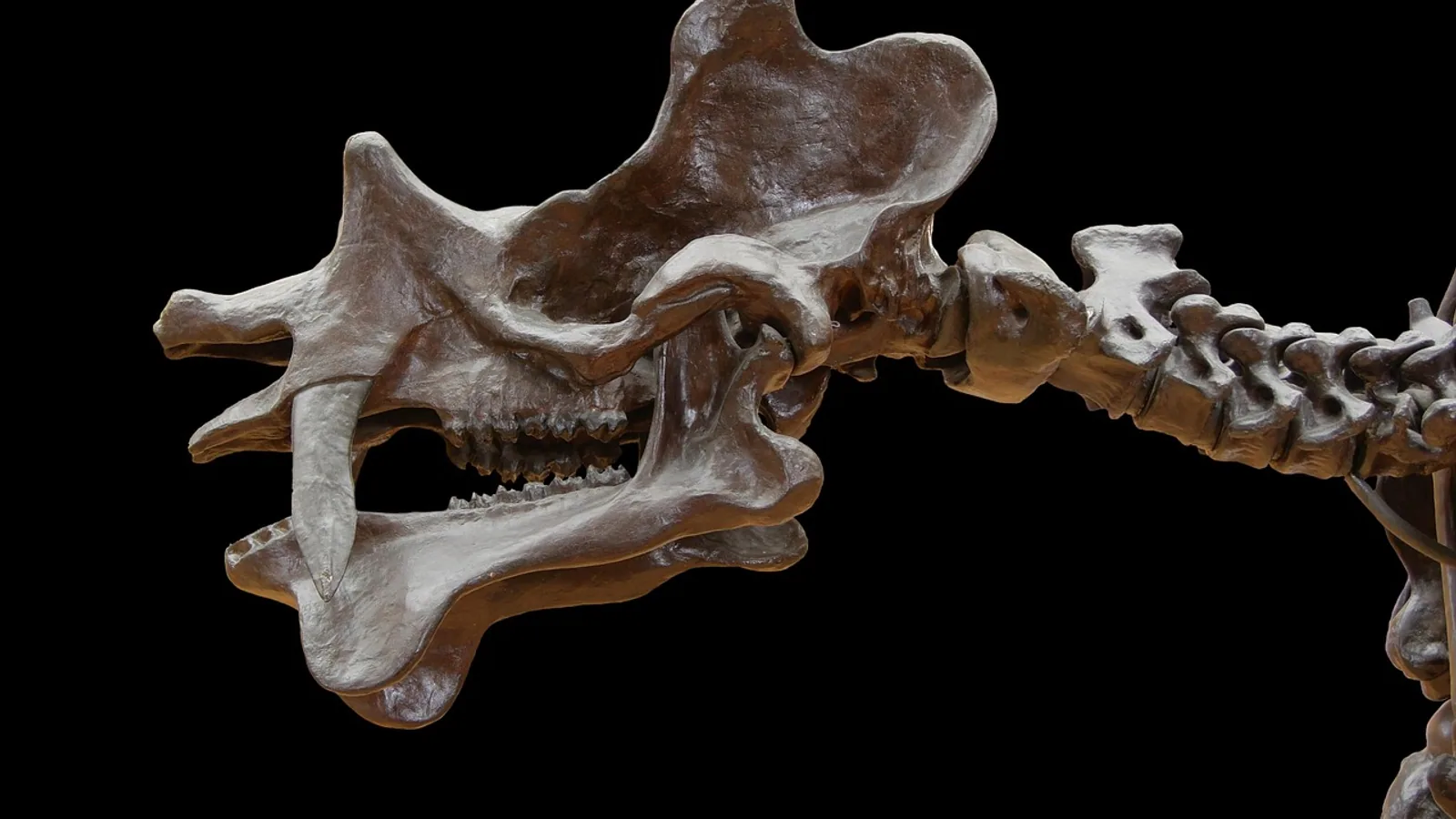
The spinal column is the segmented, dorsal axial skeleton that surrounds the spinal cord and transmits body weight from the head and trunk to the limbs. Each segment, a vertebra, is built from a body, an arch, and a set of processes that vary in shape and number by region and species.
This article is educational and is not a substitute for veterinary diagnosis or treatment.
Why the Vertebral Column Matters
The vertebral column is the mechanical backbone of the body and the bony protector of the spinal cord. Every neurologic examination you perform in a dog or cat begins with a mental map of the vertebral regions, because the clinical signs of a spinal lesion depend on which cord segments sit at which vertebral levels. In the Korean water deer and Siberian roe deer, researchers found that the cervical enlargement of the spinal cord spans C6 to T1 and the lumbar enlargement spans L4 to S1, with the spinal cord and vertebral column growing at different rates so that the cord ends well cranial to the end of the vertebral canal [1]. That mismatch between cord and bone is the reason a vertebral fracture at L4 can injure cord segments that are physically located at a different level.
Vertebral anatomy also drives surgical planning. A lesion of the C2 vertebral body, for example, sits next to the vertebral artery, the spinal cord, and the atlantoaxial joint, so surgeons must preserve the odontoid process when it is intact and use it as an anchor for reconstruction [2]. At the other end of the column, a collapsed L5 vertebral body creates spinopelvic malalignment that changes how the whole column bears load [3]. The same regional logic applies to every species you will examine.
The Five Vertebral Regions
Vertebrae are grouped into five regions, named from cranial to caudal: cervical, thoracic, lumbar, sacral, and coccygeal (caudal). The number of vertebrae in each region is written as a vertebral formula, conventionally in the order C-T-L-S-Cd. Humans have the formula C7 T12 L5 S5, with the sacral vertebrae fused into a single sacrum and a variable number of coccygeal vertebrae. Dogs and cats share the formula C7 T13 L7 S3. Horses have C7 T18 L6 S5. All of these counts describe the number of vertebrae, not the number of spinal cord segments, and the two do not always match.
Cervical Vertebrae
The cervical region sits between the skull and the first rib-bearing vertebra. Mammals almost universally have seven cervical vertebrae, regardless of neck length, so a giraffe and a mouse both have C7. The two exceptions among mammals are the sloths (genera Bradypus and Choloepus), which have more, and the manatees, which have fewer than seven. This conservation is one of the most reliable rules in comparative anatomy and is the reason a long-necked horse still has the same cervical count as a short-necked cat.
The first two cervical vertebrae are specialized and do not look like the rest. The atlas (C1) is a ring-shaped vertebra with wide transverse processes and no distinct body. The axis (C2) carries the dens, or odontoid process, which projects cranially into the atlas and forms the pivot for head rotation. The atlas and axis are responsible for neck flexion, extension, and rotation and provide insertion points for muscles and tendons [4]. In the Persian cat, radiographic study showed that the atlas has the greatest transverse process width of all cervical vertebrae, while C3 has the shortest spinous process and C7 the tallest [5]. Those regional differences matter when you are reading cervical radiographs and deciding whether a process looks abnormal.
The remaining cervical vertebrae, C3 through C7, are shorter and more uniform. Their transverse processes are perforated by the transverse foramen, which transmits the vertebral artery. This is why a cervical pedicle lesion adjacent to the vertebral artery is a surgical problem: the artery runs through the bone itself [6]. In primates, the levator scapulae muscle attaches to the cervical vertebrae and is innervated by branches of C3 through C8, with C4 through C7 consistently contributing [7]. That innervation pattern is a reminder that cervical vertebrae are not just structural, they are the origin points for the muscles that move the shoulder.
Thoracic Vertebrae
Thoracic vertebrae are the rib-bearing vertebrae. Each one articulates with a pair of ribs through facets on the body and the transverse process. The thoracic region is the least mobile part of the column in most domestic mammals because the ribs and sternum form a rigid box around the heart and lungs. Thoracic vertebrae have long, overlapping spinous processes that slope caudally in the cranial thoracic region and then reverse direction near the anticlinal vertebra, which is the vertebra whose spinous process is vertical and marks the transition toward the lumbar pattern.
The number of thoracic vertebrae varies more than the cervical count. Dogs and cats have 13, horses have 18, and humans have 12. The extra thoracic vertebrae in the horse reflect the long trunk and the large thoracic cavity needed for a running animal with a big heart and lungs.
Lumbar Vertebrae
Lumbar vertebrae are the workhorses of the caudal trunk. They have large, blocky bodies, broad transverse processes that are actually fused ribs in some species, and stout spinous processes that point cranially. The lumbar region is the most flexible part of the column in the sagittal plane, which is why dogs and cats can arch and extend their backs so dramatically. Dogs and cats have seven lumbar vertebrae, horses have six, and humans have five.
The lumbosacral junction is a high-stress region. In the spiny mouse, researchers found that the lumbosacral formula varies within a single species, with two main patterns described as VL5-VS5 and VL6-VS4, and that shifting the first sacral vertebra into a sixth lumbar vertebra changes the internal organization of the spinal cord even when the total number of lumbosacral vertebrae stays the same [8]. The L6 segment in the VL6-VS4 group took on intermediate characteristics between L5 and S1, and the gray matter showed a smoother morphological transition. This is a clean demonstration that vertebral formula and spinal cord anatomy are coupled, not independent.
Sacral Vertebrae
Sacral vertebrae are fused into a single sacrum in most mammals. The sacrum articulates with the ilium of the pelvis through the sacroiliac joint and transmits the weight of the trunk into the hind limbs. Dogs and cats have three sacral vertebrae, horses have five, and humans have five. The sacrum is the anchor point for the pelvic girdle and the origin of much of the musculature of the hind limb and tail.
Coccygeal (Caudal) Vertebrae
Coccygeal vertebrae form the tail. Their number is highly variable even within a species, and they progressively lose their arch and processes as they move caudally until the terminal vertebrae are little more than small rods of bone. In the salt marsh harvest mouse, researchers found that the third caudal vertebra is deeper, the longest tail vertebra is located more caudally, and the tail vertebrae are longer craniocaudally than in the western harvest mouse, features associated with scansorial (climbing) locomotion that help reduce body rotation and improve contact with the substrate [9]. Tail vertebrae are therefore not vestigial, they are functionally tuned to the animal's locomotor ecology.
Summary Table: Vertebral Regions and Counts
| Region | Human | Dog | Cat | Horse | Distinguishing features |
|---|---|---|---|---|---|
| Cervical | 7 | 7 | 7 | 7 | Atlas and axis specialized. Transverse foramina transmit vertebral artery. |
| Thoracic | 12 | 13 | 13 | 18 | Rib-bearing. Long overlapping spinous processes. Anticlinal vertebra. |
| Lumbar | 5 | 7 | 7 | 6 | Large bodies, broad transverse processes, cranially directed spinous processes. |
| Sacral | 5 (fused) | 3 (fused) | 3 (fused) | 5 (fused) | Fused into sacrum. Articulates with ilium. |
| Coccygeal | Variable | Variable | Variable | Variable | Tail vertebrae. Lose arch and processes caudally. |
Typical Vertebral Structure
Every vertebra, regardless of region, is built from the same basic parts. Learn these once and you can describe any vertebra in any species.
The Vertebral Body (Centrum)
The body, or centrum, is the thick, cylindrical mass on the ventral side of the vertebra. It is the weight-bearing element and the site of the intervertebral disc attachment. Bodies are shortest and most mobile in the cervical region, largest and most rigid in the lumbar region, and fused in the sacrum. In the Luristan newt, a micro-CT study found that vertebral body height peaked at trunk vertebra T10 and vertebral body length peaked at T12, showing that even within one region the body dimensions follow a gradient [10]. The same principle applies in mammals: body size and shape change gradually along the column.
The Vertebral Arch
The arch sits dorsal to the body and encloses the vertebral foramen, the hole through which the spinal cord passes. The arch is made of two pedicles and two laminae. The pedicles are the short, thick pillars that connect the arch to the body. The laminae are the flat plates that meet in the midline dorsally to close the arch. In a cervical pedicle lesion, the pathology sits in the pillar that connects the body to the arch, which is why it is close to both the vertebral artery and the spinal cord [6].
The Spinous Process
The spinous process projects dorsally from the junction of the two laminae. It is the lever arm for the epaxial muscles and the attachment point for the supraspinous and interspinous ligaments. Spinous process height varies enormously by region. In the Persian cat, C7 has the tallest spinous process of the cervical vertebrae and C3 the shortest [5]. In the Luristan newt, spinous process height peaked at caudal vertebra Cd3 [10]. The general rule is that spinous processes are tallest where the muscles need the most leverage, which is the cranial thoracic region in mammals.
The Transverse Processes
Transverse processes project laterally from the junction of the body and the arch. In the cervical region they are perforated by the transverse foramen. In the thoracic region they carry facets for the ribs. In the lumbar region they are broad and plate-like. The atlas has the widest transverse processes of any cervical vertebra in the Persian cat, significantly wider than the others [5]. Transverse processes are the attachment points for muscles that rotate and bend the column.
The Articular Facets
Each vertebra has cranial and caudal articular facets, also called zygapophyses, that form the synovial joints between adjacent vertebrae. The orientation of these facets determines the direction of movement allowed at each level. Cervical facets are oriented to permit rotation and flexion. Thoracic facets are oriented to limit movement. Lumbar facets are oriented to permit flexion and extension while limiting rotation. The facet joints are the reason the column bends more easily in some directions than others at each level.
How Vertebral Structure Is Studied
Vertebral anatomy is studied by gross dissection, radiography, computed tomography (CT), and micro-CT. Plain radiography remains the first-line imaging method in practice because it is fast and widely available. In the Persian cat study, researchers used dorsoventral, ventrodorsal, and left and right lateral recumbency radiographs to establish a normal reference range for cervical vertebral measurements [5]. CT and micro-CT add cross-sectional detail and are used when the cortical bone, the spinal canal, or the internal structure of the vertebra needs to be assessed. In the basilosaurid whale vertebra study, high-quality CT scans were used to differentiate morphotypes and to describe the inner structure, which consisted of a multi-layered cortex of periosteal bone surrounding two cones of endosteal bone [11]. That internal architecture is not visible on plain film.
Morphometric analysis is the quantitative side of vertebral study. Researchers measure vertebral body height, vertebral body length, spinous process height, spinal canal width, and spinal canal height, then compare those values across regions and species [10]. In the Syrian hamster, renal position was assessed relative to the second lumbar vertebra, and the length of the L2 body was used as an internal scale for organ size [12]. Vertebral landmarks are therefore used as reference points for structures far from the spine itself.
Comparative and Clinical Relevance
The vertebral formula is the first thing to check when you suspect a congenital or developmental spinal anomaly. A dog with a transitional vertebra at the lumbosacral junction may have a formula that does not match the textbook C7 T13 L7 S3, and that variation can change the clinical picture. The spiny mouse work shows that a shift from VL5-VS5 to VL6-VS4 alters the spinal cord itself, with the L6 segment taking on intermediate characteristics and the gray matter showing a smoother transition [8]. A transitional vertebra is therefore not just a radiographic curiosity, it can be associated with real differences in cord organization.
Species differences in vertebral count also affect how you interpret spinal cord lesions. The Korean water deer and Siberian roe deer study found that the cervical enlargement spans C6 to T1 and the lumbar enlargement spans L4 to S1, and that the positional relationship between cord segments and vertebrae shifts around these enlargements [1]. In a dog with a T13 vertebral fracture, the cord segment at risk may be a lumbar segment because the cord ends cranial to the end of the vertebral canal. This is the anatomical basis for the neurologic rule that the lesion is often one to two vertebrae cranial to the site of the bony injury.
Surgical anatomy is region-specific. At C2, the odontoid process is the key landmark. When the tip of the dens is preserved despite partial destruction of the C2 body, it remains a reliable anchor point for reconstruction, and surgeons can place an anterior bone graft and supplement it with posterior fixation [2]. At the lumbosacral junction, the biomechanical demands are high, and a pathological fracture at L5 can require anterior column reconstruction plus spinopelvic fixation from L3 to the second sacral alar-iliac level [3]. The vertebral regions are not interchangeable surgical targets.
Vertebral morphology also tracks ecology. In delphinids, vertebral shape is influenced by allometry, ecology, and phylogeny, with the relative importance of each signal differing along the column. The anterior thorax, posterior thorax, and synclinal point diversify mainly with size and habitat, while the mid-torso and tail stock retain strong phylogenetic signals [13]. In other words, the same vertebra can be shaped by different pressures depending on where it sits in the column.
Clinical Relevance, Limitations and Common Mistakes
The most common mistake is assuming that vertebral number equals spinal cord segment number. It does not. The cord is shorter than the column, and the mismatch increases caudally. Another frequent error is treating the vertebral formula as fixed within a species. Intraspecific variation is real and has been documented in the lumbosacral region of the spiny mouse [8]. A third mistake is ignoring the transverse foramen when planning a cervical approach. The vertebral artery runs through it, and a lesion of the cervical pedicle sits directly adjacent to that vessel [6].
A fourth mistake is reading a single radiograph without a reference range. Normal vertebral dimensions vary by region, by breed, and by sex. In the Syrian hamster, sex had a significant effect on the radiographic and anatomical length of the second lumbar vertebral body, with females generally showing higher values than males [12]. Without a size-matched reference, a normal variant can look like pathology.
A fifth mistake is forgetting that the sacrum is a fusion of multiple vertebrae. In the dog and cat it is three, in the horse and human it is five. Counting the sacrum as one vertebra is correct for the formula but wrong for understanding its development and its articulations.
Individual cases need a veterinarian. The anatomy in this article is a framework for understanding, not a diagnostic protocol.
Quick Review
- The five vertebral regions are cervical, thoracic, lumbar, sacral, and coccygeal.
- Every vertebra has a body, an arch (pedicles and laminae), spinous and transverse processes, and articular facets.
- All mammals have seven cervical vertebrae except sloths and manatees.
- Vertebral formulas: human C7 T12 L5 S5, dog C7 T13 L7 S3, cat C7 T13 L7 S3, horse C7 T18 L6 S5.
- The atlas (C1) and axis (C2) are specialized for head movement, and the dens of C2 is a key surgical landmark.
- The spinal cord is shorter than the vertebral column, so cord segments do not align one-to-one with vertebrae.
- Vertebral formula can vary within a species, and that variation can be coupled to spinal cord organization.
Frequently Asked Questions
How many vertebrae does a dog have?
A dog has 7 cervical, 13 thoracic, 7 lumbar, and 3 sacral vertebrae, plus a variable number of coccygeal vertebrae. The standard formula is C7 T13 L7 S3.
Do all mammals have seven cervical vertebrae?
Almost all do. The exceptions are sloths, which have more than seven, and manatees, which have fewer than seven. Neck length does not change the count.
What is the difference between the atlas and the axis?
The atlas (C1) is a ring-shaped vertebra with no distinct body and wide transverse processes. The axis (C2) has the dens, or odontoid process, which projects into the atlas and forms the pivot for head rotation.
Why does the spinal cord end before the vertebral column does?
The spinal cord and vertebral column grow at different rates, so the cord ends cranial to the end of the vertebral canal. This is why a bony lesion and the cord segment it injures are often at different levels.
What is a vertebral formula?
A vertebral formula is the count of vertebrae in each region, written in the order cervical, thoracic, lumbar, sacral, coccygeal. Examples include C7 T13 L7 S3 for the dog and C7 T18 L6 S5 for the horse.
Can vertebral number vary within a species?
Yes. Intraspecific variation has been documented in the lumbosacral region of the spiny mouse, where two main formulas (VL5-VS5 and VL6-VS4) occur and are associated with differences in spinal cord organization.
Related Articles
- Affinity Column Chromatography: Principles and Applications
- Affinity Chromatography for Protein Purification: Principles and Column Selection
- Canine Spinal Cord Anatomy and Segmental Innervation
- Anesthesia for Patients with Neurologic Disease: Intracranial and Spinal
- RNA Extraction from Cells: Choosing Between Column-Based and TRIzol Methods
- Dachshund Health Problems: IVDD Grade 1-5 Management and Spinal Surgery
- Vertebral Column: Vertebra Anatomy and Regions Explained
- Condyloid Joint: Structure and Movement
- Papillary Dermis: Structure and Function Explained
Sources
- Spinal Segmental Position Relative to the Vertebrae in Korean Water Deer and Siberian Roe Deer.
- Partial C2 corpectomy with iliac/fibula bone graft and posterior instrumented fusion for C2 vertebral body lesions: An innovative approach.
- Stentoplasty-assisted Anterior Column Reconstruction with Navigation-guided Spinopelvic Fixation in an L5 Pathological Fracture Secondary to Vertebral Hemangioma - A Case Report.
- Comparative anatomy and evolution of the atlantoaxial complex in the fossorial lineage Amphisbaenia (Squamata: Lacertoidea).
- Evaluation of Radiological and Anatomical Features of Cervical Vertebrae in Adult Persian Cat.
- O-arm Navigation-assisted Excision of Cervical Osteoid Osteoma Adjacent to the Vertebral Artery: Technical Note and Case Report.
- Comparative anatomy of the levator scapulae, serratus anterior and rhomboid muscles in primates: Morphological adaptations related to the primate locomotion evolution.
- Vertebral formula variability is coupled with spinal cord anatomy changes in the spiny mice.
- Comparative skeletal anatomy of salt marsh and western harvest mice in relation to locomotor ecology.
- Morphometrical study of Luristan newt (Neurergus kaiseri) vertebral column with micro-CT scan.
- A vertebra of a small species of Pachycetus from the North Sea and its inner structure and vascularity compared with other basilosaurid vertebrae from the same site.
- Assessment of Renal Measurements and Position in the Syrian Hamster (Mesocricetus auratus) Using Survey Radiography and In Situ Macroscopic Anatomy.
- Evolutionary trends in the vertebral morphology of extant Delphinidae.